696
Journal of Chinese Pharmaceutical Scienceshttp://www.jcps.ac.cn
Synthesis of
N
-
(phenylcarbamothioyl)
-
benzamide derivatives and their
cytotoxic activity against MCF
-
7 cells
GGGGGGGGGGGG
Dini Kesuma
1,2*, Siswandono
2, Bambang Tri Purwanto
2, Marcellino Rudyanto
2GGGG
GGG
GGG
1. Faculty of Pharmacy, University of Surabaya, Jalan Kalirungkut, Surabaya, East Java 60293, Indonesia 2. Faculty of Pharmacy, University of Airlangga, Jalan Dharmawangsa, Surabaya, East Java 60286, IndonesiaG
Abstract: Cancer is one of the leading causes of death both in developing countries and across the globe. In Indonesia, cancer ranks as the fifth primary cause of death following heart disease, stroke, respiratory tract and diarrhea. Therefore, studies on thiourea derivative compounds as anticancer agents have been profoundly conducted but still require further continuous development. In the present study, we aimed to synthesize new anticancer compounds of N-(phenylcarbamothioyl)-benzamide derivatives, namely N-(phenylcarbamothioyl)-4-bromobenzamide and N-(phenylcarbamothioyl)-4-fluorobenzamide compounds and assess their activities against MCF-7 breast cancer cells. The initial step was to predict the drug-receptor activity through docking between the tested compounds using epidermal growth factor receptor (EGFR) (PDB code: 1M17). The compounds were futher synthesized from the reactions between benzoyl chloride derivatives and N-phenylthiourea. The structures of the new compounds were identified using FTIR, 1H NMR, 13C NMR and mass spectra. The cytotoxic activities (IC50) to breast cancer
cells of MCF-7 N-(phenylcarbamothioyl)-4-bromobenzamide compound and N-(phenylcarbamothioyl)-4-fluorobenzamide were 0.27 mM and 0.31 mM, respectively. These two new compounds had better cytotoxic activities than those of the current hydroxyurea-based anticancer drugs (the reference compound) with an IC50 value of 9.76 mM. Furthermore, these two new compounds were not toxic to Vero normal cells. Therefore, they possessed tremendous potentials as the candidates for new drugs against breast cancer.
GG
Keywords: N-(phenylcarbamothioyl)-benzamide derivatives; Cytotoxic; MCF-7 cells; EGFR
G
GGCLC number: R916 Document code: A Article ID: 1003–1057(2018)10–696–07
Received: 2018-06-12, Revised: 2018-08-15, Accepted: 2018-09-20. Foundation items: Directorate General of Resources for Science, Technology and Higher Education of Ministry of Research, Technology and Higher Education (KEMRISTEK DIKTI) with scheme of scholarship funding for PhD program at University of Airlangga, Surabaya, Indonesia.
*
Corresponding author. Tel.: +62312981110 E-mail: dinikesuma@gmail.com
http://dx.doi.org/10.5246/jcps.2018.10.071
1. Introduction
The International Agency for Research on Cancer
(IARC) has found that there were 14 067 894 new
cases of cancer and 8 201 575 cancer-related deaths
worldwide in 2012. Both lung and breast cancer are
the leading causes of death compared with other types
of cancer, with breast cancer ranks the top, especially
in women. Ironically, it has been proposed that the
prevalence of breast cancer is increased, and it becomes
one of serious problems in healthcare systems globally[1]. Thiourea is an organic compound consisting of
carbon, nitrogen, sulfur and hydrogen atoms. The
compound shares similarities to urea except for the
oxygen atoms, which are then replaced by sulfur.
Hydroxyurea, nitrosourea and 5-fluorouracil are urea
compounds still used today as anticancer drugs[2–4]. However, it has been widely reported that patients,
particularly those with essential thrombocythaemia,
have some adverse drug reactions when treated using
hydroxyurea[5,6]. This results in the diminishing numbers of the clinical use of hydroxyurea, although, as a
matter of fact, it is still used as a DNA replication
inhibitor in biochemical research and development of
Copyright © 2018 Journal of Chinese Pharmaceutical Sciences, School of Pharmaceutical Sciences, Peking University http://www.jcps.ac.cn
www.jcps.ac.cn
moth uorobenzami he drug-receptor acticeptor ac ) (PDB code: 1M17). The cde: 1M17). The N
-Nphenylthiourea. nylthiourea. The structures oe struc tra. The cytotoxic activities (ICotoxic activities ( 50)
d and NN-(phenylcarbamothioyl)henylcarbamot -4 fl nds had better cytotoxic activitiesbetter cytotoxic ound) with an ICth an IC5050value of 9.76 mvalue of efore, they possessed tremendous po efore, they possessed tremen
zamide derivatives; Cytotoxic; MCFatives; Cytotoxic; M
ocument code:code:A A ArticleA
ww
697
the anticancer drugs[7]. Findings of research data have suggested that the hydrophilic properties of hydroxyurea
are associated with the less optimal activity of this
compound due to its poor membrane penetration ability.
Therefore, it can be suggested that development of new
anticancer drugs of urea and thiourea derivatives which have
hydrophobic properties will result in better membrane
penetration ability[8–11]. Li[9] has synthesized urea and thiourea derivatives and proved that phenylthiourea
derivatives, N-(5-chloro-2-hydroxybenzyl)-N-(4
-hydroxybenzyl)-N'-phenylthiourea, have cytotoxic activity
on MCF-7 cells by inhibiting EGFR and HER-2.
Nakisah[10] has also shown that the compounds of
2-[3-(2-methyl benzoyl)-thioureido]-acetic acid and
2-[3-(4-methyl benzoyl)-thioureido]-acetic acid have
cytotoxic activity against MCF-7 cells as well.
In this present study, we synthesized N-(phenyl
-carbamothioyl)-4-bromobenzamide/4-Br-BPCT and
N-(phenylcarbamothioyl)4-fluorobenzamide/4-F-BPCT.
The presence of the substituents of bromo and fluoro at
the benzoyl ring could enhance the lipophilic and
electronic properties of these two compounds compared
with their lead compound (BPCT). As a result, the drug
and receptor bonds were improved, thus leading to the
increased activities among these two compounds[12–14]. This study was different from the study of Li[9] in regards to the different modification substrates. Study
by Li has modified the benzyl groups, while our
present study modified the benzoyl groups. This study
was initiated with activity predictions using molecular
modeling in silico and docking test compound with EGF
(epidermal growth factor) Receptor PDB code: 1M17
of Protein Data Bank (PDB). Molecular modeling
was analyzed using Molegro Virtual Docker (MVD)
program 5.5[15]. The activity prediction was carried out using EGFR receptor (1M17) because its ligand is
Erlotinib[9], an anticancer drug which inhibits the EGFR pathway[4,9,16]. The test compounds were synthesized from
N-phenylthiourea with R-benzoyl chloride (R = 4-Br and
4-F) using an acyl nucleophilic substitution reaction[17,18].
The structures of the synthesized compounds were
then identified with IR spectrophotometers, 1H NMR
Spectrometers, 13C NMR and mass spectrometers[19]. The cytotoxic activities of the two test compounds
were observed through cytotoxic assay using MTT method
(3-(4,5-dimetylthiazole-2-yl)-2,5-diphenyltetrazolium
bromide) in vitro on breast cancer cells MCF-7 and
Vero normal cells[20]. After the identification of
cytotoxic activity test on MCF-7 cells, the IC50 was
then compared with hydroxyurea. Ourstudy might
provide candidates of anticancer drugs from new
thiourea derivatives, which have potent cytotoxic
activity in breast cancer MCF-7 cells.
2. Materials and methods
2.1. Materials and instruments
Materials for synthesis included phenylthiourea,
4-bromobenzoyl chloride, 4-fluorobenzoyl chloride
(Sigma Aldrich), tetrahydrofuran (THF), triethylamine
(TEA), acetone, ethyl acetate, n-hexane, chloroform
and ethanol. Materials for activity test included test
compounds and HU, cell cultures of MCF-7 and Vero,
culture medium DMEM and M199, buffer saline
phosphate (PBS), FBS (fetal bovine serum), trypsin,
penicillin-streptomycin, fungizon, DMSO, 0.5 mg/mL
MTT (3-(4,5-dimetylthiazol-2-yl)-2,5-diphenyltetrazolium
bromide) and SDS 10% in HCl 0.01 N. Glass tools for
synthesis were Corning hot plate P351, Fisher-John
Electrothermal Mel-Temp, Jasco FT-IR 5300
Spectrophotometer, 1H NMR Spectrometer and 13C NMR Agilent 500 MHz with DD2 console system at 500 MHz
(1H) and 125 MHz (13C) and mass Spectrometer (Waters). Tools for cytotoxic test included 5% CO2 incubator,
LAF, micropipet with blue and yellow tip, test tube,
Dini Kesuma et al. / J. Chin. Pharm. Sci. 2018, 27 (10), 696–702
Copyright © 2018 Journal of Chinese Pharmaceutical Sciences, School of Pharmaceutical Sciences, Peking University http://www.jcps.ac.cn
www.jcps.ac.cn
yl
-PCT and
mide/4-FF--BPCBPCT.
ts of bromo and fluoro at omo and fluoro at
uld enhance the lipophilic andance the lipophilic a
erties of these two compounds cof these two comp
ead compound (BPCT). As acompound (BPCT)
bonds were improvonds wer
ies amon
cn
xyurea.
f anticancer druncer d
atives, which have potenhich have pot
in breast cancer MCFcancer MCF--7 cells.7 c
2. Materials and me 2. Material
698
vortex, 96-well microplate, conical tube, inverted
microscope, hemocytometer and ELISA-reader. Molecular
modeling: ChemBioDraw Ultra 15.0, Molegro Virtual
Docker (MVD) 5.5.
2.2. Methods
2.2.1. Molecular modeling
Activities of new compounds were predicted using
molecular modeling in silico, and docking of two test
compounds with EGFR (epidermal growth factor receptor)
PDB code: 1M17 of Protein Data Bank (PDB) was
carried out using computer program Molegro Virtual
Docker (MVD) 5.5. The EGFR receptor (1M17) was
chosen because its ligand is Erlotinib[9], an anticancer
drug, which inhibits EGFR pathway[4]. Hydroxyurea
was used as a reference compound.
2.2.2. Synthesis of 4-Br-BPCT and 4-F-BPCT
compounds
N-Phenylthiourea was mixed with THF and TEA
in a round flask, and a solution of R-benzoyl chloride
(R = 4-Br; 4-F) in THF was added into the mixture
over the ice bath through a dropping funnel using a
magnetic stirrer. The mixture was refluxed and stirred
on top of a water bath. The reaction was terminated
when the stain in the TLC formed a single stain. After
the termination, THF was evaporated in the rotary
evaporator. Then recrystallization was carried out[21]. The structures of new compounds were identified using
spectroscopy: infrared, 1H NMR, 13C NMR and HRMS[19].
2.2.3. Cytotoxicity test of MTT assay method
MCF-7 and M199 cells were seeded into 96-well
plates and then incubated for 24 h in 5% CO2
incubators. Furthermore, test solutions, positive and
negative controls of various concentrations were added.
Each concentration was replicated for three times. Wells
containing no cells and only filled with medium were used
as medium controls. At the end of incubation, each well
was added with 100 μL of 0.5 mg/mL MTT, followed
by incubation for 3 h, and then the MTT reaction was
discontinued by adding 100 μL of 10% SDS in 0.01 N
HCI into each well. The microplate was wrapped
in paper and incubated at 37 ºC for 24 h. The live
cells converted MTT into a dark blue formazan.
Elisa reader was utilized to identify the absorption at
λ = 595 nm. The IC50 values of the two test compounds
and the reference compound were obtained by using
probit analysis[20,21].
3. Results and discussion
Drug activity was predicted in silico, and the RS value
was used as the indicator. According to the result of in silico
test (Table 1), the values of RS BFTU, 4-Br-BPCT,
4-F-BPCT, RS HU were –76.9757, –85.4741, –83.5488
and –38.4495, respectively. The smaller RS value
indicated the more stable bonds between drug-receptors,
leading to better activities[22]. The RS values of the two test compounds were smaller than those of the lead
compound and the reference compound. The smaller
values suggested their better activities.
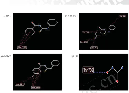
The numbers and types of amino acids involved are
listed in Table 2 and Figure 2. Based on the bonding of
drugs and amino acids, it was predicted that the greater
number of hydrogen bonds and steric bonds (Van der
Waals and Hydrophobic) resulted in the more stable
bonding between drugs and receptors, which might further
affect the greater biological activity. The 4-Br-BPCT
compound produced the largest number of bonds with
amino acids at the EGFR receptor. Therefore, it was
predicted that its activity as cytotoxic agents would
be better than 4-F-BPCT, BFTU and HU. The better
activity of the 4-Br-BPCT could be attributed to the
higher lipophilic value of 4-Br (0.86) compared with
4-F (0.14). Therefore, the membrane penetration of
4-Br-BPCT was better than 4-F- BPCT, and the activities
of 4-Br-BPCT were also improved[13,14].
Dini Kesuma et al. / J. Chin. Pharm. Sci. 2018, 27 (10), 696–702
Copyright © 2018 Journal of Chinese Pharmaceutical Sciences, School of Pharmaceutical Sciences, Peking University http://www.jcps.ac.cn
www.jcps.ac.cn
CT
th THF and TEA F and TEA
ion of R-benzoyl chloride benzoyl chloride
THF was added into the mixturs added into the
ath through a dropping funnelrough a droppin
The mixture was reflThe mixtu
bath. The rth
T
predicted in silico, and then silico, and th
e indicator. According to the result oor. According to the
Table 1), the values of RS BFT, the values of R
4-F-BPCT, RS HU were PCT, RS HU we –76.9
and
and –38.4495, respect38.4495,
indicated the mo indicated
leadingadingt
699
Dini Kesuma et al. / J. Chin. Pharm. Sci. 2018, 27 (10), 696–702
Compounds BPCT 4-Br-BPCT 4-F-BPCT HU
RS (kkal/mol) –76.9757 –85.4741 –83.5488 –38.4495
Cl O
N H H2N
S
Addition R
O Cl NH
S N H
Elimination
N H O
N H S
R R + HCl
R = 4-Br R = 4-F R = 4-Br
R = 4-F +
.. ..
..
..
Figure 1. Ligand interaction with amino acids at the EGFR binding sites where hydrogen bonds are indicated by blue dashed-lines, while steric interruptions are indicated by red dashed-lines: (a) BPCT compound, (b) 4-Br-BPCT compound, (c) 4-F-BPCT compound and (d) HU reference compound.G
Table 1. Rerank score (RS) value.
G
Compounds Amino acids
Thr 766 Val 702 Lys 721 Leu 764
BPCT 3S – – –
4-Br-BPCT 3S 1S 3S 1S
4-F-BPCT 4S – 1S –
HU 1H – – –
Table 2. Chemical and amino acid bonds involved in the interaction of 4-Br-BPCT and 4-F-BPCT compounds with EGFR (1M17).
G
Description: H: Hydrogen bond and S: Steric bond (Van der Waals and Hydrophobic).G
Figure 2. Reaction mechanism underlying the synthesis of 4-Br-BPCT and 4-F-BPCT.
G
(a) BPCT (b) 4-Br-BPCT
(c) 4-F-BPCT (d) HU
Copyright © 2018 Journal of Chinese Pharmaceutical Sciences, School of Pharmaceutical Sciences, Peking University http://www.jcps.ac.cn
www.jcps.ac.c
BPCT
BPCT
kal/mol) –76.9757
ww
j
ww
ww
sites where hydrogen bonds are indicatre hydrogen bonds a pound, (b) 4) 4--BrB-BPCT compound, (c) 4BPCT comp
ue.
700
The 4-Br-BPCT and 4-F-BPCT compounds were
synthesized from R-benzoyl chloride (R = 4-Br and 4-F)
with N-phenylthiourea in one stage. The two compounds
were yellow light crystals luster and insoluble in water.
The structure of the synthesized compounds was
identified by IR, 1H NMR, 13C NMR, and HRMS
spectroscopy as follows:
N-(Phenylcarbamothioyl)-4-bromobenzamide: It was
obtained as a yellow crystal, yield 67%, m.p. 132–133 ºC.
1H NMR (DMSO-d
Aromatic); 3328 and 1596 (NH strech sec. amides);
1077 and 830 (C=S). HRMS (m/z): C14H10N2OSBr
(M-H)– = 332.9687 and Calc. Mass = 332.9697.
N-(Phenylcarbamothioyl)-4-fluorobenzamide: It was
obtained as a yellow crystal, yield 45%, m.p. 123–124 ºC.
1H NMR (DMSO-d
(C=O amide), 1663 and 1498 (C=C Aromatic), 3269
and 1598 (NH strech sec. amides), 1108 and 805 (C=S).
HRMS (m/z): C14H10N2OSF (M-H)– = 273.0507 and
Calc. Mass = 273.0498.
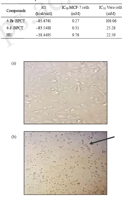
Table 3 presents the cytotoxic test results (IC50) of
the two test compounds in MCF-7 cancer cells and
Vero normal cells. IC50 values of two test compounds
were better than reference compound of hydroxyurea.
The IC50 value of the 4-Br-BPCT compound was better
than that of the 4-F-BPCT compound. This finding was
similar to the activity predictions performed using
in silico (Table 3). The smaller value of RS (stable
bonding of drugs and receptors) could result in the
better cytotoxic activities. In addition, the 4-Br-BPCT
compound also had a better lipophilic value (0.86)
compared with the 4-F-BPCT (0.14)[13,14].
Dini Kesuma et al. / J. Chin. Pharm. Sci. 2018, 27 (10), 696–702
Compounds RS
Figure 3. MCF-7 cells before administration of a test compound (4-Br-BPCT): living cells condition (a) and black arrow shows: MCF-7 cells after administration of a test compound (4-Br-BPCT) with a dose of 1000 μg/mL: the presence of dead cells after administration of a test compound (4-Br-BPCT) (b).
G
Copyright © 2018 Journal of Chinese Pharmaceutical Sciences, School of Pharmaceutical Sciences, Peking University http://www.jcps.ac.cn
www.jcps.ac.
r),
KBr), r), νmaxx and 1479 (C=C 79 (C=C
NH strech sec. amides); ech sec. amides);
. HRMS (S (m/zm/ ): C C1414HH100N2OSBrB
9687 and Calc. Mass = 332.969Calc. Mass
bamothioyl)amothioyl-4-fluoro
701
4. Conclusions
In this study, we synthesized two new compounds,
namely N-(phenylcarbamothioyl)-4-bromobenzamide and
N-(phenylcarbamothioyl)-4-fluorobenzamide. They had
in vitro cytotoxic activities against human breast
cancer cells (MCF-7), which were higher than those of
hydroxyurea-based anticancer drugs. The in silico
prediction results proved that the RS values of the two
new compounds were lower than those of the lead
compound and HU. The RS of 4-Br-BPCT compound
was lower than of 4-F-BPCT compound. The IC50
values of N-(phenylcarbamothioyl)-3-bromobenzamide
and N-(phenylcarbamothioyl)-4-fluorobenzamide were
0.27 mM and 0.31 mM, respectively, and both were
more active than hydroxyurea (IC50 = 9.76 mM). The
cytotoxic effect of 4-Br-BPCT was higher than that of
4-F-BPCT, and it may be related to 4-Br lipophilic
values, which were higher than 4-F. These two new
compounds were more suitable for binding enzymes
compared with hydroxyurea, as they had better
inhibitory activities. Collectively, these two new
compounds could be used as new targets because they
had toxic effects on cancer cells but not Vero normal
cells. Further studies are required to examine the
molecular mechanisms on EGFR receptor of these two
new compounds.
Acknowledgements
This study was supported by funding from Directorate
General of Resources for Science, Technology and Higher
Education of Ministry of Research, Technology and
Higher Education (KEMRISTEK DIKTI) with scheme
of scholarship funding for PhD program at University
of Airlangga, Surabaya, Indonesia.
References
[1] World Health Organization. Cancer. 2014. Retrieved from www.who.int on March 13, 2017.
[2] Bell, F.W.; Cantrell, A.S.; Hogberg, M.; Jaskunas, S.R.; Johansson, N.G.; Jordan, C.L; Kinnick M.D.; Lind P.; Morin Jr. J.M.; Noréen R.; Öberg B.; Palkowitz J.A.; Parrish C.A.; Pranc P.; Sahlberg C.; Ternansky R.J.;
Vasileff R.T.; Vrang L.; West S.J.; Zhang H.; Zhou, X.X. Phenethylthiazolethiourea (PETT) compounds, a new class of HIV-1 reverse transcriptase inhibitors 1. Synthesis
and basic structure-activity relationship studies of PETT analog. J. Med. Chem. 1995, 38, 4929–4936.
[3] Mutschler, E. Dynamics of Pharmacology and Toxicology
Drugs. Bandung. ITB. 1999, 56–62.
[4] Avendaño, C.; Menéndez, J.C. Medicinal Chemistry of
Anticancer Drugs. 2nd ed. Amsterdam: Elsevier. 2015,
15–19, 396–406.
[5] Barosi, G.; Besses, C.; Birgegard, G.; Briere, J.; Cervantes, F.; Finazzi, G.; Gisslinger H.; Griesshammer M.; Gugliotta L.; Harrison C.; Hasselbalch H.; Lengfelder E.; Reilly J.T.; Michiels J.J.; Barbui T. A unified definition
of clinical resistance/intolerance to hydroxyurea in essential thrombocythemia: results of a consensus
process by an international working group. Bioorg. Med. Chem. Lett. 2009, 19, 755–758.
[6] Tibes R.; Mesa R.A. Blood consult: resistant and
progressive essential thrombocythemia. Blood. 2001,
118, 240–242.
[7] Koç, A.; Wheeler, L.J.; Mathews, C.K.; Merrill, G.F.
Hydroxyurea arrests DNA replication by a mechanism that preserves basal dNTP pools. J. Biol. Chem. 2004,
279, 223–230.
Dini Kesuma et al. / J. Chin. Pharm. Sci. 2018, 27 (10), 696–702
Copyright © 2018 Journal of Chinese Pharmaceutical Sciences, School of Pharmaceutical Sciences, Peking University http://www.jcps.ac.cn
www.jcps.ac.cn
n that of
4-Br lipophilic lipophilic
an 4-F. These two new These two new
e suitable for binding enzymes le for binding enzym
h hydroxyurea, as they had oxyurea, as th
activities. Collectively, thivities. Collective
ould be used as neuld be us
s on can
riptase inh
activity relationship relationship Med. Chem. 199595, 38, 4929, 4929–4936
] Mutschler, E. Dynamics of Pharmaer, E. Dynamics of
Drugs. Bandung. ITB. Drugs. Bandung 19
[4] Avendaño, C4] Aven
702
[8] Bielenica, A.; Stefańska, J.; Stępień, K.; Napiórkowska, A.; Augustynowicz-Kopeć, E.; Sanna, G.; Madeddu S.; Boi S.;
Giliberti G.; Wrzosek M.; Struga M. Synthesis, cytotoxicity and antimicrobial activity of thiourea derivatives incorporating 3-(trifluoromethyl)phenyl moiety. Eur. J.
Med. Chem. 2015, 101, 111–125.
[9] Li, H.Q.; Yan, Y.; Shi, L.; Zhou, C.F.; Zhu, H. Synthesis and structure-activity relationships of N-benzyl-(X-2
-hydroxybenzyl)-Nʹ-phenylureas and thioureas as antitumor agents. Bioorg. Med. Chem. 2010, 18, 305–313.
[10] Nakisah, Tan, J.W.; dan Shukri, Y.M. Anti-Cancer Activities of Several Synthetic Carbonylthiourea Compounds on MCF-7 Cells, UMTAS. Malaysia. 2011.
[11] Song, D.Q.; Du, N.N.; Wang, Y.M.; He, W.Y.; Jiang,
E.Z.; Cheng, S.X.; Wang, Y.X.; Li, Y.H.; Wang, Y.P.; Li, X.; Jiang, J.D. Synthesis and activity evaluation of phenylurea derivatives as potent antitumor agents.
Bioorg. Med. Chem. 2009, 17, 3873–3878.
[12] Kar, A. Medicinal Chemistry, 4th ed. New Delhi: New
Age International Ltd Publishers. 2007, 794–810. [13] Siswandono, Development of New Drugs. Surabaya:
Airlangga University Press. 2014.
[14] Topliss, J.G. Utilization of Operational Schemes for Analog Synthesis in Drug Design. J. Med. Chem. 1972,
15, 1006–1009.
[15] Manual Software Molegro Virtual Docker. http:// www.molegro/mvd-technology.php. 2011.
[16] Tartarone A.; Lazzari, C.; Lerose, R.; Conteduca, V.; Improta, G.; Zupa, A.; Bulotta, A; Aieta, M; Gregorc, V. Mechanisms of resistance to EGFR tyrosine kinase inhibitors gefitinib/erlotinib and to ALK inhibitor crizotinib. Lung Canc. 2013, 81, 328–336.
[17] Clayden, J.; Greeves, N.; Warren, S.; Wothers, P. Organic
Chemistry, 2nd ed. New York: Oxford University Press.
2012, 279–289.
[18] McMurry, J.M. Fundamental of Organic Chemistry, 7th ed., Belmont: Brooks/Cole. 2011, 349.
[19] Pavia, D.L.; Lampman, G.M.; Kriz, G.S.; James R.; Vyvyan, J.R. Spectroscopy. 4th ed. Belmont: Brooks/Cole.
2009, 851–886.
[20] Cancer Chemoprevention Research Center Faculty of Pharmacy UGM (CCRC-UGM) Fixed procedure Cytotoxic Test Method MTT. 2012.
[21] Widiandani T.; Arifianti L.; Siswandono. Docking,
Synthesis, and Cytotoxicity Test Human Breast Cancer Cell Line T47D of N-(Allylcarbamothioyl)benzamide.
Int. J. Pharm. Clin. 2016, 8, 372–376.
[22] Hinchliffe, A. Molecular Modelling for Beginners, 2nd ed. Chichester: John Wiley and Sons Ltd.
2008.
Dini Kesuma et al. / J. Chin. Pharm. Sci. 2018, 27 (10), 696–702
Copyright © 2018 Journal of Chinese Pharmaceutical Sciences, School of Pharmaceutical Sciences, Peking University http://www.jcps.ac.cn
www.jcps.ac.cn
n of mor agents. agents. –3878.
istry, 4thed. New Delhi: New ed. New Delhi: New
l Ltd Publishers. blishers. 20072 , 794794–810. o, Development of New Drugvelopment of New
iversity Press. ersity Pre 2014
tilization
al of Organg
oks/Cole. e. 20112011, 349., 349. L.; Lampman, G.M.; Kriz, G.S.; Jpman, G.M.; Kriz, yvyan, J.R. R. Spectroscopy. Spectroscopy 44ththed. Belm
200909, 851, –886.86 [20
[20] Cancer Chemoprev] Cancer Che of Pharma
of

