Improved language performance subsequent to low-frequency
rTMS in patients with chronic non-fluent aphasia post-stroke
C. H. S. Barwood
a, B. E. Murdoch
a, B.-M. Whelan
a, D. Lloyd
a,b, S. Riek
b, J. D. O
Õ
Sullivan
c,
A. Coulthard
dand A. Wong
ca
Centre for Neurogenic Communication Disorders Research, School of Health and Rehabilitation Sciences, University of Queensland, Australia;bSchool of Human Movement Studies, University of Queensland, Australia;cDepartment of Neurology, Royal Brisbane and WomenÕs Hospital, Australia; anddDepartment of Medical Imaging, Royal Brisbane and WomenÕs Hospital, Australia
Keywords:
aphasia, language, stroke rehabilitation, transcranial magnetic stimulation (TMS)
Received 7 August 2010 Accepted 28 October 2010
Background: Low-frequency repetitive transcranial magnetic stimulation (rTMS) has emerged as a potential tool for neurorehabilitation and remediation of language in chronic non-fluent aphasia post-stroke. Inhibitory (1 Hz) rTMS has been applied to homologous language sites to facilitate behavioural language changes. Improvements in picture-naming performance and speech output over time have been reported. Methods: Low-frequency (1 Hz) rTMS was applied to six real stimulation and six sham placebo patients for 20 min per day, for 10 days, and behavioural language outcome measures were taken at baseline (pre-stimulation) and 2 months post-stim-ulation.
Results: The findings demonstrate treatment-related changes observed in the stimu-lation group when compared to the placebo control group at 2 months post-stimu-lation on naming performance as well as other aspects of expressive language and auditory comprehension.
Conclusions: These findings provide considerable evidence to support the theory of rTMS modulating mechanisms of transcallosal disinhibition in the aphasic brain and highlight the potential clinical applications for language rehabilitation post-stroke.
Introduction
Recent experimental trials of low-frequency repetitive transcranial magnetic stimulation (rTMS) applied to patients with chronic non-fluent aphasia have demon-strated its capacity to modulate and inhibit extraneous levels of right hemisphere (RH) activation in homolo-gous language sites, which may impede language recovery in some patients [1]. Overactivation in RH frontal regions including the inferior frontal gyrus (IFG) and the motor cortex has previously been iden-tified using functional neuroimaging in populations of persons with aphasia post-stroke. [1–4].
There is now considerable evidence to suggest that rTMS to the apical portion of BrocaÕs area homologue, pars triangularis (dorsal and posterior), can result in improved language function for patients with non-fluent aphasia. [5,6]. For example, improvements in behavioural language performance following 10 days of
low-frequency rTMS (1 Hz, inhibitory) has been dem-onstrated in a small group of patients with non-fluent aphasia [5]. Language improvements to date are reported primarily for functions related to picture naming in case studies and small patient samples (e.g. four patients) at 8 months post-stimulation and up to 43 months post-stimulation [2,5,7–9]. Hamilton et al. [6] suggest that the application of rTMS to the intact contralateral hemisphere may induce effects that gen-eralize beyond naming to propositional speech includ-ing spontaneously elicited speech usinclud-ing picture stimuli. With reference to the theory of transcallosal disin-hibition proposed by Heiss and Theil [10], rTMS has been applied to a language homologue in the RH to inhibit extraneous activity identified on previous func-tional neuroimaging studies in patients with non-fluent aphasia in the IFG [11,12] and in patients with left hemisphere (LH) brain tumours [13]. High levels of activation found in homologous language centres post-stroke suggest an interhemispheric shift in neural lan-guage networks to recruit undamaged neural resources in the unaffected hemisphere [14,15]. Recruitment of RH sites post-stroke represented by increased cortical activation is postulated to be less efficient in language
recovery than LH regions [10]. It is possible that mechanisms of transcallosal disinhibition may facilitate downregulation of overactive homologous language sites and upregulation of LH language networks, yielding functional language improvements over time. The theory of mutual inhibition of LH language sites in the lesioned brain has provided the framework for previous investigations into the effects of rTMS in chronic aphasia [2,5,6,9,16].
The present study aimed to elucidate the effects of rTMS on the aphasic brain at 2 months post-stimula-tion, with respect to behavioural language measures and to address some of the inadequacies of existing research in this field by improving on sample size numbers, utilizing state of the art neuronavigational techniques and including a placebo control group as a comparative measure of treatment group language changes over time with double-blinded assessments. This research aims to test current hypotheses regarding the ability of low-frequency rTMS to inhibit overacti-vation in the homologue to BrocaÕs area and induce behavioural language improvements in picture-naming function over time. It is proposed that the suppression of the right pars triangularis using rTMS will modulate prefrontal and temporo-parietal neural connections responsible for naming processes to facilitate behavio-ural improvements in naming performance and prop-ositional speech.
Materials and methods
Patients
Twelve (12) right-handed patients with chronic aphasia received rTMS. Handedness was assessed using the Edinburgh Handedness Inventory (Oldfield, 1971). All patients had suffered a left middle cerebral artery (MCA) stroke between 2–6 years previously and had residual language impairments. A double-blind meth-odology was employed where patients were randomly assigned to one of two groups either receiving real conditionTMS [n= 6, aged between 54 and 67 years; mean age (SD) = 60.8 (5.98) years; mean time post-stroke (SD) = 3.49 (1.27) years] or a placebo TMS (using a SHAM coil) [n= 6, aged between 51 and 85 years; mean age (SD) = 67 (13.11) years, mean time post-stroke (SD) = 3.46 (1.53) years]. Table 1 provides detailed biographical information about the patients who participated in the present study.
Patient recruitment and screening
Patients were recruited through two major metropoli-tan hospitals and university research databases in the
Greater Brisbane region, Australia. Ethical clearance was granted from the Royal Brisbane and WomenÕs Hospital, Princess Alexandra Hospital and the Uni-versity of Queensland ethical review boards. All patients gave informed consent to participate in this research, and procedures conformed with the declara-tion of Helsinki. Patient exclusion criteria for rTMS were derived from guidelines published by Wasserman [17]. Participants did not receive speech therapy services during their participation in this research study.
Neuroimaging and cortical target identification
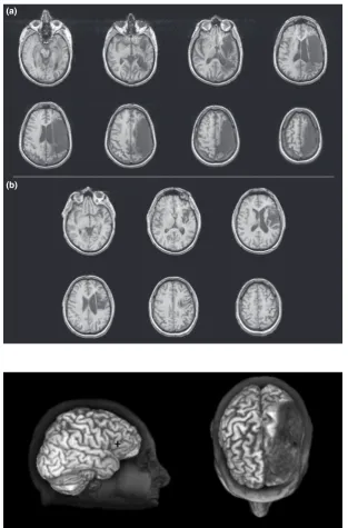
Prior to stimulation, all subjects were required to undertake a 3-Tesla structural magnetic resonance imaging (MRI) scan to aid target localization and to provide additional information on the size, extent and composition of the lesion. Figure 1 shows examples of MRIs from two of the stimulated patients. Acquisition parameters were as follows: TR = 19 msec, TE = 4.92 msec, matrix size = 256·256, number of slices = 128, slice thickness = 2 mm. The study neu-roradiologist provided a radiological report to specify affected neural regions. A computerized MRI visuali-zation and analysis system [18] was then employed to visually mark target areas [homologue to Brodmann area 45 (BA 45) and the right motor hand knob] with a crosshair (i.e. +).
The language-related cortical area in the contralat-eral (right) hemisphere (i.e., homologue to BA 45) was established as the stimulation target. The foot of the third frontal convolution represents the classical defi-nition of BrocaÕs area [19]. Within the left IFG, this region incorporates the structures pars triangularis and pars opercularis, also known as BA 45 and BA 44, respectively. The cytoarchitecture of BrocaÕs area was informed by papers from Amunts et al. [20,21]. Pre-vious studies have revealed TMS of BA 45 (pars triangularis) and not BA 44 (pars opercularis) signifi-cantly increases the accuracy and decreases reaction time of picture naming in patients with aphasia [5,8]. The apical portion of right BA 45 was therefore selected as the specific stimulation point for the present study, delimited ventrally by the horizontal ramus of the Sylvian fissure [22] and laterally by the vertical ramus of Sylvian fissure. Figure 2 shows the stimu-lation target marked on 3D reconstruction of the patientÕs brain.
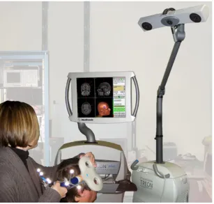
Neuronavigational techniques
guidance system designed for neurosurgical application but adapted for this research via several custom soft-ware modifications. Figure 3 shows the setup of the neuronavigational system. Within the Stealthstation, theÔhot spotÕwas converted to a target plan. Reflective
markers attached to the subjectÕs head were detected via an infrared tracking camera, subsequently logging the position of the head in space. The software then co-registered the position of the subjectÕs head with the MRI data set.
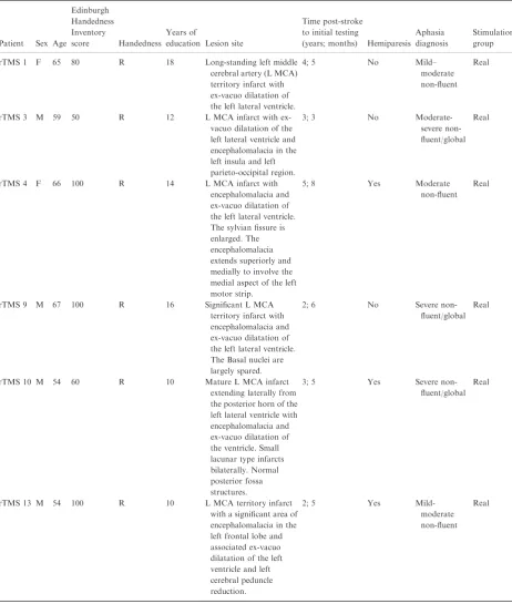
Table 1Patient biographical information
Patient Sex Age
Edinburgh Handedness Inventory
score Handedness Years of
education Lesion site
Time post-stroke to initial testing
(years; months) Hemiparesis Aphasia diagnosis
Stimulation group rTMS 1 F 65 80 R 18 Long-standing left middle
cerebral artery (L MCA) territory infarct with ex-vacuo dilatation of the left lateral ventricle.
4; 5 No Mild– moderate non-fluent
Real
rTMS 3 M 59 50 R 12 L MCA infarct with ex-vacuo dilatation of the left lateral ventricle and encephalomalacia in the left insula and left parieto-occipital region.
3; 3 No Moderate-severe non-fluent/global
Real
rTMS 4 F 66 100 R 14 L MCA infarct with encephalomalacia and ex-vacuo dilatation of the left lateral ventricle. The sylvian fissure is enlarged. The encephalomalacia extends superiorly and medially to involve the medial aspect of the left motor strip.
5; 8 Yes Moderate non-fluent
Real
rTMS 9 M 67 100 R 16 Significant L MCA territory infarct with encephalomalacia and ex-vacuo dilatation of the left lateral ventricle. The Basal nuclei are largely spared.
2; 6 No Severe non-fluent/global
Real
rTMS 10 M 54 60 R 10 Mature L MCA infarct extending laterally from the posterior horn of the left lateral ventricle with encephalomalacia and ex-vacuo dilatation of the ventricle. Small lacunar type infarcts bilaterally. Normal posterior fossa structures.
3; 5 Yes Severe non-fluent/global
Real
rTMS 13 M 54 100 R 10 L MCA territory infarct with a significant area of encephalomalacia in the left frontal lobe and associated ex-vacuo dilatation of the left ventricle and left cerebral peduncle reduction.
2; 5 Yes Mild-moderate non-fluent
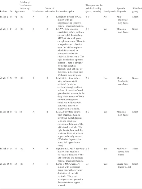
Table 1(Continued)
Patient Sex Age
Edinburgh Handedness Inventory
score Handedness Years of
education Lesion description
Time post-stroke to initial testing
(years; months) Hemiparesis Aphasia diagnosis
Stimulation group rTMS 2 M 72 100 R 14 L inferior division MCA
infarct with an accompanying tempero-parietal encephalomalacia.
4; 0 No Mild-moderate non-fluent
Sham
rTMS 5 F 51 100 R 13 L CVA, total anterior circulation infarct with an extensive left hemisphere MCA/stroke with gross encephalomalacia. There is a hyperintense collection over the left hemisphere which is assumed to represent a subacute subdural haematoma. The right hemisphere appears normal. There is atrophy of the left cerebral peduncle and left side of the pons, in keeping with Wallerian degeneration.
3; 4 Yes Moderate non-fluent
Sham
rTMS 8 M 77 100 R 13 L MCA territory infarct with subacute right occipital (posterior cerebral artery) territory infarct. A couple of small globular foci are seen in the deep white matter of both cerebral hemispheres consistent with chronic ischaemia related to microvascular disease.
2; 2 No Mild-Moderate non-fluent
Sham
rTMS 11 M 66 80 R 12 L MCA territory infarct with encephalomalacia involving the left frontal lobe and moderate ex-vacuo dilatation of the left lateral ventricle. The right hemisphere and the posterior fossa structures appear relatively normal (Wallerian degeneration noted left upper brain stem)
2; 3 Yes Moderate non-fluent
Sham
rTMS 14 M 71 100 R 10 Significant L MCA territory infarct with moderate ex-vacuo dilatation of the left ventricle and tempero-parietal encephalomalacia
2; 9 Yes Moderate-severe non-fluent
Sham
rTMS 15 M 63 100 R 15 Large L MCA territory infarct with significant tissue loss with ex-vacuo dilatation of the left ventricle. The right hemisphere and posterior fossa structures appear normal
6;3 Yes Severe non-fluent/global
Sham
Resting Motor Threshold (rMT)
The TMS coil (bi-phasic stimulation) was placed on the motorÔhot spotÕfor the first dorsal interosseous (FDI) muscle in the left hand (i.e. knob within pre-central gyrus) in the contralateral hemisphere as determined by MRI marking and Stealthstation software (Medtronic, USA). Motor-evoked potentials (MEPs) were then elicited via TMS and surface EMG recorded from FDI via Ag/AgCl electrodes (1 cm in diameter), positioned over the belly of the muscle and the metacarpo-pha-langeal joint of the index finger, respectively. rMT was
defined as the minimum stimulus intensity eliciting five responses of about 50lV of 10 consecutive trials (50% successful MEPs) in the relaxed contralateral FDI [23]. For the present research study on stroke patients, stimulation was applied at 90% of the rMT attained [5,8,16]. Patient stimulation intensities ranged between 35–55% of maximum stimulator output.
Stimulation protocol
Low frequency, 1 Hz rTMS was applied to patients for 20 min per day (1200 pulses), for 10 days (10 sessions)
(a)
(b)
Figure 1 Structural 3T magnetic reso-nance imaging scans for two patients of the twelve patient cohort, demonstrating the range of lesion severities (a) Partici-pant rTMS 15 (b) ParticiPartici-pant rTMS 13.
as defined by Naeseret al.[5] The stimulation protocol aimed to downregulating RH overactivation in homologous language sites. The TMS target was the anterior portion of homologue to right pars triangularis (BA 45) in BrocaÕs area as cited in previous rTMS studies as that location consistently results in the best response to rTMS [2,5,16]. A figure of eight, 70 mm diameter rTMS coil and identical sham coil were uti-lized (Magstim, Carmarthenshire, Wales, UK).
Sham stimulation
The sham stimulation group in this study served as a placebo control condition. The sham coil employed (Magstim, Carmarthenshire, Wales, UK) was identical in shape and size to the real stimulation coil and pro-duced the same audible click as the real coil without the production of a magnetic field. As the sham TMS coil does not activate the cortex, stimulation in this condi-tion would not induce neuro-physiological changes that could influence behavioural language function. Sham stimulation was used to verify the effects of the real stimulation protocol as a comparative control measure.
Language outcome measures
Standardized language assessments, administered by a speech-language pathologist who was blinded as to
whether the participant had received real TMS stimu-lation or sham stimustimu-lation, were utilized to measure changes in behavioural language function associated with rTMS treatment. One week prior to stimulation (baseline) and 2 months post-stimulation, patients were tested using the standard form of the Boston Naming Test (BNT) [24] and selected subtests of the Boston Diagnostic Aphasia Examination (BDAE) [25]. Sub-tests were administered according to a protocol out-lined by Naeser et al. [5]. Receptive and expressive language abilities were tested. The subtests adminis-tered were the following: picture description (Cookie Theft Picture) (analysis of complexity index and longest number of words per phrase length), word compre-hension, word comprehension by category, commands, complex ideational materials, repetition (single words, non-words and sentences), responsive naming, naming screening of special categories, naming colours, naming actions, naming animals, naming tools and implements. Additionally, a set of 144 black and white line drawings of objects, taken from a previously published common object picture inventory [26], was used to measure changes in picture naming. Pictures were administered in lists of 48 words, balanced for aver-age response latency, syllable length, frequency and visual complexity and percentage agreement. Norms pertaining to these variables were obtained from the online International Picture Naming Database. Verbal
responses were recorded via a microphone positioned approximately 10 cm from the subjectÕs mouth.
A series of two-way repeated measures analyses of variance (ANOVA) were conducted for all BDAE subsets
including the BNT, picture-naming accuracy and latency, by time of testing (two levels, baseline, prior to TMS vs. 2 months post-TMS) and group (real stimu-lation vs. sham). Post hoc analyses were conducted between the two groups using an independent samples t-test at baseline and 2 months post-rTMS. A repeated measurest-test was used to measure within-group per-formance, baseline to 2 months.
Results
The individual patient assessment results on the com-prehensive language battery are presented in Appen-dixes S1–S4. Appendix S5 outlines mean values, standard deviations, significant interactions and effect size. Significant interactions of group (rTMS, sham)·time (baseline, 2 months) were identified for the following subtests: BDAE naming actions (P < 0.01), BDAE naming tools and instruments (P < 0.05), BDAE repetition of sentences (P< 0.05), Cookie Theft picture description complexity index (P < 0.05), Cookie Theft picture description longest words per phrase (P< 0.01), Commands (P< 0.05), the overall score calculated from the BDAE subtests administered (P < 0.01), The BNT (P< 0.05), Snod-grass and Vanderwart [26] picture-naming latency (P < 0.05) and Snodgrass and Vanderwart [26] picture-naming accuracy (P < 0.05).
Post hocanalyses
Post hoc analyses revealed no significant differences between the real stimulation and sham conditions at baseline for all language subtests, (P> 0.05).
At 2 months post-stimulation, significant differences between the stimulation and sham group were found for a number of language subtests. The real stimulation group scored significantly higher on BDAE naming actions (t= 4.16,P< 0.01, df = 10), BDAE naming tools and instruments (t= 3.00, P< 0.05, df = 10), Cookie Theft picture description complexity index (t= 4.05, P< 0.05, df = 10), BDAE overall score (t= 3.7, P< 0.05, df = 10) and picture-naming accuracy [26] (t= 3.21,P< 0.05, df = 10). The real stimulation group performed significantly lower on the Snodgrass and Vanderwart [26] picture-naming latency (t= 3.6,P< 0.05, df = 10).
Within the real stimulation group, significant differences across time (baseline to 2 months post-stimulation) were found. Performance at 2 months
post-stimulation was higher on BDAE naming actions (t= 2.609,P< 0.05, df = 5), BDAE naming of tools and instruments (t= 3.796,P< 0.01, df = 5), BDAE overall score (t= 4.145, P< 0.01, df = 5), Com-mands (t= 3.371, P< 0.05, df = 5) and picture-naming accuracy [26] (t= 3.162, P< 0.05, df = 5). Performance at 2 months post-stimulation was signifi-cantly lower for picture-naming latency [26] (t= 3, P< 0.05, df = 5). No significant results within the sham group across time were found. As can be seen in Table 1, the age variance for the sham group was higher than for the participants receiving real stimulation. However, further statistical analysis failed to determine a significant age·time interaction in the sham group for any language subtest (P> 0.05).
Discussion
provide valuable evidence to show that rTMS effects in the aphasic brain may be wider reaching within the bilateral language system than first proposed.
Although the majority of rTMS studies present improved expressive language abilities, the present study reports significant differences and improvements in receptive language performance on an auditory commands subtest at 2 months post-stroke when com-pared to baseline measures. A study by Naeseret al.[9] treated a single patient using a protocol of continuous positive airway pressure (CPAP) combined with rTMS-documented significant improvements in auditory commands; however, it is unclear whether these are as a result of rTMS or increased oxygenation facilitated by CPAP. Additionally, Martin et al. [2] reported improvements in auditory comprehension on com-mands and complex ideational materials in a poor responding speech output patient to 6 months post-rTMS. Whilst the effects of rTMS on expressive language appear to be more concrete, the effects on receptive language remain inconclusive and require further investigation.
The present outcomes are in agreement with the theories of mutual inhibition of LH language sites in chronic non-fluent aphasia and the induction of plastic neural changes when rTMS is applied to RH homolo-gous sites. Indeed, heterogeneity within the present aphasic sample may be a confounding factor when interpreting the significant differences identified in behavioural language between real stimulation and sham groups. Differences between the groups and within the groups in variables such as age, time post-stroke, lesion size and affected neural regions must all be taken into consideration. It is hypothesized that age and lesional variations may provide the greatest influ-ence on rTMS responses within both groups.
Conclusion
This research provides favourable indication that rTMS can be applied to the unaffected RH in persons with aphasia to facilitate brain reorganization in accordance with the theories of transcallosal disinhibition. The results demonstrate significant treatment effects on behavioural language outcome measures of picture naming, spontaneous speech and auditory comprehen-sion between the real stimulation and placebo control groups. With the emerging evidence regarding the positive effects on aspects of auditory comprehension and other aspects of linguistic output, supplementary investigations would be highly beneficial. Further lon-gitudinal investigations are crucial to expand the evidence base and forge a path for rTMS as a clinical tool for the remediation of language deficits in aphasia.
Acknowledgements
We acknowledge and thank the Communication Dis-ability CentreÕs Aphasia Registry for their assistance in recruiting participants for this project.
Supporting Information
Additional Supporting Information may be found in the online version of this article:
Appendix S1.Receptive language subtests. Appendix S2.Repetition and picture description. Appendix S3.Naming subtests.
Appendix S4. Snodgrass and Vanderwart Picture naming.
Appendix S5.Results of statistical analyses.
Please note: Wiley-Blackwell is not responsible for the content or functionality of any supporting materials supplied by the authors. Any queries (other than missing material) should be directed to the corre-sponding author for the article.
References
1. Belin P, Van Eeckhout P, Zilbovicius M,et al.Recovery from nonfluent aphasia after melodic intonation therapy: a PET study.Neurology1996;47:1504–1511.
2. Martin PI, Naeser MA, Ho M,et al.Overt naming fMRI pre- and post-TMS: Two nonfluent aphasia patients, with and without improved naming post-TMS. Brain Lang 2009;111:20–35.
3. Perani D, Cappa SF, Tettamanti M,et al.A fMRI study of word retrieval in aphasia. Brain Lang 2003; 85: 357–368.
4. Rosen HJ, Petersen SE, Linenweber MR,et al. Neural correlates of recovery from aphasia after damage to left inferior frontal cortex.Neurology2000;55:1883–1894. 5. Naeser MA, Martin PI, Nicholas M, et al. Improved
picture naming in chronic aphasia after TMS to part of right BrocaÕs area: an open-protocol study. Brain Lang 2005;93:95–105.
6. Hamilton RH, Sanders L, Benson J, et al. Stimulating conversation: enhancement of elicited propositional speech in a patient with chronic non-fluent aphasia fol-lowing Transcranial magnetic stimulation. Brain Lang 2010;113:45–50.
7. Martin PI, Naeser MA, Ho M, et al. Research with transcranial magnetic stimulation in the treatment of aphasia.Curr Neurol Neurosci Rep2009;9:451–458. 8. Naeser MA, Martin PI, Baker EH,et al.Overt
proposi-tional speech in chronic nonfluent aphasia studied with the dynamic susceptibility contrast fMRI method. Neu-roimage.2004;22:29–41.
9. Naeser MA, Martin PI, Lundgren K, et al. Improved language in a chronic nonfluent aphasia patient after treatment with CPAP and TMS.Cogn Behav Neurol2010; 23:29–38.
11. Ohyama M, Senda M, Kitamura S, Ishii K, Mishina M, Terashi A. Role of the nondominant hemisphere and undamaged area during word repetition in poststroke aphasics. A PET activation study.Stroke1996;27:897– 903.
12. Schlo¨sser R, Hunsche S, Gawehn J, et al. Characteriza-tion of BOLD-fMRI signal during a verbal fluency par-adigm in patients with intracerebral tumors affecting the frontal lobe.Magn Reson Imaging2002;20:7–16. 13. Thiel A, Herholz K, Koyuncu A,et al.Plasticity of
lan-guage networks in patients with brain tumors: a positron emission tomography activation study.Ann Neurol2001; 50:620–629.
14. Kim Y-H, Ko M-H, Parrish TB, Kim H-G. Reorganiza-tion of cortical language areas in patients with aphasia: a functional MRI study.Yonsei Med J2002;43:441–445. 15. Thulborn KR, Carpenter PA, Just MA. Plasticity of
language-related brain function during recovery from stroke.Stroke1999;30:749–754.
16. Naeser MA, Martin PI, Nicholas M, et al. Improved naming after TMS treatments in a chronic, global aphasia patient–case report.Neurocase2005;11:182–193. 17. Wassermann EM. Risk and safety of repetitive
transcra-nial magnetic stimulation: report and suggested guidelines from the International Workshop on the Safety of Repetitive Transcranial Magnetic Stimulation, June 5-7, 1996. Electroencephalogr Clin Neurophysiol 1998; 108: 1–16.
18. Singh K. Mri3dx.Aston University. http://www.aston.ac. uk/lhs/staff/singhkd/mri3dx/features (accessed 21/11/ 2007).
19. Broca P. Remarques sur le sie‘ge de la faculteÕdu langage articule, suivies d_une observation d_apheÕmie (perte de la parole).Bulletins de la SocieÕteÕanatomique de Paris1861; 6:330–357.
20. Amunts K, Schleicher A, Bu¨rgel U, Mohlberg H, Uylings HB, Zilles K. BrocaÕs region revisited: cytoarchitecture and intersubject variability. J Comp Neurol 1999; 412: 319–341.
21. Amunts K, Weiss PH, Mohlberg H, et al. Analysis of neural mechanisms underlying verbal fluency in cytoar-chitectonically defined stereotaxic space–the roles of Brodmann areas 44 and 45.Neuroimage.2004;22:42–56. 22. Devlin JT, Matthews PM, Rushworth MFS. Semantic processing in the left inferior prefrontal cortex: a com-bined functional magnetic resonance imaging and trans-cranial magnetic stimulation study.J Cogn Neurosci2003; 15:71–84.
23. Rossini PM, Barker AT, Berardelli A,et al.Non-invasive electrical and magnetic stimulation of the brain, spinal cord and roots: basic principles and procedures for rou-tine clinical application. Report of an IFCN committee. Electroencephalogr Clin Neurophysiol1994;91:79–92. 24. Kaplan E, Goodglass H, Weintraub S.The Boston
Nam-ing Test. Philadelphia: Lippincott, Williams and Wilkins, 2001.
25. Goodglass H, Kaplan E, Barresi B. The assessment of aphasia and related disorders, 3rd edn. Philadelphia: Lippincott, Williams and Wilkins, 2001.
26. Snodgrass JG, Vanderwart M. A standardized set of 260 pictures: norms for name agreement, image agreement, familiarity, and visual complexity. J Exp Psychol Hum Learn.1980;6:174–215.
27. Cappa SF, Sandrini M, Rossini PM, Sosta K, Miniussi C. The role of the left frontal lobe in action naming: rTMS evidence.Neurology2002;59:720–723.