Effect of purified gambir leaves extract to prevent atherosclerosis in rats
Nanang Yunarto, Nurul Aini
Center for Biomedical and Basic Technology of Health, National Institute of Health Research and Development, Ministry of Health Republic of Indonesia
Corresponding address: Nanang Yunarto, M.Si, Apt e-mail: [email protected]
Received: September 7, 2015; Revised: September 20, 2015; Accepted: September 25, 2015
Abstrak
Latar belakang: Aterosklerosis merupakan faktor risiko timbulnya penyakit jantung koroner (PJK). Senyawa katekin memiliki aktivitas antioksidan tinggi yang dapat menghambat terjadinya aterosklerosis. Ekstrak daun gambir (Uncaria gambir, Roxb.) mengandung katekin tinggi sehingga berpotensi menghambat terjadinya
aterosklerosis. Penelitian ini bertujuan untuk menguji efek ekstrak terpurifikasi daun gambir dalam menghambat
aterosklerosis pada tikus.
Metode: Desain penelitian ini adalah eksperimental laboratorium yang dilakukan di Laboratorium Farmasi
dan Laboratorium Hewan, Badan Litbangkes, Kemenkes RI pada tahun 2014. Ekstrak gambir dipurifikasi untuk
diperoleh kandungan katekin yang optimal, selanjutnya dilakukan uji aktivitas antioksidan menggunakan pereaksi 2,2-difenil-1-pikrilhidrazil (DPPH) dengan pembanding asam askorbat. Penelitian ini menggunakan 36 ekor tikus putih jantan galur Sprague Dawley berusia 2,5 bulan yang dibagi secara acak ke dalam enam kelompok yaitu kelompok normal, kontrol negatif (akuades), kontrol positif (atorvastatin 2 mg/200 g bb), ekstrak dosis I (20 mg/200 g bb), dosis II (40 mg/200 g bb) dan dosis III (80 mg/200 g bb). Tikus diinduksi dengan makanan yang mengandung lemak tinggi dan perlakuan pengobatan sesuai kelompoknya selama 60 hari, kecuali kontrol normal.
Hasil: Kadar katekin dalam ekstrak gambir terpurifikasi diperoleh sebesar 92,69%. Dari uji aktivitas antioksidan diperoleh IC50 11,76 µg/mL. Hasil pengukuran aktivitas antiateroskelrosis menunjukkan bahwa jika
dibandingkan dengan kontrol negatif, ketiga dosis ekstrak gambir terpurifikasi mampu mencegah terjadinya
aterosklerosis dengan menghambat penebalan dinding aorta dan pembentukan sel busa (p<0,05). Aktivitas antiaterosklerosis meningkat dengan bertambahnya dosis.
Kesimpulan: Ekstrak gambir terpurifikasi mempunyai efek mencegah penebalan dinding dan pembentukan sel busa aorta tikus. (Health Science Journal of Indonesia 2015;6:105-10)
Kata kunci: gambir, katekin, antiaterosklerosis
Abstract
Background: Atherosclerosis is a risk factor for coronary heart disease (CHD). Catechin have high antioxidant activity that can prevent atherosclerosis. Gambir (Uncaria gambir, Roxb.) leaves extract have high catechin content thereby potentially inhibiting atherosclerosis. This research was aimed to examine
effect of purified gambir leaves extract to prevent atherosclerosis in rats.
Methods: The experimental laboratory study was conducted in Pharmacy Laboratory and Animal Laboratory, National Institute of Health Research and Development, Ministry of Health, Republic of Indonesia in 2014.
Gambir leaves extract were purified to gain optimum catechin. Afterwards, antioxidant activity was tested
using 2.2-diphenyl-1-picrylhydrazyl (DPPH) method, with ascorbic acid as positive control. Thirty six white male Sprague Dawley rats aged 2.5 months were randomly divided into six groups, i.e. normal control group, negative control group (aquadest), positive control group (atorvastatin 2 mg/200 g bw),extract dose I (20 mg/200 g bw), dose II (40 mg/200 g bw) and dose III (80 mg/200 g bw). The rats were given high fat diet and treatment according to their group for 60 days, except for normal control group.
Results:Catechin content in the purified gambir leaves extract was 92,69%. From antioxidant activity test, IC50 was found to be 11,76 µg/mL. Anti-atherosclerotic activity study shown that compared to negative control, all three doses
of purified gambir leaves extract were able to prevent atherosclerosis through inhibition of aortic wall thickening and
foam cell formation due to high fat diet (p<0.05). Anti-atherosclerotic activity increased with increasing dose.
Conclusion:Gambir leaves purified extract had the effect of preventing the thickening of the walls and foam cell formation rat aorta. (Health Science Journal of Indonesia 2015;6:105-10)
In the last decade, cardiovascular disease caused by atherosclerosis develop into a major killer in Indonesia. It has become a national health problem, thus we need an effective prevention efforts in overcoming atherosclerosis. Management therapy often used nowadays, especially in the acute phase, is administration
of fibrinolytic or management of intervention, followed
by drug therapy. However, such management should be supported by preventive measures in order to prevent the incidence of new patients or the emergence of similar attacks in older patients in the future.1-2
Atherosclerosis is accumulation of fat in the matrix of the tunica intima, followed by the formation of connective tissue in blood vessel walls. High level of total cholesterol, low density lipoprotein (LDL) and very low density lipoprotein (VLDL) in the blood will intensify the process of atherosclerosis.3
Synthetic drugs that are usually used to prevent the atherosclerosis are cholesterol-lowering drugs such as simvastatin and atorvastatin. However, these drugs have side effects such as myopathy, hepatotoxicity, peripheral neuropathy, dizziness, diarrhea, allergies and increase diabetes mellitus risk. Today, several research investigate potential medicinal plants that have similr effect with synthetic drugs, but with more tolerable side effects. Catechins are
flavonoid derivatives secondary metabolites that
have antioxidant and anti-atherosclerotic activity. Catechin compounds have cholesterol-lowering activity through inhibition of pancreas cholesterol esterase, bile acid binding, and reduction of cholesterol solubility in micelles that can delay the absorption of cholesterol. Administration of catechins for six weeks can reduce the average area
of atherosclerotic lesions by 32% in rat aorta.4-6
One of the native plants in Indonesia which contains large amount of catechins is Gambir (Uncaria gambir
Roxb.).Gambir leaf extract contains catechins as a major component as well as some other components such as catechutannic acid, quercetin, red catechu,
gambir fluorescent, fats and waxes.7 The presence of a
high content of catechins in Gambir leaves makes it a potential plant to be used as traditional medicine. This
study aimed to examine effect of purified gambier
leaves extract to prevent atherosclerosis in rats.
METHODS
This study was an experimental laboratory design and was conducted in Pharmacy laboratory and Animal Laboratory, Center for Biomedic and Basic
Technology of Health, National Institute of Health Research and Development, Ministry of Health Republic of Indonesia in 2014.
Preparation and purification of extract
The leaves of gambir was procured from Herbal Plant, Lima Puluh Kota District in West Sumatra and it was authenticated by Herbarium Laboratory, Andalas University, Padang. Gambir extract was prepared by boiling the leaves and pressing them.
The liquid extract was filtered and then concentrated
using vaccum oven at 80°C to get dried extract.
Extract purification process is done through
following steps. The gambir extract was grinded into powder, suspended in n-hexane and homogenized using sonicator for 10 minutes. The suspension was
filtered using filter paper. The residue is then dissolved
in ethyl acetate and homogenized using sonicator for 10 minutes. After that, the solution was partitioned by adding distilled water, then shaken in a separator funnel and allowed to stand for 30-60 minutes until two layers were formed (layer of ethyl acetate on top and a layer of distilled water at the bottom). Both layer formed is then separated. Ethyl acetate layer is evaporated using rotary evaporator (Buchi) until viscous extract
is gained. Furthermore, viscous extract is evaporated
in a fumehood, and then dried using vacuum oven
at 40-50 °C until a fixed weight is obtained. Finally, characterization of the purified extract is conducted.
This include organoleptic inspection, moisture content, loss on drying, and ash content.8,9
Catechin assay in purified extract
Catechin standard calibration curve was done by plotting six concentrations, i.e. 25; 50; 100; 150; 200
and 300 ppm. Standards and purified extract samples
were analyzed utilizing High Performance Liquid Chromatography/HPLC (Waters), with C18 column
sized 4.6 × 150 mm, 0.45 mL/min flow rate, 1.0 mL
injection volume and UV detection at 280 nm using PDA detector (Waters). The mobile phase gradient
is used with the mobile phase A consist of 0.03% acetic acid in a mixture of acetonitrile: water (5:95) and mobile phase B consist of 0.1% triflouroasetat
acid in acetonitrile. Mobile phase gradient conditions
was 100% A at minutes 0 to 4, 71.5 A and 28.5 B at minutes 4 through 20, and 100% B until minutes 30.10 Antioxidant activity test
solution of DPPH. The solution was then pipetted to spectrophotometer cuvette and incubated at 27°C for 20 minutes. Control blanks were made from ethanol, meanwhile positive control used was ascorbic acid. Absorbance was measured using a UV-VIS spectrophotometer (Hitachi) at 515 nm.11
Aortic thickness measurement and foam cell observation
The protocol of animal study has received the ethical clearance from Health Research Ethics Commitee
Faculty of Medicine University of Indonesia number 365/H2.F1/ETIK/2014.
Thirty six male Sprague Dawley strain rats aged 2,5 months old were randomly divided into six groups: normal group, negative group, positive group (atorvastatin), dose I (20 mg/200 g bw), II (40 mg/200 g bw) and dose III (20 mg/200 g bw) groups. Rats were induced with high cholesterol and saturated fat feeds for 60 days, except for normal group. After that, test animals were decapitated.
For histopathological treatment using hematoxylin
and eosin stain (HE), 5 cm abdominal aortic tissue were taken. Observation of the aorta thickness was conducted using microscope equipped (Nikon E
2000) with a micrometer at 100 times magnification.
The thickness of the aorta were measured on 8 view zone clockwise: 12:00, 13:30, 15:00, 16:30, 18:00,
19:30, 21:00 and 22:30. The thickness of the aorta
was then calculated using the following formula:
Observations of foam cell formation in histopathology preparations of rat aorta is performed using a microscope at 400 times magnification.12
Data analysis
Data from the animal experiments were expressed as
mean ± SD. The statistical significance of differences
between the groups were analyzed with one-way ANOVA, followed by LSD post-hoc test analysis using SPSS software 17.0 version, p values of less
than 0.05 were considered to indicate significant
differences.
RESULTS
Extract purification process was done twice. Each purification process used 200 g gambir leaves
extract. Purified extract obtained respectively were 129.48 g and 128.72 g. The average yield of purified extract obtained is 64.55%. This indicates a more optimal purification process using ethyl acetate as the
solvent. The characterization results showed in Table
1 proved that the purified extract met all parameters
required in The Indonesian Herbal Pharmacopoeia.13
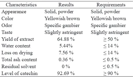
Table 1. Several characteristics of purified extract
Characteristics Results Requirements
Appearance Solid, powder Solid, powder
Color Yellowish brown Yellowish brown Odor Specific gambier Specific gambier Taste Slightly astringent Slightly astringent Yield of extract 64.88 % ≥ 50 %
Antioxidant activity test of purified extract using
DPPH method came with IC50 value of 11.56 mg/ mL. This value is smaller than ascorbic acid used as comparator (IC50 of 24.72 mg/mL). The smaller the IC50 value means the higher the antioxidant activity.
These data show the antioxidant activity of purified
extract of gambir leaves is very strong.
Table 2. Aortic thickness
Significantly different: # p<0.05 compared with normal group;
*p<0.05 compared with negative group, one way Anova
Aortic thickness shown that compared to negative
control, all three doses of purified gambir leaves
extract were able to prevent atherosclerosis through inhibition of aortic wall thickening. (p<0.05).
After being observed in microscope at 400 times x
magnification, the formation of foam cells in negative
DISCUSSION
The purified extract gained has a moisture content of 5.44%, which complies to Indonesia Herbal Pharmacopoeia (IHP), i.e. below 14%. The less water
content in the material ingredients can reduce the risk of microbial and fungal growth, or damage caused by insects. Loss on drying (LOD) examination aims to determine water and volatile components lost when
heated at 105°C. The purified extract was found to have LOD of 7.56%. LOD value that is bigger than
water content explains that in addition to water, there are also volatile components. Determination of total ash content was conducted to determine the amount of material remains after burning at 700°C. The total ash
content obtained was 0.36%, which also met Herbal Pharmacopoiea requirements i.e. less than 0.5%. Small
ash content value indicates that the material is left slightly. The remaining materials include physiological ash, derived from plant tissue itself as well as non-physiological ash which is a residue of foreign material on the surface of plant, such as sand and soil. It can be said that smaller value of ash content means smaller
impurity in the purified extract.9-13
Total catechin content in the purified extract derived from HPLC analysis was 92.69%. The catechin content which is more than 90% met the requirement of IHP. These result was consistent with Kurniatri (2015) that purification
process can improve the purity and levels of catechin.14
Catechins are flavonoid of polyphenol groups that
function as a free radical scavenger to provide the hydrogen atom. The structure that allows the radical scavenging activity of polyphenols is the presence of 3,4-dihydroxyl (catechol structure) in the B ring, which acts as an electron donor and became the target of radical. The 3-OH structure of ring C is also
beneficial to the activity of antioxidant polyphenols.
Conjugation bond at C2-C3 with 4-keto group plays a role for the electron delocalization of the B ring, which in turn increases free radical scavenging capacity. Besides that, the 3-OH group and 5-OH, in combination with 4-carbonyl function and C2-C3 double bond, also raise antioxidant activity. In the absence of o-dihydroxy structure in the B ring, the hydroxyl substituents on the catechol ring A can be compensated and the ability antiradikal activity of polyphenols is increased.15,16
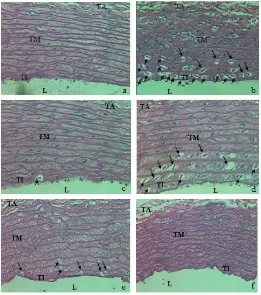
Figure 1. Formation of foam cells in aortic rats
The third dose of purified extract was able to reduce
the aortic thickness of rats under the mechanism of inhibition of LDL oxidation. Catechin as the largest
component in the purified extract has antioxidant activity
that is able to reduce LDL oxidation which can induce the formation of several cytokines that can activate pathways stimulated by mitogen-activated protein kinase (MAPK) leading to high expression of the enzyme
matrix metalloproteinase-9 (MMP 9). Antioxidants also
reduce the toxicity of oxidized LDL on endothelial cells, smooth muscle cells and macrophages as well as reduce degradation oksidatif.17 Catechins increase HDL levels.
HDL has a role to transport cholesterol away from the arteries and back to the liver so it may lower the amount of cholesterol in plaque ateroma.17-18
The observation of microscopic atherosclerosis can be seen from the formation of foam cells in the
aorta. Foam cells was formed of many macrophages
and dendritic cells such as membrane-bound lipids in the cytoplasm. Cells containing the lipid looks like a bubble. The cells’ formation begin when phagocytic cells ingest Apolipoprotein-B. The observations of foam cell would appear more clearly with microscope
at 400× magnification. Purified extract was able to prevent the formation of foam cells on rats aorta. From
microscopic observations, it can be seen that higher
dose of purified extract can reduce the formation of
foam cells better. Hartayo’s in vitro study showed that catechin phenolic group, either in extract or single-isomer form, can protect LDL from oxidation triggered by free radical.19 Furthermore, according to Yang and Koo, catechins have an activity to lower the number of lipid peroxidation products and is able to inhibit LDL oxidation in the endothelium.
MMP 9 enzyme contribute a role in stabilizing the
atherosclerotic plaque so those plaques would not
rupture which in turn may flow through bloodstream
and cause blood clots. Decreased expression of
MMP 9 shows that catechins can be used as an agent
to prevent plaque rupture in the foam cells of aorta.20
These results are consistent with research from Auclair et al which explain that oral administration of catechins for six weeks can reduce the average area of atherosclerotic lesions in the rat aorta.6
In conclusion,gambir leaves purified extract had the effect of preventing the thickening of the walls and foam cell formation rat aorta.
Acknowledgment
The authors would like to thank Prof. Bastaman Basuki, Prof. Agus Suwandono, and Dr. M. Wien Winarno for technical assistance in preparing this article.
REFERENCES
1. Moore KJ, Tabas I. Macrophages in the pathogenesis of atherosclerosis. Cell. 2011;145:341-55.
2. World Health Organization. Global atlas on cardiovascular disease prevention and control. Geneva. The Organization. 2011.
3. Tomkin GH, Owens D. LDL as a cause atherosclerosis. The Open Atherosclerosis & Thrombosis Journal. 2012;5:13-21.
4. Fadecko J, Singh RB, Chaithiraphan S, et al. Clinical manifestations of adverse effects of statins, oxidative stress and possible role of antioxidants in prevention. The Open Nutraceuticals Journal. 2010;3:154-65. 5. Ngamukote S, Mäkynen K, Thilawech T, et
al. Cholesterol-lowering activity of the major polyphenols in grape seed. Molecules. 2011;16:5054-61.
6. Auclair S, Milenkovic D, Besson C, et.al. Catechin reduces atherosclerotic lesion development in apo
E-deficient mice: a transcriptomic study. Atherosclerosis. 2009;204:21-7.
7. Amos L. Catechin content of gambir production center in Indonesia. Jurnal Standardisasi. 2010;12:149-55.
8. Yunarto N, Elya B, Konadi L. Potency of ethyl acetate fraction of gambir leaves extract (Uncaria gambir Roxb.) as antihyperlipidemia.The Indonesian Pharmaceutical Journal. 2015;5:1-10.
9. Ministry of Health, Republic of Indonesia. Indonesia Herbal pharmacopeia. Jakarta. 2008.
10. Dionex. Sensitive determination of catechins in tea by high performance liquid chromatography. The Organization. 2011.
11. Kassim MJ, Hussin MH, Achmad A, et al. Determination
of total phenol, condensed tannin and flavonoid contents
and antioxidant activity of Uncaria gambir extracts.
Indonesia Pharmacy Journal. 2011;22:50-9.
12. Maliya A. Differences in serum lipid profile and development of abdominal aortic atherosclerosis lesions between the given freshly Momordica charantia and control groups [thesis]. Semarang. Universitas Diponegoro; 2006. Indonesian.
13. World Health Organization. Quality control methods for herbal materials. 2nd Edition. Geneva. The Organization. 2011.
14. Kurniatri AA, Adelina R, Setyorini H, et al.
Formulation of gambir extract film coated tablet. The Indonesian Pharmaceutical Journal. 2015;5:9-15.
15. Amic DD, Beslo D, Trinajstic. Structure-radical
scavenging activity relationship of flavonoids.
Journal of Croatia Chemica Acta. 2003;76:55-61. 16. Rahmawati N, Fernando A, Wachyuni. Phenolic
content and antioxidant activity of leaf extract of dried gambir (Uncaria gambir, Roxb). Indonesia Chemica Acta. 2013;4:1-6.
18. Brewer HB. High-Density Lipoproteins: A new potential therapeutic target for the prevention of cardiovascular disease. Arteriosclerosis Thrombosis
and Vascular Biology.2004;24:387-91.
19. Hartoyo A, Astuti M. Antioxidative activity and Hipocholesterolimic of green tea extract and
tea fragrance in rats were given rations rich in polyunsaturated fatty acids. Jurnal Teknologi dan Industri Pangan. 2002;12:1-6.