MODE OF DISPERSAL AND VARIATION IN POPULATION OF
WHITE ROOT FUNGUS
RIGIDOPORUS MICROPORUS
AS
REVEALED BY MYCELIAL INCOMPATIBILITY
Suwandi
Department of Plant Pests and Diseases Faculty of Agriculture, Sriwijaya University
Abstract
Management of the white root disease in Indonesia and other tropical regions has been developed without any knowledge of population of the causal fungus. We determined 33 mycelial compatibility groups (MCGs) out of 62 isolates collected from 11 plant species in South Sumatra and Bangka Island. Mycelial incompatibility in R. microporus is characterized by formation of demarcation line and/or sparse aerial mycelial growth along the zone of interaction of genetically different mycelia. Failure to anastomosis between incompatible mycelia induced abnormal, spiral-like of hyphal growth. Most of isolates colonizing the stumps were somatically compatible with isolates from either a nearby stump or living trees, which suggested the evidence of stump-to-tree or stump-to-stump clonal growth of the fungus. Each study site was occupied by at least one MCG which indicated that local population of R. microporus consists of different genotypes (genetic individual) which spread clonally to form patches of mycelial networks. A territorial clone covered large area (up to 52 m across) that encompassed both old trees (Hevea brasiliensis, Artocarpus integra and Arenga pinata) and recent non-woody plants (Languas galangal and Musa paradisiaca). All of isolates occurring on different location have different MCG. This indicated that no evidence of clonal (vegetative) spread between separate locations. Most of isolates of R. microporus were strongly pathogenic on rubber seedlings. These isolates belonged to different MCG and showed strongly laccase activity as compared to the less pathogenic isolates.
INTRODUCTION
Rigidoporus microporus (Sw.) Overeem synonym R. microporus (Klotzsch) Imazeki is white root rot fungus that cause significant losses in industrial plantations including rubber (Liyanage, 1997; Semangun, 2000) and black pepper (Suwandi, 2003).The fungus attacks roots and collar region of taproot causing white root disease and eventually kills trees at any growth stage. The disease attacks increase cumulatively as increased of rubber planting generations or even for other crops generated from rubber plantations, such as black pepper, cassava and acacia. In Bangka Island, where the black pepper and cassava are grown in ex-rubber area (field generated from rubber trees), the white root disease may cause total loss on those plants (Suwandi, 2003).
sustainable strategy and tactics in management of plant disease (McDonald, 1997). However, no studies have been reported dealing with population structure of R. microporus in any countries including Indonesia. Studying fungal population biology requires appropriate markers for identifying individual genotypes (clones or genets) in a population of the fungus (Anderson and Kohn, 1995). Mycelial incompatibility has been reported to be successfully used as a maker for identification of individual genotype in polypore fungi, i.e. Ganoderma boninense (Miller et al., 1999), Helicobasidium mompa (Aimi et al., 2002), and Heterobasidion annosum (Lygis et al., 2004). In this paper, macro- and microscopic of mycelial incompatibility in R. microporus, using of MCG to describe mode of dispersal, and relatedness of MCG with variation in pathogenicity and laccase activity are discussed.
MATERIALS AND METHODS
Collection, isolation and determination of mycelial compatibility
The study involved isolates represented population of R. microporus from rubber and non-rubber hosts in South Sumatra and Bangka Island (Table 1). Isolations were conducted as routinely performed in our laboratory (Phytopathology Laboratory, Sriwijaya University). Freshly collected basidiomes, rhizomorphs or decayed wood were cleaned from soil particles and wood debris by running tap water and wrapped in a paper towel wetted with water containing 30 mg/L benomyl and 100 mg/L streptomycin. The sample then was enclosed in a sealed PE plastic bag and keep in room temperature. An active growing mycelial strands out of the wrapped samples were planted on MEA (malt extract agar) supplemented with 30 mg/L benomyl and 100 mg/L streptomycin. Pure cultures were maintained at room temperature in slant of MEA embedded with rubber wood.
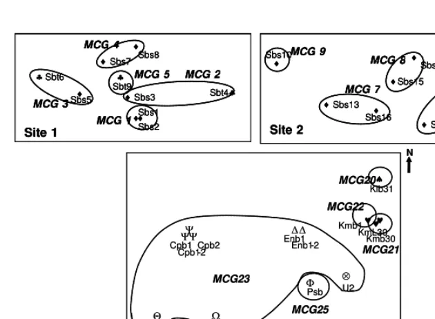
To determine mycelial incompatibility, isolates from the same sites were paired in all possible combinations. For each incompatibility group determined, 1-2 isolates were selected for pairings with isolates from other sites. Self-crosses were performed as controls. Isolates are grouped into the same mycelial compatibility group (MCG), when paired isolates grow together and merge to form a single culture. The interaction is determined as incompatible when paired culture produce a lysis zone and incompatible isolates then are grouped as different MCG. Each pairing was repeated at least twice. The number of SIGs and area it occupied in a field describes the role of basidiospores dissemination (sexual population) and the extent of clonal spread through rhizomorph in the fungus population.
Cytological characteristic of incompatible pairings
Pathogenicity assay
Pathogenicity of isolates was tested on rubber seedlings in a pot system as routinely performed in our laboratory. Rubber seeds collected from plantings clone GT-1 were sown in sand culture and watered every day until germinate for 2 weeks. Seedlings then were transferred to clear plastic pot (volume 250 ml) and filled with mixture (in equal volume) of autoclaved soil and sand. Seedlings were grown in shading area for one month.
Inocula were prepared as colonized wood sticks. Autoclaved rubber wood sticks (1 X 0.5 X 5 cm) were inoculated with a-quarter plate size of 7-days-old mycelia of tested isolates and allowed to be colonized by the mycelia for 2 months. Inoculation was done by carefully inserting two sticks closely to taproot. Each isolate was inoculated on 10 seedlings. Assay was repeated at least twice. Disease severity was assessed 3 months after inoculation based on 9 scale of disease severity (Nandris et al., 1987). Pathogenicity is rated as weak, moderate and strong, when the average of severity value was less than 3.5, between 3.5 to 5.5, and more than 5.5, respectively.
Laccase activity
Laccase activity was assessed indirectly by dye decolorization method (Kiiskinen et al., 2004). Tested isolates were grown on malt extract agar (2%) containing 0.04% dye Remazol Brilliant Blue R (RBBR) (catalog no. R-8001, Sigma). Each isolate was grown on four plates. After 7 days incubation, the enzyme activity was examined by scoring the decolorization intensity. Test was repeated twice.
RESULTS AND DISCUSSIONS
In this study, we successfully isolated 62 field isolates of R. microporus associated with white root disease on 11 host plants from 4 locations in South Sumatra and 6 locations in Bangka Island (Table 1). Some isolates were found on non woody plants such as pineapple, banana and galangal (Languas galangal) and this finding is the first report on infection of white root fungus on non-woody, herbaceous plants.
Observation of hyphae after stained with DAPI using fluorescence microscopy showed that all hyphae including the hyphal tip of the fungus were composed by two nuclei (Fig. 2C), except for the oldest hyphae that have 4 or more nuclei (Fig. 2D). No difference in number of nuclei of incompatible pairings as well as were found in self and non-paired isolates. In incompatible pairings, we usually found abnormal or spiral-like hyphae, which typically produced at zone of interaction between two different hyphae (Fig.2A, 2B). Fusion between incompatible hyphae was not observed in all ten slide cultures tested.
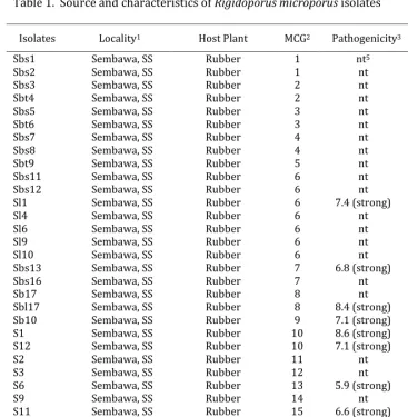
Table 1. Source and characteristics of Rigidoporus microporus isolates
Isolates Locality1 Host Plant MCG2 Pathogenicity3 Laccase4
Sbs1 Sembawa, SS Rubber 1 nt5 nt
Sbs2 Sembawa, SS Rubber 1 nt nt
Sbs3 Sembawa, SS Rubber 2 nt nt
Sbt4 Sembawa, SS Rubber 2 nt nt
Sbs5 Sembawa, SS Rubber 3 nt nt
Sbt6 Sembawa, SS Rubber 3 nt nt
Sbs7 Sembawa, SS Rubber 4 nt nt
Sbs8 Sembawa, SS Rubber 4 nt nt
Sbt9 Sembawa, SS Rubber 5 nt nt
Sbs11 Sembawa, SS Rubber 6 nt nt
Sbs12 Sembawa, SS Rubber 6 nt nt
Sl1 Sembawa, SS Rubber 6 7.4 (strong) +++
Sl4 Sembawa, SS Rubber 6 nt +++
Sl6 Sembawa, SS Rubber 6 nt +++
Sl9 Sembawa, SS Rubber 6 nt +++
Sl10 Sembawa, SS Rubber 6 nt +++
Sbs13 Sembawa, SS Rubber 7 6.8 (strong) nt
Sbs16 Sembawa, SS Rubber 7 nt nt
Sb17 Sembawa, SS Rubber 8 nt +++
Sbl17 Sembawa, SS Rubber 8 8.4 (strong) +++
Sb10 Sembawa, SS Rubber 9 7.1 (strong) +++
S1 Sembawa, SS Rubber 10 8.6 (strong) nt
S12 Sembawa, SS Rubber 10 7.1 (strong) nt
S2 Sembawa, SS Rubber 11 nt nt
S3 Sembawa, SS Rubber 12 nt nt
S6 Sembawa, SS Rubber 13 5.9 (strong) nt
S9 Sembawa, SS Rubber 14 nt nt
S11 Sembawa, SS Rubber 15 6.6 (strong) nt
M32 Muara Enim, SS Rubber 16 6.6 (strong) nt
M34 Muara Enim, SS Pineapple 16 6.3 (strong) nt
M35 Muara Enim, SS Rubber 16 6.6 (strong) nt
M36 Muara Enim, SS Rubber 16 6.7 (strong) nt
Isolates Locality1 Host Plant MCG2 Pathogenicity3 Laccase4
Dd1 Inderalaya, SS Erythrina variega 17 7.6 (strong) +++ Ddb1 Inderalaya, SS Erythrina variega 18 7.6 (strong) nt Ddb2 Inderalaya, SS Erythrina variega 18 nt nt Dd4 Inderalaya, SS Erythrina variega 19 6.7 (strong) nt Dd5 Inderalaya, SS Erythrina variega 19 nt nt Klb31 Balunijuk, Bangka Coconut 20 7.9 (strong) nt Kmb30 Balunijuk, Bangka Vernicia montana 21 6.2 (strong) +++ Kml30 Balunijuk, Bangka Vernicia montana 21 nt ++ Kmb1 Balunijuk, Bangka Vernicia montana 22 7.5 (strong) +++ Cpb1 Balunijuk, Bangka Artocarpus integra 23 nt ++ Cpb1-2 Balunijuk, Bangka Artocarpus integra 23 4.7 (moderate) +++ Cpb2 Balunijuk, Bangka Artocarpus integra 23 4.6 (moderate) nt Ldb Balunijuk, Bangka Black pepper 23 5.1 (moderate) ++ U2 Balunijuk, Bangka Cassava 23 3.9 (moderate) + Enb1 Balunijuk, Bangka Sugar palm 23 5.1 (moderate) nt Enb1-2 Balunijuk, Bangka Sugar palm 23 4.6 (moderate) nt LB Balunijuk, Bangka Galangal 23 6.1 (strong) +++ Krb Balunijuk, Bangka Rubber 24 5.3 (moderate) ++ Psb Balunijuk, Bangka Banana 25 5.1 (moderate) nt Ldpb Payak Benua, Bangka Black pepper 26 3.9 (moderate) + Ubpb Payak Benua, Bangka Cassava 27 6.0 (strong) nt Ldca Cengkong, Bangka Black pepper 28 7.2 (strong)
Ubca Cengkong, Bangka Cassava 29 4.9 (moderate) +++
Krkc Kace, Bangka Rubber 30 6.1 (strong) nt
Ubkc Kace, Bangka Cassava 31 7.0 (strong) nt
1 SS : South Sumatra
2 MCG : Mycelial compatibility group
3 Pathogenicity on rubber seedlings, based on 9 scale disease severity,
0 = no rhizomorph on root and 9 = plant died after 3 month inoculation.
Weak = disease severity less than 3.5; moderate = disease severity 3.5-5.5; strong = disease severity more than 5.5.
4 Measured as decolorization intensity of 0.04% Remazol Brilliant Blue R (RBBR),
+ = less intense and +++ = more intense.
5 nt = not tested.
This phenomenon was different with incompatibility in other polypore fungi such as H. mompa that formerly fused before cell death due to failure of anastomosis (Aimi et al., 2002).
Figure 1. Macroscopic mycelial interaction between isolates of Rigidoporus microporus
Figure 2. Hyphae of Rigidoporus microporus after stained with DAPI (400 X) A and B : formation of abnormal braching system in incompatible pairing. C : binucleate of young hyphae.
All of isolates occurring on different location have different MCG (Table 1). This indicated that no evidence of clonal (vegetative) spread between separate locations. Therefore, dissemination of the fungus through mycelia colonized rootstocks was not unlikely occurs.
Isolates of R. microporus collected from South Sumatra were strongly pathogenic on rubber seedlings (Table 1) as they caused necrotic on most part of taproot and killed the test plants. Some moderately pathogenic isolates were found on either stumps or diseased plants in ex-rubber fields planted with black pepper in Bangka Island. Interestingly, most of these isolates belong to MCG 23, the oldest and the most widely distributed MCG in this study. A moderately pathogenic isolate of this MCG, U2 grew slower than strongly pathogenic LB. It was hypothesis that reduced pathogenicity in this isolates may be associated with infection of dsRNA mycovirus as found in other polypore, H. mompa (Ikeda et al., 2003). Our attempts to detect dsRNA elements in these isolates were not success (data not shown).
Laccase activity of isolates of R. microporus as qualitatively determined by RBBR decolorization intensity was varied regardless their MCG. Most of strongly pathogenic isolates decolorized RBBR more intense than the less pathogenic ones. RBBR decolorization by moderately pathogenic isolates was varied, but generally less intense than strongly pathogenic isolates (Table 1). As our future study will focus on exploration hypovirulence in the white root fungus, therefore, RBBR decolorization method may be useful in preliminary selection of reduced pathogenic isolates.
REFERENCES
Aimi, T., Yotsutani, Y. , and Morinaga, T. 2002. Cytological analysis of anastomoses and vegetative incompatibility reactions in Helicobasidium mompa. Current Microbiology 44:148–152.
Anderson, J.B. and Kohn, L.M. 1995. Clonality in soilborne, plant-pathogenic fungi. Annu. Rev. Phythopathol. 33:369-391.
Ikeda, K., Nakamura, H., & Matsumoto, N. 2003. Hypovirulent strain of the violet root rot fungus Helicobasidium mompa. J. Gen. Pathol. 69:385-390.
Kiiskinen, L.L., Ratto, M., & Kruus, K. 2004. Screening for novel laccase-producing microbes. J. Appl. Microbiol. 97:640-646.
Liyanage, A.de S. 1997. Rubber. In Hillocks, R.J. and Waller, J.M. (Eds.). Soilborne diseases of tropical crops. CAB International. Pp.331-347.
Lygis, V., Vasiliauskas, R., and Stenlid, J. 2004. Planting Betula pendula on pine sites infested by Heterobasidion annosum: disease transfer, silvicultural evaluation, and community of wood-inhabiting fungi. Can. J. For. Res. 34:120–130.
McDonald, B.A. 1997. The population genetics of fungi: Tools and Techniques. Phytopathology 87:448-453
Miller, R. N. G., Holderness, M., Bridge, P. D., Chung, G. F., and Zakaria, M. H. 1999. Genetic diversity of Ganoderma in oil palm plantings. Plant Pathology 48:595–603.
Nandris, D., Nicole, M., Geiger, J.P. 1987. Variation in virulence among Rigidoporus lignosus
and Phellinus noxius isolates from West Africa. Eur. J. For. Path. 17: 271-281.
Semangun, H. 2000. Diseases of plantation crops in Indonesia. Gadjah Mada University Press, Yogyakarta. (in Indonesian).