Corresponding author: Ayunda Dwi R, Department of Conservative Dentistry Faculty of Dentistry Universitas Padjadjaran Jl. Sekeloa Selatan No.1 Bandung, West Java-Indonesia, Ph./Fax.: +6222-2504985/2532805
Evaluation of the mummiication treatment success at
Conservative Dentistry Clinics of Faculty of Dentistry
Universitas Padjadjaran in February 2011-2012
Ayunda Dwi Rahmawati*, Wazillah Nasserie*, Rahmi Alma Farah Adang*
*Department of Conservative Dentistry Faculty of Dentistry Universitas Padjadjaran, Indonesia
ABSTRACT
Indonesia is one of many developing countries with high caries prevalence which needs more attention regarding the countermeasures as well as the preventive treatment. Dental problems or pulp injury was able to treated with an endodontic procedure such as pulpotomy. The success of the
mummiication treatment was able to evaluated through subjective and objective examinations. The purpose of this study was to describe an evaluation of the success of the mummiication treatment at Conservative Dentistry Clinics of Faculty of Dentistry Universitas Padjadjaran by examining the condition of the teeth that have received mummiication treatment. This study was a descriptive study with
purposive sampling technique. The number of samples was as much as 38 teeth from patients who have
completed mummiication treatment. The results showed the success of mummiication treatment was
as much as 67% in less than three months, 44% in the range of time 3-6 months, and 29% in more than 6
months. The conclusion of this study was the mummiication treatment success at Conservative Dentistry
Clinics of Faculty of Dentistry Universitas Padjadjaran was high in less than three months and decreasing
in more than six months after treatment.
Keywords: Mummiication treatment, treatment success evaluation.
INTRODUCTION
Healthy teeth will function well in mastication, verbal communication as well as an aesthetic support.1 Oral hygiene is one of the most
important things to be taken care of to maintain the good function of teeth and avoid any dental problems. Based on Indonesia Health Survey (SKRT-Surkesnas) in 2001, as much as 60% of Indonesian population was having dental problems, which includes tooth decay (dental caries).2
According to the studies conducted by Joelimar and Mandel in 1985 and Sundoro in 2005, the increase of caries prevalence occurred in most of developing countries. Indonesia is one of many developing countries with high caries prevalence which needs more attention regarding the countermeasures as well as the preventive treatment.3 A study held by Faculty of
Dentistry Universitas Indonesia showed that 80%
of Indonesian population sufered dental caries.
(SKRT) in 2004 discovered that the prevalence of caries in Indonesia was as much as 90.05%.4
One of the factors causing the defect in the dental hard tissue is the plaque accumulation that is also initiated dental caries. Bacteria invasion caused acid formation on the tooth surface causing demineralization of the tooth enamel which progressively reaching the dentine.
Bacteria accumulation formed calciication inside
the pulp and tubule causing direct contact with
the pulp thus causes inlammation (Eccles, 1994).
Dental problems or pulp injury can be treated with endodontic procedure such as pulpotomy.5
American Association of Endodontists
divides pulpotomy/pulp amputation into vital amputation and mortal/devitalization/
mummiication amputation. Mummiication is
the removal of a devitalized pulpal tissue inside the pulp. The pulp tissue inside the root canal
was left in a sterile condition and mummiied by mummiication medicament.6
A study conducted by Zeng at China in 2004,
had explained the advantage of mummiication and low complexity of the procedure, fast and simple treatment with afordable cost, and
popular for pulp treatment in few hospitals of the remote areas. As one of the developing countries,
Indonesia uses mummiication as a permanent
endodontic treatment due to many factors, which includes time and money, despite the incomplete instruments especially in the Community Health Center (Puskesmas) located in the remote areas.
Mummiication used as an emergency procedure in
many developing countries to decrease pain until the advanced endodontic treatment can be done.7
A study conducted by Xie and Hong at
China in 2010 by mummiied the patient’s teeth
in the age range above 70 years old. This research
revealed that this procedure was efective within
one month of treatment, while the percentage of success was as much as 86.9% in one year, and 76.2% in 2 years after treatment. Criteria for a successful treatment can be seen through
subjective and objective examination.7 Pulpotomy
was successful if the patient did not complaining
any pain in the subjective examination while
functioning the teeth (masticating). However, the
objective examination resulted in negative results
on the percussion testing, with no periapical abnormalities in radiography results.5 The soreness
was signiicantly decreased after mummiication.
Thus clinicians were assuming the treatment was successful,8 without any radiographic examination
for evaluation.
Conservative Dentistry Clinics of Faculty of Dentistry Universitas Padjadjaran and Dental Polyclinic of Hasan Sadikin Hospital, Bandung,
Indonesia, were still performing mummiication
treatment to prepare their medical graduates for dental practices in the remote areas with limited instruments compared to developed countries
with a middle economic population. Zeng’s study in China stated that the mummiication procedure
was still popular in the hospital of remote areas in
China. Xie and Hong also performed mummiication
on patients with the age range of above 70 years old, although this procedure has already been eliminated in several developing countries and most recent literature. The purpose of this study was to describe an evaluation of the success of
the mummiication treatment at Conservative
Dentistry Clinics of Faculty of Dentistry Universitas
Padjadjaran by examining the condition of the teeth that have received mummiication treatment
in 3 months, 3-6 months, and more than 6 months after obturation control.
METHODS
This research type is descriptive, which gives a picture or description regarding an event as clear as possible, without any treatment on objects under study.9
The population of this research was the
teeth of patients that treated with mummiication
procedure in the Conservative Dentistry Clinics of Faculty of Dentistry Universitas Padjadjaran. The sample was taken using the purposive sampling technique, which gathers samples within
criteria according to researcher’s consideration.9
Inclusion criteria included less than 3 months,
3-6 months, and more than 6 months mummiied
teeth; patients willing to do a post-treatment
radiographic examination; agreed to participated
in this research. The instruments and materials
used were patient’s status, radiographic photo,
treatment diagnosis, mouth mirror, dental
explorer, and dental tweezers.
explanation regarding the research procedures and the objective of a radiographic examination; illing and signing informed consent; examining patient’s tooth as a subjective examination by asking patient’s complaints, and an objective examination by doing percussion testing and
radiographic interpretation.
RESULTS
Research results were obtained from 38
mummiied teeth that have been mummiied.
Sample distribution was based on the time range
from the obturation control until the next control
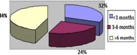
treatment, as seen in Figure 1.
Figure 1 showed that the amount of
mummiied teeth in the range of less than 3 months since obturation control until the next
control was as much as 12 teeth (32%), the range of 3-6 months was 9 teeth (24%), and the range of more than 6 months was as much as 17 teeth (44%).
Sample distribution based on time range
diference showed the treatment success. The
treatment success determined according to the
subjective and objective examination. From the subjective examination by the patient’s complaint
obtained the results that the teeth were
functioning, while from the objective examination
by the percussion testing and radiographic interpretation obtained only one positive results
indicated unsuccessfulness of the mummiication
treatment.
Treatment success based on the time range
since obturation control until the next control
treatment was seen in Table 1.
Table 1 showed that in the range of less than 3 months, 8 teeth (67% successful treatment) of which the patient did not complaining any pain while functioning the teeth, a negative result on percussion test and no signs of periapical abnormality. As much as 4 teeth (33% unsuccessful treatment) showing abnormality in radiographic results and positive response in percussion test. In the range of 3-6 months, as much as 4 teeth (44%) were successfully treated, and 5 teeth (56%) were unsuccessfully treated. In the range of more than 6 months, as much as 5 teeth (29%) were successfully treated, and 12 teeth (71%) were unsuccessfully treated.
DISCUSSION
This research showed higher success percentage in the range of less than 3 months. This result was consistent with clinical research conducted by Berger in 1965 and Redig in 1968 that showed higher success percentage in a short-range period.8 However, the result did not consistent
with Guelmann’s research in 2002 that showed a low success in the irst three months of treatment
due to diagnostic error and abnormality before the treatment choice was performed, where the tooth was having a periapical abnormality. This
condition was restricted for the mummiication
treatment as written in Bence (1990)5 that
explaining the condition for mummiication were
irreversible pulpitis teeth without any periapical abnormality.
This research results showed that treatments
in the range of more than six months resulted in
low success percentage. This result was consistent
with Wang’s research in 1995 that showed a high unsuccessfulness in mummiication when
evaluated in a long period. A study conducted by Xie in 2010 also suggested that the treatment was
efective only in the irst month after treatment.
In 2011, a study conducted by Cohen
explained that the unsuccessfulness of this
Successful Unsuccessful
N Total Percentage Total Percentage
<3 months 12 8 67% 4 33%
3-6 months 9 4 44% 5 56%
>6 months 17 5 29% 12 71% Figure 1. Sample distribution based on the time range from
obturation control until the next control
Table 1. Percentage of mummiication treatment succes based on the time range since obturation control until the
treatment might caused by the wrong consideration
in diagnosis, patient’s bad health condition causing
decrease in immunity thus weakens the body towards bacteria, where bacterial virulence was
stronger in the host’s low immunity.10 Inadequate
debridement during cavity preparation may cause entrance of saliva into the dried cavity, operator error may cause perforation, and inadequate sterilization may increase the amount of bacteria in the sterilized cavity.
Endodontic treatment was successful if the
tooth was able to functioning without pain, and showed negative responses to percussion test, with no radiolucent interpretation found in radiography results.8 This statement was consistent with the study held by Guelmann in 2002 that stated no pain complaint on the treated tooth indicated the treatment success.
Operator error in ensuring the diagnosis might become the reason of an unsuccessful treatment in less than three months period, as well as error in deciding whether the infection was only localized in the pulp or had also infected the periapical. A periapical abnormality was
only seen after six months of obturation, as an
indicator of the treatment success. This condition
was consistent with Guelmann’s study in 2002
that showed unsuccessfulness of treatment in the
irst three months of the procedure due to the
operator error when diagnosing the tooth and pulp infection condition.11
Unsuccessfulness in a long range of time might cause by the pulp tissue left inside the root canal in a sterilized and non-vital condition that may cause focal infection. Grossman stated
this statement in 1998 regarding mummiication deinition as the procedure of removing devitalized
pulp tissue from the pulpal room leaving the tissue inside the root canal in a sterilized condition and
mummiied by mummiication medicament.
A post-treatment evaluation should be followed up within 6-12 months after obturation control to observed any changes in the periapical tissue. This condition was consistent with Walton and Torabinejad (2008) recommended for
re-examination within six months to 4 years after mummiication, as six months considered as a rational
interval of control treatment for most patient. Table 1 showed as much as 12 teeth (71%) were having unsuccessful treatment in more
than six months period. This unsuccessfulness
was indicated by the positive response in the
objective examination (complaining pain during
percussion test) and also supported by periapical abnormalities showing the widening periodontal ligament and the presence of radiolucent lesion surrounded by a radiopaque linear lesion in the
apex area. This condition was consistent with the
research conducted by Wang in 1995. The research performed by Pudjonirmolo in 1993 stated that the treatment was clinically successful if the tooth was able to function normally without any pain, no complaint during percussion testing, and no periapical abnormalities.
No lesion since the beginning until the end of the treatment proofed the treatment success.7
A study conducted by Ingle in 2002 showed a decrease up to 20% in treatment success that might caused by a microleakage restoration that may caused by bacteria. This result was also
consisted by Guelmann’s research in 2002 stated
that the low percentage of successful treatment after three months might caused by incorrect
diagnosis and inlamed pulp condition, while
unsuccessfulness in a long period might relate to the microleakage of its restoration.11
Table 1 showed that as much as 17 teeth had completed the treatment for more than 6 months, with 12 of them showed unsuccessfulness indicated by the soreness feeling during percussion testing and the presence of periapical abnormality showing a widening periodontal ligament and the presence of radiolucent lesion surrounded by a
radiopaque linear lesion of the apex area in the
post-treatment radiography. This condition was consistent with research by Huth in 2011 that stated clinically, one of the indications of an unsuccessful treatment was the soreness feeling during the percussion test. Unsuccessfulness shown in radiographic results were radiolucent in the periapical region and the widening periodontal membrane.
CONCLUSION
The mummiication treatment success
at Conservative Dentistry Clinics of Faculty of Dentistry Universitas Padjadjaran was high in less
than three months and decreasing in more than six
REFERENCES
1. Guyton AC, Hall JE. Buku Ajar Fisiologi
Kedokteran. 5th ed. Jakarta: EGC; 1983.
2. National Institute of Health Research and Development (NIHRD). Indonesia National Health Survey – Round 1 2001. Jakarta: Ministry of Health Republic of Indonesia; 2001.
3. Sundoro EH. Serba-serbi Ilmu Konservasi Gigi. Jakarta: UI Press; 2005.
4. National Institute of Health Research and Development (NIHRD). Indonesia National Health Survey – Round 2 2004. Jakarta: Ministry of Health Republic of Indonesia; 2004.
5. Bence R, Pinsky LD, Meyers RD, Sundoro EH.
Buku Pedoman Endodontik Klinik. Jakarta:
UI-Press; 1990.
6. Grossman LI, Oliet S, Del Rio CE. Endodontics Practice. 11th ed. Philadelphia: Lea & Febiger; 1988. p. 102-10, 228-32, 266-8.
7. Walton RE, Torabinejad M. Prinsip & Praktik
Ilmu Endodonsia. 3rd ed. Jakarta: EGC; 2008.
77, 373-384, 430.
8. Kennedy, D.B. 1992. Konservasi Gigi Anak.
Jakarta: EGC; 2003. p. 213, 260-1.
9. Kountur R. Metode Penelitian untuk penulisan Skripsi dan Tesis. Jakarta: Penerbit PPM; 2007. 10. Hargreaves KM, Cohen S. Pathways of The
Pulp. 10th ed. St. Louis: Mosby-Elsevier; 2011.
p. 2-20, 36-7, 40-1, 628-9.