D. Ramadhani et al. / Atom Indonesia Vol. 42 No. 2 (2016) 71 - 77
Comparison of Radiosensitivity of Human
Chromosomes 1, 2 and 4 from one Healthy Donor
D. Ramadhani
*1, S. Purnami
1and M. Yoshida
2 1Center forRadiation Safety Technology and Metrology, National Nuclear Energy Agency Jl. Lebak Bulus Raya No. 49, Jakarta 12070, Indonesia
2
Department of Radiation Biology, Institute of Radiation Emergency Medicine, Hirosaki University, Japan
A R T I C L E I N F O A B S T R A C T
Article history:
Received 29 September 2015
Received in revised form 29 March 2016 Accepted 31 March 2016
Keywords: Chromosome FISH
Ionizing radiation Radiosensitivity Translocation
In general, it was assumed that the chromosome aberration induced by ionizing radiation is proportional to the chromosome size. From this viewpoint, the higher chromosome size, the more resistant to radiation. However, different opinions, in which chromosomes are particularly sensitive or resistant to radiation, are also still followed until now. Here in this research, we compared the chromosome sensitivity between chromosomes number 1, 2, and 4 using the FISH (fluorescence in situ hybridization) technique. From this research, we expect that the information obtained could show clearly whether a longer chromosome is more frequently involved in translocations and also more resistant to radiation than a shorter one. The type of chromosome aberration considered was limited only to translocation and we used one sample donor in order to avoid donor variability. The whole blood
from a healthy female was irradiated with γ-rays with doses of 1, 3 and 5 Gy, respectively. Isolated lymphocytes from the whole blood were then cultured for 48 hours. After the culture process was completed, preparations of harvest and metaphase chromosomes were carried out. Chromosomes 1, 2, and 4 were stained with different fluorochromes. The translocation of each chromosome at each dose point was subsequently evaluated from 50 images obtained from an automated metaphase finder and capturing system. An additional analysis was performed to identify which chromosome arm was more frequently involved in translocation. Further analyses were also conducted with the aim of determining which chromosome band had a higher frequency of radiation-induced breakage. The experimental results showed that chromosome number 4 was more frequently involved in translocations compared to chromosomes 1 and 2 at 5 Gy. In contrast, at doses of 1 and 3 Gy translocations involving chromosomes number 1 and 2 were more numerous compared to the ones involving chromosome 4. However, if the number of translocation was accumulated for all the doses applied, the chromosome number 4 was the chromosome most frequently involved in translocations. Breakpoint analysis revealed that in chromosome 1, chromosome 2, and chromosome 4, the highest chromosome bands as break position were in band q32, p13, and q21, respectively. It can be concluded that chromosome 4 is more sensitive to radiation in all doses point, despite having less DNA content than chromosomes 1 and 2. Thus, it was showed that our research cannot support the general assumption about chromosome aberration induced by radiation being proportional to DNA content.
© 2016 Atom Indonesia. All rights reserved
INTRODUCTION
The chromosomal radiosensitivity is
representative of the individual radiosensitivity [1].
Corresponding author
E-mail address: [email protected] DOI: http://dx.doi.org/10.17146/aij.2016.505
It can be measured using several cytogenetic techniques, and one of the most useful method for assessing it is fluorescence in situ hybridization (FISH). FISH has been shown to be a useful method for assessment of chromosomal radiosensitivity [2]. A differential susceptibility of chromosomes for aberration induction will be discernible through a
Atom Indonesia Vol. 42 No. 2 (2016) 71 - 77
Atom Indonesia
Journal homepage: http://aij.batan.go.id
D. Ramadhani et al. / Atom Indonesia Vol. 42 No. 2 (2016) 71 - 77
systematic and extensive analysis. In general the chromosome aberration induced by radiation
will be proportional to deoxyribonucleic acid (DNA) content that is proportional to the chromosome size [3].
Pandita et al. [4], using premature chromosome condensation-fluorescence in situ hybridization (PCC-FISH) in human lymphocytes at G0 stage, has shown that there is a direct
relationship between chromosome size and
aberration frequency. Luomahaara et al. [5] examined the distribution of breakpoints that were
induced by radiation chromosomes 1, 2, and 4. In their study, they obtained their samples from
donors from radiation accident victims in Estonia in 1994 and from in vitro irradiated lymphocytes. irradiated lymphocytes, in vitro. However, Wojcik and Streffer [6] showed that, in general, chromosome 1 was more frequently involved in translocations compared to chromosome 2, even though this result was not always reproducible.
The translocation is a type of chromosomal aberration in which a large segment of one chromosome breaks off and attaches to a different chromosome. Chromosome translocations are now considered as a valuable biomarker of radiation exposure and cancer risk [7,8]. Several studies
showed that chromosome translocations in
peripheral blood lymphocytes can be considered as the most reliable biomarker for measurement of low-dose radiation effects and for retrospective biodosimetry [9-12]. Another study also revealed that the analysis results of translocations using FISH after in vitro irradiation correlated with clinical response to radiation in prostate cancer patients. Based on that result, the authors suggested that the cytogenetic assay should be considered as a potential predictor of radiosensitivity [13]. Even now, there are techniques that can be used as
radiosensitivity predictors, such as the γ-H2AX
assay. Djuzenova et al. showed that the γ-H2AX assay shows the potential for use in screening the individual radiosensitivities of breast cancer patients [14].
Until now, there has been no agreement on to which extent the chromosomes are particularly sensitive or resistant to radiation. However, several
studies supported the assumption that the higher the chromosome size, the more resistant the
chromosome is to radiation. It seems that donor
variability is probably a contributing factor to the disagreement, as most studies were performed using
lymphocytes of a homogeneous donor [15,16]. The works of Wojcik and Streffer [6] and of
Sommer et al. [16] seem to support this argument. Their works show that the types of aberration studied affected the radiation sensitivity of chromosomes; for example, human chromosome number 1 was more susceptible to translocations than that of chromosome 2, while Wojcik and Streffer [6] showed that chromosome number 2 was more prone to deletions than that of chromosome 1.
The aim of this research was to compare the sensitivities of chromosomes number 1, 2 and 4 using FISH technique. The aberration type analyzed was limited only to translocation, and only one sample donor was used in this research to avoid donor variability. In this work, we assumed that the higher the chromosome size is, the more prone the chromosome is to be involved in the translocation and also the more radiation-resistant it is. Additional analyses were also conducted to identify the chromosome arms that more involved in the translocation and to detect the higher frequency bands as breakpoint position induced by radiation.
EXPERIMENTAL METHODS
Subjects and irradiation
Eighty milliliters of blood was collected by venipuncture from one 41-year-old healthy female donor without a history of ionizing radiation
exposure beyond routine diagnostic exposures.
The whole blood was then irradiated with γ-rays
from 137Cs in doses of 1, 3, and 5 Gy, respectively, with a dose rate of 0.649 Gy/min at the Institute for Environmental Sciences in the Rokkasho village, Aomori prefecture, Japan.
Blood culture
Lymphocytes to be cultured were isolated from whole blood using Vacutainer CPT Tube (BD Biosciences USA). Cultures were set up in Roswell Park Memorial Institute 1640 (RPMI 1640) culture medium supplemented with
4-(2-hydroxyethyl)-1-piperazineethanesulfonic acid (HEPES) and L-glutamine, 20% fetal bovine serum (FBS),
kanamycin, colcemid 0.05 µg/mL, and
phytohaemagglutinin (PHA). The culture was maintained in a 5%-humidified CO2 incubator at 37°C for 48 hours. Cells were then treated with
D. Ramadhani et al. / Atom Indonesia Vol. 42 No. 2 (2016) 71 - 77
Fluorescence in situ hybridization (FISH)
The slides that were kept overnight in the
previous procedure were hardened for 1 hour at 65°C. Commercial chromosome DNA probes
(MetaSystems, Altlussheim, Germany) were used
to directly label chromosome 1 (Texas Red), 2 (fluorescein isothiocyanate/FITC), and 4
(FITC/Texas Red) following the manufacturer's
recommended protocol. The chromosome (SSC) buffer at 73°C for two min. Subsequently, the
slides were treated in 0.5× SSC / 0.05% of Tween 20 at room temperature for 30 s. Finally, they were rinsed twice briefly in distilled water to prevent salt crystal formation. Air-dried slides were then
embedded and counterstained with 15 µL of 4’,
6-diamidino-2-phenylindole (DAPI) and mounted with cover glass; nail polish was used to prevent the DAPI from leaving the area in cover slip.
Fluorescence image capture and analysis
The automatic metaphase finder was
performed with a Zeiss Axioplan 2 Imaging epifluorescent microscope connected to a Cool Cube
(MetaSystems, Altlussheim, Germany) and the Metafer 4 imaging system (MetaSystems,
Altlussheim, Germany). Fluorescent images of metaphase spreads were captured and analyzed with the ISIS software from MetaSystems and ImageJ translocation was carried out manually for chromosomes number 2 and 4. Since the chromosomes 2 and 4 were submetacentric, the
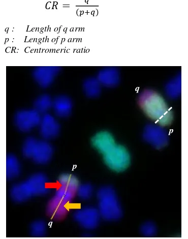
shortest arm was identified as the p arm. For example, in Fig. 1 it can be seen clearly that the
centromere in chromosome 4 was in the dashed line
and the shorter arm is defined as the chromosome’s
D. Ramadhani et al. / Atom Indonesia Vol. 42 No. 2 (2016) 71 - 77
original chromosome one through observation of the short arm. The area loss percentage was converted to chromosome length, and then it was plotted to chromosome image obtained from International System for Human Cytogenetic Nomenclature (ISCN) 2009 from the Atlas of Genetics and Cytogenetics in Oncology and Haematology website (http://atlasgeneticsoncology.org/index.html) [18]. A detailed process of this method is available in additional file of this paper. The method used in this paper was a modified form of Schilling et al. [19].
RESULTS AND DISCUSSION
Many studies have previously been conducted for many years up to present to identify the chromosomes that are particularly sensitive to ionizing radiation. Here in this research, it was
clearly found that for the highest dose used (5 Gy), chromosome number 4 underwent more
translocations than did chromosomes 1 and 2, while at 1 and 3 Gy, the number of translocations of chromosomes number 1 and 2 was higher than that of chromosome 4, as shown in Fig. 2. However, for the total number of translocation for all doses,
Table 1. Translocation number at doses of 1, 3, and 5 Gy
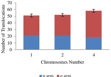
Radiation chromosome arms underwent more translocation that did the p arms. In total, for all doses, q chromosome arms underwent 101 translocations, while p arms underwent 60. Moreover, at 1 Gy, there was no translocation for the p arm of
chromosome 4. At 3 and 5 Gy, the q arms of chromosome 4 underwent three times as many translocations as did the p arms (Table 1). Possibly, a factor that causes q arms to get translocated more frequently is the larger size of the q arm compared to the p arm. As can be seen in Table 1 and Fig. 2, for all doses, the q arms of chromosome 2 and 4 underwent more translocations than the p arms. The p arms of chromosome 1 also underwent fewer translocations than the q arms for all doses, even though the difference was smaller. It is possible that the difference is smaller because chromosome 1 is metacentric; it has equal-sized p and q arms.
Fig. 2. Number of translocations in chromosomes 1, 2, and 4 at
doses of 1, 3, and 5 Gy.
Fig. 3. Total number of translocations in chromosomes 1, 2,
and 4 for all applied doses.
In our study, it has been shown that the most frequent break position of chromosome 1 was in band q32. This experimental result is in good agreement with the results of Barrios et al. that examined the lymphocytes from cancer patients after radiotherapy [20]. Barrios et al. argued that the band q32 is a hot point for clastogenic effects (causing chromosomal breakage) of ionizing radiations. For chromosome 2, the break position was depicted in band p13, while for chromosome 4 was in band q21. This research only considered the break positions that are located in light bands, as only those bands contain the active genes.
D. Ramadhani et al. / Atom Indonesia Vol. 42 No. 2 (2016) 71 - 77
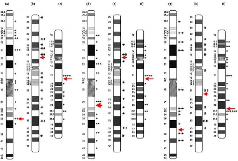
Fig. 4. Breakpoint positions in chromosome 1 at doses of (a) 1 Gy; (d) 3 Gy; and (g) 5 Gy; chromosome 2 at doses of (b) 1 Gy;
(e) 3 Gy; and (h) 5 Gy; and chromosome 4 at doses of (c) 1 Gy; (f) 3 Gy; and (i) 5 Gy.
Our experimental results also showed that the ionizing radiation induced breakpoints in the chromosomes were not random. Figure 4 of our experimental data shows that the q21 breakpoint on chromosome 4 seems to have a high breakage
frequency for all doses. Breakages were more frequent in light bands compared to dark
bands in all chromosome number at all doses. Possible explanation for these finding is that the light bands contain more active genes
than do the dark bands. Our findings also support other several studies that found that breakages
induced by radiation were more frequent in light bands that are considered as gene-rich regions [21,22].
From previous studies it was known that
chromosomal radiosensitivity based on the translocation in lymphocytes can be proposed as a predictive assay for detection of radiosensitive individuals [23]. Several studies used chromosome translocations to identify the cancer patients with higher radiosensitivity. A study by Huber et al. showed that translocations can be used as a test to identify breast tumour patients with potentially elevated radiosensitivities [23].
In biodosimetry using painting of several chromosomes, there was an assumption that the frequencies of chromosomal aberrations were
proportional to their size, which is important
because it will extrapolate to the whole genome [24]. In contrast, our experimental research found that the frequencies of chromosome
aberrations were not proportional to their size. Based on these findings, for a biodosimetry
purpose, it is possible that biodosimetry using painting of all chromosomes (multicolor
FISH) is better than with painting only several chromosomes. Multicolor FISH is also
considered as the best method for assessment
of a chromosome’s structural damage because it
allows unstable and stable aberrations to be detected [25].
Other methods such as telomeric and pancentromeric probes combined with chromosome paint probes can also be use in order to accurately discriminate between translocations and dicentrics [26]. This technique can also overcome the problem of the high cost of the probes for several laboratories [27]. A study by M’kacher . showed that using the telomere and centromere (TC) staining probes it can provides the most precise cytogenetic biological dosimetry currently available. Another advantage of TC staining method is that it does not require a high level of expertise to identify the chromosome aberrations induced by radiation exposure [28].
D. Ramadhani et al. / Atom Indonesia Vol. 42 No. 2 (2016) 71 - 77
CONCLUSION
Based on the experimental results, it can be concluded that chromosome 4 was more sensitive to radiation in all doses point despite having less DNA content than chromosome 1 and 2. The general assumption about radiation-induced chromosomal aberration being proportional to DNA content cannot be supported by our experimental results. For a biodosimetry purpose it is possible that multicolor FISH will give a better result than
painting only several chromosomes. A comparison of the radiation dose estimates using multicolor FISH and three-color FISH should be conducted in the next experiment to validate or invalidate this assumption.
ACKNOWLEDGMENT
The authors gratefully acknowledge the Institute of Radiation Emergency Medicine at Hirosaki University and National Nuclear Energy Agency of Indonesia (BATAN) via the Center for Technology of Radiation Safety and Metrology (PTKMR) for supporting this research. This
8. Anonymous, Cytogenetic Dosimetry:
Applications in Preparedness for and Response Genet. Toxicol. Environ. Mutagen. 784 (2015) 10. DOI: 10.1016/j.mrgentox. 2015. 04. 005. Res. 37 (2011) 25. DOI: 10.1667/RR2433.1.
D. Ramadhani et al. / Atom Indonesia Vol. 42 No. 2 (2016) 71 - 77
26. E.A. Ainsbury, E. Bakhanova, J.F. Barquinero et al., Radiat. Prot. Dosimetry 147 (2011) 573 DOI: 10.1093/rpd/ncq499.
27. R. M’Kacher, E. El Maalouf, G. Terzoudi
et al., Int. J. Radiat. Oncol. Biol. Phys. 91
(2015) 640. DOI: 10.1016/j.ijrobp.2014. 10.048.
28. R. M’kacher, E.E.L. Maalouf, M. Ricoul
et al., Mutat. Res. 770 (2014) 45. DOI : 10.1016/j.mrfmmm.2014.09.007.
1.