New bioactive metabolites produced by
Colletotrichum
sp., an
endophytic fungus in
Artemisia annua
Hong Lu
a, Wen Xin Zou
a, Jun Cai Meng
a,c, Jun Hu
b, Ren Xiang Tan
a,*
aState Key Laboratory of Pharmaceutical Biotechnology,School of Life Sciences,Nanjing,Uni6ersity,Nanjing210093,PR China bState Key Laboratory of Co-ordination Chemistry,Nanjing Uni6ersity,Nanjing210093,PR China
cNational Laboratory of Applied Organic Chemistry,Lanzhou Uni6ersity,Lanzhou,PR China
Received 30 April 1999; received in revised form 24 September 1999; accepted 28 September 1999
Abstract
In addition to ergosterol (I), 3b,5a,6b-trihydroxyergosta-7,22-diene (II), 3b-hydroxy-ergosta-5-ene (III), 3-oxo-ergosta-4,6,8(14),22-tetraene (IV), 3b-hydroxy-5a,8a-epidioxy-ergosta-6,22-diene (V), 3b-hydroxy-5a,8a-epidioxy-ergosta-6,9(11),22-triene (VI) and 3-oxo-ergosta-4-ene (VII), a plant hormone indole-3-acetic acid (IAA) and three new antimicrobial metabolites were characterized from the culture ofColletotrichumsp., an endophyte isolated from inside the stem ofArtemisia annua. The structures of the new metabolites were elucidated by a combination of spectroscopic methods (IR, MS, 1H and 13C NMR) as
6-isoprenylindole-3-carboxylic acid (1), 3b,5a-dihydroxy-6b-acetoxy-ergosta-7,22-diene (2) and 3b,5a-dihydroxy-6b -phenylacety-loxy-ergosta-7,22-diene (3), respectively. The compounds1–3andIII–Vinhibited the growth of all the tested bacteria (Bacillus subtilis,Staphylococcus aureus,Sarcina luteaandPseudomonassp.) with minimal inhibitory concentrations (MICs) ranging from 25 to 75 mg/ml. Moreover, metabolites2 and3, together with the known sterolsIII andV, were inhibitory against the fungi
Candida albicansandAspergillus nigerwith MICs between 50 and 100 mg/ml. At 200 mg/ml, compounds 1–3,IIIandIVwere shown to be fungistatic to the crop pathogenic fungiGaeumannomyces graminisvar.tritici,Rhizoctonia cerealis,Helminthosporium sati6umandPhytophthora capisici. This is the first report on the endophytic fungus fromA.annuaand the bioactive metabolites thereof. © 2000 Elsevier Science Ireland Ltd. All rights reserved.
Keywords:Artemisia annua; Asteraceae; Endophyte; Metabolites; Auxin; Antimicrobial
www.elsevier.com/locate/plantsci
1. Introduction
Endophytes are microorganisms that live in the intercellular spaces of stems, petioles, roots and leaves of plants causing no discernible manifesta-tion of their presence and have typically gone unnoticed [1]. The symbiosis between plant and endophyte was ascertained, namely, the former protects and feeds the latter which produces ‘in return’ bioactive (plant growth regulatory, an-tibacterial, antifungal, antiviral, insecticidal, etc.) substances to enhance the growth and competitive-ness of the host in nature [2]. Accordingly, some
endophytes could be reliable sources of materials of the agricultural and/or pharmaceutical potential as exemplified by taxol [3], subglutinol A and B [4], and peptide leucinostatin A [5] (all could be produced by both endophytes and the hosts).
Artemisia annua (A. annua) L. (Asteraceae), a traditional Chinese medicinal herb well recognized for its synthesis of artemisinin (an antimalarial drug), was found to be a widespread species that can thrive in many geographically different areas. In addition to the remarkable ecological adapt-ability, this plant is strongly resistant to insects and pathogens. The study was thus undertaken in order to ascertain the presence of endophytes in-side the plant, and if any the potential for synthe-sizing bioactive compounds. We wish hereby to
report that Colletotrichum sp., an endophyte inA.
* Corresponding author. Tel.:+86-25-359-3201; fax:+ 86-25-359-3201.
E-mail address:rxtan@netra.nju.edu.cn (R.X. Tan)
annua can produce in vitro metabolites that were shown to be antimicrobial. Others are known to be plant growth regulatory.
2. Materials and methods
2.1. General
IR spectra were recorded in KBr disks on a Perkin – Elmer 577 instrument. All NMR experi-ments were performed on a Bruker AM 500 FT-NMR spectrometer using TMS or solvent signals as the internal standard. Mass spectra were run on a VG-ZAB-HS mass spectrometer. Silica gel (200 –
300 mesh) for column chromatography and GF254
(30 – 40 mm) for TLC were produced by Qingdao
Marine Chemical Factory, Qingdao, China. Sep-hadex LH-20 was purchased from Pharmacia Bio-tech, Sweden. All other chemicals used in this study were of analytical grade.
2.2. Source and selection of Colletotrichum sp.
Fresh stems of A. annua were collected from
apparently healthy plants from May to October 1997 in the suburb of Nanjing, China. The stems were cut into rods (: 10 cm in length), and rinsed in running water. After successive surface steriliza-tion in 75% ethanol and 40% formalin (3 min each) [6], the stem rods were rinsed three times in sterilized distilled water, and cleaved aseptically into small segments. The effectiveness of the steril-izing procedure was reinforced by the vitality test as described elsewhere [6]. The flat sides of the segments were carefully placed onto potato dex-trose agar (PDA) plates (supplemented with 100 mg/l ampicilin and 150 mg/l streptomycin sulphate to suppress the bacterial growth), and incubated at 28°C until the outgrowth of endophytes was dis-cerned. Hyphal tips originating from segments were transferred to petri dishes containing PDA medium free of antibiotics. Each isolate was then grown and examined to ascertain that it originated from a single organism. Thus, a total of 178 fungal strains belonging to 32 taxa were obtained.
All of the isolated filamentous endophytic fungi fromA.annuawere inoculated on PDA, corn meal agar (CMA), oatmeal agar (OA), water agar (WA) and Czepak – Dox agar. Incubation under different conditions induced sporulation followed by
iden-tification according to the morphology of the fun-gal culture, the mechanism of spore production, and the characteristics of the spores. Among these
fungal strains, Colletotrichum sp. was more
fre-quently isolated. According to the accepted defini-tion of endophyte [7], the result of the vitality test and the higher isolation frequency (18 out of 178 fungal strains), the microorganisms were
consid-ered as endophytes. A strain of theColletotrichum
sp. designated as B501 was selected for further study because of its greater potential for produc-ing antimicrobial and plant growth regulatory
substances. Living culture of Colletotrichum sp.
B501 has been deposited under the number AF99008 in China Center for Type Culture Col-lection (CCTCC). Other strains were stored presently on PDA slants at 4°C and in 40%
glyc-erol at −70°C in the Herbarium of Nanjing
University.
2.3. Culti6ation
The fresh mycelium grown on PDA medium at 28°C for 5 days was inoculated into 500 ml Erlen-meyer flasks containing 100 ml PD medium. After 2 days of the incubation at 28°C on rotary shaker at 150 rpm, a 40-ml culture liquid was transferred as seed into each of a total of 250 1000-ml Erlen-meyer flask containing 400-ml PD medium. The cultivation that followed was kept for 10 days at 28°C and 150 rpm on a rotary shaker.
2.4. Extraction and fractionation
The culture filtrate (total volume 110 l) and mycelium were extracted exhaustively with ethyl acetate. Evaporation of the solvent from the ex-tract in vacuo gave a residue (35 g) which was chromatographed on a silica gel column (700 g) eluting successively with petroleum ether (1.5 l)
and a petroleum ether – acetone gradient (50:1
1:50, 5 l) and acetone (1.5 l). Based on the TLC monitoring, the collected fractions (300 ml each) were combined into six parts (E-1: 28 g, E-2, 2.3 g; E-3, 0.5 g; E-4, 1.0 g; E-5, 0.7 g; E-6, 2.5 g). E-1 contained mainly lipids of no biological interest. CC (column chromatography) of E-2 over silica gel (150 g) with petroleum ether – ethyl acetate
(50:11:1, 4 l) gave I (72 mg), and two mixtures
ether – ethyl acetate (20:1, 2 l) mixture yielding V
(50 mg) andVI (6 mg) as needles. Gel filtration of
E-3 over Sephadex LH-20 with CHCl3– MeOH
(1:1) gave acid1 (10 mg). Preparative TLC of E-4
with petroleu methyl acetate mixture (15:1,
devel-oped twice) yieldedIV (15 mg) and a fraction that
gave VII (7 mg) by gel filtration over Sephadex
LH-20 with CHCl3– MeOH (1:1). E-2/2 was
com-bined with E-5, and the mixture was separated by
CC over silica gel (60 g) with CHCl3– MeOH
gradient (50:11:1, 1.2 l) to give 2 (15 mg) and
two mixtures (E-5/1 and E-5/2). Gel filtration of
E-5/1 over Sephadex LH-20 with CHCl3to yield3
(20 mg). E-6, combined with E-5/2, was subjected
to further CC fractionation over silica gel (130 g)
with CHCl3– MeOH gradient (30:11:1, 1.7 l) to
yield III (10 mg) as white needles, and two gums
(E-6/1 and E-6/2). Repeated gel filtration of E-6/1
over Sephadex LH-20 with CHCl3– MeOH
mix-ture (1:1) afforded IAA (Indole acetic acid) (55
mg), and treatment of E-6/2 in the same manner
gave II (20 mg).
2.5. Antimicrobial acti6ity
The minimal inhibitory concentrations (MICs) were determined by paper – disk assay on LB (yeast extract 5, peptone 10, NaCl 5 and agar 20 g/l, pH 7.0) and/or PDA plates seeded with 106
cells (and/or spores)/ml suspension of tested bacte-ria and fungi, followed by incubation at 37°C for bacteria (48 h) and at 28°C for fungi (96 h), respectively. All metabolites isolated from the cul-ture were dissolved in ethanol and applied to disks at different concentrations. The test microorgan-isms were Bacillus subtilis (B. subtilis); Staphylo-coccus aureus (S. aurens); Sarcina lutea (S. lutea); Pseudomonas sp.; Candida albicans (C. albicans); Aspergillus niger (A. niger); Trichophyton rubrum (T. rubrum) and Cunninghamella elegans (C. elegans).
The fungistatic action was evaluated on separate PDA plates containing metabolites at concentra-tions 0 (control), 50, 100 and 200 mg/ml,
respec-tively. The crop pathogenic fungi
Gaeumannomyces graminis var. tritici, Rhizoctonia cerealis, Helminthosporium sati6um and Phytoph-thora capisiciwere inoculated thereon. Each treat-ment had three replicates. After 5 days of incubation at 28°C, colony diameter was measured and the effect of each compound was evaluated based on fungal growth.
3. Results
3.1. Identification of the endophytic fungus
Colletotrichum sp. was isolated frequently from
older stems of A. annua. Newly isolated mycelium
grew well on PDA, and easily produced fruiting bodies. Colonies with a regular margin attained 40 – 50 mm in diameter after incubation on PDA at 28°C for 5 days. The aerial mycelia where white to grey, and grey to darkish green on the reverse side of the plate. Conidia form in armeniaceous masses from abundant dark acervuli. Sclerotia are black, globose and sparse. Setae, grown out of the conidiophores, are dark brown with five septa (3.7×114.8 mm). Ellipsoid to cylindrical conidia are straight, nonseptate, and rounded at ends, 4.0 – 5.1×11.8 – 15.2 mm in size. Appressoria,
typi-cal of the genus Colletotrichum, are unicellular,
dark brown, irregular, often lobed, and sized 6.4 –
6.8×12.8 – 16.3 mm. These morphological
charac-teristics led to the identification of the endophytic
fungus as a Colletotrichum sp. [8].
3.2. Analyses of the metabolites
The IAA produced by the endophytic fungus was readily identified by co-TLC and -HPLC with
the authentic sample, and by comparing its H%H
NMR and EIMS data with those in the literature
[9]. The molecular formula of the new metabolite1
was analyzed to be C14H15O2N by its spectral data
(EIMS, DEPT, 1H and 13C NMR). The IR
ab-sorption band at 1661 cm−1indicated the presence
of a carboxyl group conjugating presumably to an
aromatic nucleus. In the 1H-NMR spectrum of 1,
the presence of an isoprenyl group was revealed by
typical signals at d1.77, 1.78 (each 3H, s), 3.48
(2H, d, J=7.6 Hz) and 5.39 (1H, t, J=7.6 Hz).
Furthermore, a set of signals at d7.16 (1H, br d,
J=8.0 Hz), 7.24 (1H, br s) and 8.13 (1H, d,
J=8.0 Hz), 8.54 (1H, br s), and 7.97 (1H, d,
J=2.7 Hz) suggested that it was an isoprenylated
indole-3-carboxylic acid [10]. This proposal was
reinforced by its13C NMR spectrum, in which the
isoprenyl group gave characteristic carbon reso-nance lines atd17.9 (CH3), 25.8 (CH3), 34.4 (CH2),
123.5 (CH) and 132.5 (C). All 1H and 13C NMR
data of compound 1 were assigned by comparing
iso-prenyl group on C-6 was required by splitting pattern of H-4 atd8.13 (1H, d,J=8.0 Hz) and the
signal of C-7 at d110.6 [10,11]. The structure of
metabolite 1 was thus established as
6-isoprenyl-indole-3-carboxylic acid.
The identification of ergosterol (I) was
estab-lished on its spectral data (1H and 13C NMR,
DEPT, and 1H-1H COSY) [12]. So identified were
3b,5a,6b-trihydroxyergosta-7,22-diene (II) [13], 3b-hydroxy-ergosta-5-ene (III) [14], 3-oxo-ergosta-4,6,8(11),22-tetraene (IV) [15], 3b-hydroxy-5a,8a -epidioxy-ergosta-6,22-diene (V) [16], 3b -hydroxy-5a,8a-epidioxy-ergosta-6,9(11),22-triene (VI) [16] and 3-oxo-ergosta-4-ene (VII) [17].
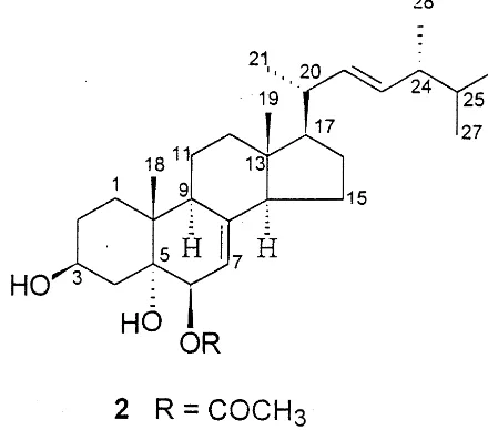
The 1H and 13C NMR spectra of compound 2
were closely similar to those ofIIsuggesting that it was presumably a derivative of 3b,5a,6b -trihy-droxyergosta-7,22-diene [13]. A pair of
three-pro-ton singlets at d0.46 and 0.91, along with four
methyl doublets (J=6.4 Hz) at d0.72, 0.74, 0.81
and 0.92, and double doublets at d5.12 (J=15.4,
7.2 Hz) and 5.05 (J=15.4, 8.0 Hz) was indicative
of its ergosta-22-ene skeleton. However, an acetate
singlet atd2.03 in its 1H NMR spectrum of 2 and
an IR ester absorption band at 1738 cm−1
indi-cated the presence of an acetoxy group. Further-more, a pair of mutually coupled proton signals at
d4.80 and 5.16 (each 1H, d, J=5.0 Hz) ascribed
to H-6 and H-7 suggested that sterol 2 was
6-O-acetyl derivative of 3b,5a,6b -trihydroxyergosta-7,22-diene [18]. This proposal was confirmed by its
13C NMR data, which were assigned by
compar-ing them with those of II [13]. All these evidence
established the structure of 2 as 3b,5a
-dihydroxy-6b-acetoxy-ergosta-7,22-diene, a hitherto
unre-ported ergosterol derivative.
The 1H and 13C NMR spectra of compound 3
were similar in part to those of2 indicating that it was also a 3b,5a,6b-trihydroxy-ergosta-7,22-diene derivative. However, the acetate singlet atd2.03 in
the 1H-NMR spectrum of 2 was replaced by a set
of signals including a methylene singlet at d3.69
and five proton multiplet centered at d7.33,
pre-sumably ascribable to a phenylacetyl group. The presence of this group was further confirmed by
the base peak at m/z 91 in its EI mass spectrum,
and a group of carbon resonance lines at d170.7
(C), 41.5 (CH2), 132.8 (C), 128.6 (2×CH), 129.3
(2×CH), 127.5 (CH) in the 13C NMR spectrum
of3. Furthermore, a pair of doublets (J=5.0 Hz)
at d4.87 and 5.25 arising from H-6 and H-7
demonstrated that the phenylacetyl group was an-chored on C – 6 [18]. Therefore, the structure of the new sterol 3 was determined as 3b,5a -dihydroxy-6b-phenylacetyloxy-ergosta-7,22-diene.
3.3. Spectroscopic data of the new metabolites
3.3.1. Isoprenylindole-3-carboxylic acid (1)
White needle; C14H15O2N; m.p. 54 – 55°C; IR
131.7 (C-2), 124.1 (C-3), 121.4 (C-4), 121.5 (C-5), 137.5 (C-6), 110.6 (C-7), 129.0 (C-8), 137.5 (C-9), 34.4 (C-1%), 123.5 (C-2%), 132.5 (C-3%, 17.9 (C-4%),
25.8 (C-5%).
3.3.2. 3b,5a-Dihydroxy-6b -acetoxy-ergosta-7,22-diene (2) (C-12), 43.8 (C-13), 54.7 (C-14), 21.2 (C-15), 28.0 (C-16), 55.8 (C-17), 12.2 (C-18), 17.9 (C-19), 39.4 (C-20), 21.2 (C-21), 135.4 (C-22), 132.1 (C-23), 42.8 (C-24), 33.1 (C-25), 20.0 (C-26), 19.6 (C-27), 17.6 (C-28), 170.7 and 20.0 (acetyl).
3.3.3. 3b,5a-Dihydroxy-6b -phenylacetyloxy-ergosta-7,22-diene (3)
Colorless gum; C36H52O4; IRnmax (cm−1): 3414,
Table 1
Antimicrobial activities of metabolites from the endophyte culture
Test microbes MICs (mg/ml)
2 3 III IV
1 V
75 50
Bacillus subtilis 25 75 25 75 * 75
Pseudomonas sp. 50 75 50 50 100 75 75
Candida albicans * * 50
100 50 50 *
All the 11 metabolites isolated from cultures of
the endophyteColletotrichumsp. were subjected to
antimicrobial assay. Compounds 2, 3, III and V
exhibited antimicrobial activities against the bacte-riaB. subtilis, S. aureus,S. luteaandPseudomonas sp. (MICs: 25 – 75 mg/ml), and against the fungi C. albicans and A. niger (MICs: 50 – 100 mg/ml). But
none was active against T. rubrum and C. elegans
even at the highest concentration (200 mg/ml).
Meanwhile, the growth of those bacteria could
also be inhibited by compounds 1 and IV (MICs:
25 – 75 mg/ml) (Table 1).
On the other hand, all compounds of the en-dopyhtic origin were tested for fungistatic
activi-ties to the crop pathogenic fungi Phytophthora
capisici (Phc), Rhizoctonia cerealis (Rhc), Gaeu-mannomyces graminis var. tritici (Ggt), and Helminthosporium sati6um (Hes). As summarized
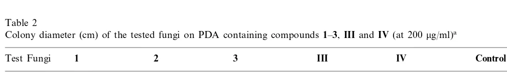
in Table 2, compounds 1, 2 and III were
fun-gistatic to Phc and Rhc, the sterols 2, 3 and IVto
Ggt, and metabolites1and 3to Hes. However, no
fungistasis could be discerned at 50 and 100mg/ml.
973; EIMS m/z (rel. int.): 548 (M+)(0.9), 412 (C-11), 39.1 (C-12), 43.7 (C-13), 54.8 (C-14), 21.1 (C-15), 28.0 (C-16), 55.8 (C-17), 12.2 (C-18), 17.9 (C-19), 40.3 (C-20), 21.1 (C-21), 135.4 (C-22), 132.2 (C-23), 42.8 (C-24), 33.1 (C-25), 19.9 (C-26), 19.6 (C-27), 17.6 (C-28), 170.7 (C-1%), 41.5 (C-2%),
Table 2
Colony diameter (cm) of the tested fungi on PDA containing compounds1–3,IIIandIV(at 200 mg/ml)a
1 2 3 III IV Control
aThe diameter of each inoculum is 0.5 cm. Ggt, Gaeumannomyces graminis var. tritici; Phc, Phytophthora capisici; Rhc,
On the other hand, the study concerned only one of the 32 endophytes isolated. Our preliminary assay also indicated that some of the other endo-phyte isolates are also capable of synthesizing bioactive substances. This observation suggested that A. annua endophytes may have pharmaceuti-cal and/or agricultural potential. Further investiga-tion concerning the topic is highly desired.
Acknowledgements
This work was financed by grants for RXT from NNSF (No.39725033 and 39670873) and for HL from CPSF (No. 1998-06-23).
References
[1] G.A. Strobel, D.M. Long, Endophytic microbes embody pharmaceutical potential, ASM News 64 (1998) 263 – 268.
[2] G.C. Carroll, Fungal endophytes in stems and leaves: from latent pathogen to mutualistic symbiont, Ecology 69 (1988) 2 – 9.
[3] A. Stierle, G.A. Strobel, D. Stierle, Taxol and taxane production byTaxomyces andreanae, an endophytic fun-gus of Pacific yew, Science 260 (1993) 214 – 216. [4] J.C. Lee, E. Lobkovsky, N.B. Pliam, G.A. Stroble, J.
Clardy, Subglutinol A and B: immunosuppressive com-pounds from the endophytic fungus Fusarium cubgluti
-nans, J. Org. Chem. 60 (1995) 7076 – 7077.
[5] G.A. Stroble, W.M. Hess, Glucosylation of the peptide leucinostatinA, produced by an endophytic fungus of European yew, may protect the host from leucinostatin toxicity, Chem. Bio. 4 (1997) 529 – 536.
[6] B. Schulz, U. Wanke, S. Draeger, H.J. Aust, Endophytes from herbaceous plants and shrubs, effectiveness of sur-face sterilization methods, Mycol. Res. 97 (1993) 1447 – 1450.
[7] D. Wilson, Fungal endophytes: out of sight but should not be out of mind, Oikos 68 (1993) 378 – 384.
[8] J.C. Wei, Manual of Fungal Identification, Shanghai Science and Technology Press, Shanghai, 1979, pp. 463 – 476.
[9] M.P. Ho, H.S. Chung, M.L. David, Pulsed high-pressure liquid injection of biological molecules into supersonic beam/mass spectrometry with resonant two-photon ion-ization detection, Appl. Spectroscopy 42 (1988) 1200 – 1206.
[10] H. Chiji, Y. Arakawa, S. Ueda, M. Kuroda, M. Izawa, 5,2%-dihydroxy-6,7-methylenedioxyisoflavone from seed balls of sugar beet, Phytochemistry 25 (1986) 281 – 282. [11] G. Burton, A.A. Ghini, E.G. Gros,13C NMR spectra of
substituted indoles, Mag. Reson. Chem. 24 (1986) 829 – 831.
[12] R.J. Cushley, J.D. Filipenko, 13C fourier transform
4. Discussion
Previously, Colletotrichum spp. such as C.
gloeosporioides [19,20], C. magna [21] have been reported as endophytes of some plants. To our
knowledge, Colletotrichum sp. was ascertained for
the first time to be an endophyte inside the repre-sentative of the family Asteraceae. Among the eleven metabolites characterized from the culture ofColletotrichumsp., the new compounds1–3and known ergosterol derivatives (III, IV and V) were inhibitory against Gram-negative and -positive
bacteria, such as Pseudomonas sp. and B. subtilis
Table 1. Among these antibacterial metabolites, the sterols 2, 3, III and V were antifungal, too, and compounds1–3, IIIandIVfungistatic to the crop pathogenic fungi at 200 mg/ml. In addition to the antimicrobial activity, sterol V was previously re-ported to be antiviral [22]. These findings suggested
the possibility that the endophyte Colletotrichum
sp. inA.annuacould protect the host by producing metabolites, which may be toxic or even lethal to phytopathogens. IAA is a well-known important auxin that can regulate plant physiological
pro-cesses at low concentrations. Sterol III was
re-ported to possess plant growth stimulating activity
[23]. That the endophyte Colletotrichum sp. can
produce plant growth regulators such as IAA, raised a possibility that the presence of endophytic fungus inA.annuacould regulate the growth of the host. In conclusion, the characterization of antimi-crobial and plant growth regulatory metabolites
from the culture ofColletotrichumsp. showed that
N.M.R. XIII-reassignment of the13C spectrum of
ergos-terol, Org. Magn. Reson. 8 (1976) 308 – 309.
[13] M. Kobayashi, M.M. Krishira, K. Ishida, V. Anji-aneyulu, Marine sterols XXIV: isolation of 24-methylenecholestane-1a,3b,5a,6b,16b-pentol from
Sinulariasp. of soft coral, Chem. Pharm. Bull. 40 (1992) 2845 – 2846.
[14] L.M. Zeng, Z Zeng, J.Y. Su, Studies on the steroids of marine sponge, Chem. Res. Chin. Univ. 11 (1995) 174 – 177.
[15] M. Kobayashi, M.M. Krishira, K. Ishida, V. Anji-aneyulu, Marine sterols XXII: occurrence of 3-oxo-4, 6, 8(14)-triunsaturated steroids in the sponge Dysidea herbacea, Chem. Pharm. Bull. 40 (1992) 72 – 74. [16] W.G. Ma, X.C. Li, D.Z. Wang, C.R. Yang, Ergosterol
peroxides from Cryptoporus 6ol6atus, Yunnan Zhiwu Yanjiu 16 (1994) 196 – 200.
[17] W.R. Abraham, G. Schmeda-Hirschmann, 24(s)-3b -hy-droxy-ergosta-5-en-6-one from Cyttaria johowii, Phyto-chemistry 36 (1994) 459 – 461.
[18] F. Cafieri, E. Fattorusso, M. Gavagnin, C. Santacroce, 3b, 5a, 6b-trihydroxysterols from the mediterranean
bry-ozoanMyriapora truncata, J. Nat. Prod. 48 (1985) 944 – 947.
[19] I. Okane, A. Nakagiri, T. Ito, Preliminary study of endophytic fungi in evergreen plants from Ishigaki and Iriomote Islands, Institute for Fermentation Research Communications (Osaka) 0 (1997) 45 – 51.
[20] C.S. Smith, T. Chand, R.F. Harris, J.H. Andrews, Colo-nization of a submersed aquatic plant, eurasian water milfoilMyriophyllum spicatum, by fungi under controlled conditions, Appl. Environ. Microbiol. 55 (1989) 2326 – 2332.
[21] S. Freeman, R.J. Rodriquez, Genetic conversion of a fungal plant pathogen to a nonpathogenic, endophytic mutualist, Science 260 (1993) 75 – 78.
[22] U. Lindequist, A. Lesnau, E. Teuscher, H. Pilgrim, An-tiviral activity of ergosterol peroxide, Pharmazie 44 (1989) 579 – 580.
[23] A.M Rashkes, A.U Kariev, A.A Umarov, M.M Kiktev, N.K Khidyrova, Ya.V Rashkes, Kh.M Shakhidoyatov, Composition and growth-stimulating activity of sec-ondary metabolites of cotton leaves, Khim. Prir. Soedin. 4 (1995) 614 – 617.