Corresponding author: [email protected]
Clinical characteristics of adult uncorrected
secundum atrial septal defect: a pilot study
Lucia Krisdinarti1, Anggoro Budi Hartopo1*, Dyah Wulan Anggrahini1, Ahmad Hamim Sadewa2, Abdus Samik Wahab1,3, Budi Yuli Setianto1,
1Department of Cardiology and Vascular Medicine, Faculty of Medicine/Dr. Sardjito Gen-eral Hospital, 2Department of Biochemistry, Faculty of Medicine, 3Department of Child Health, Faculty of Medicine/Dr. Sardjito General Hospital, Universitas Gadjah Mada, Yogyakarta, Indonesia
DOI: http://dx.doi.org/10.19106/JMedSci004802201603
ABSTRACT
Atrial septal defect (ASD) is the most frequent congenital heart disease in adulthood. Pulmonary hypertension (PH) complicating ASD compels patients seeking medical assistance because of its disabling symptom. Most adult ASD develop PH which render signiicant morbidity and mortality. The aim of the study is to characterize the clinical proiles of adult patients with ASD. The study design was cross sectional. The subjects were enrolled consecutively from outpatient clinics and inpatient wards. The demography, medical and imaging data were collected and recorded in case report form. Descriptive statistics was applied to characterize the subjects. Seventy-six subjects were enrolled. The majority were women (77.6 %) in the productive and child-bearing ages (63.2%). The most common symptoms were dyspneu on effort, fatigue, and palpitation. Most subjects had functional capacity of WHO class functional II (70.2 %). The mean oxygen
saturation was 96.4 %. Based on the echocardiography examination, 77.6% of subjects had suffered from PH. The mean longest diameter of defects were 2.7 cm. The direction of blood low was mostly left to right (77.6 %). Left and right ventricle function were within normal limit. Right heart catheterization showed mean left atrial pressure 11.5 mmHg, which conirmed the precapillary or arterial PH. Mean pulmonary artery pressure was 42.0 mmHg. The pulmonary artery resistance index mostly less than 4 Wood Unit/
m2 (63.7 %), indicating the feasibility to close the defect. Whereas 24.6 % of subjects were contraindicated for closing. Pulmonary artery hypertension (PAH) was diagnosed in
77.6 % subjects, meanwhile 13.2 % had borderline PAH. In conclusion, most adult ASD
patients had developed PAH, mostly young women in productive ages, mainly visited
hospital due to symptom of PH, the direction of low predominantly left to right side and
mostly had reduced functional capacity.
ABSTRAK
Subjek diseleksi secara berurutan dari klinik rawat jalan dan rawat inap. Data demograi, medis dan pencitraan dikumpulkan dan dicatat dalam suatu blangko laporan kasus. Penelitian deskriptif dilakukan untuk menilai karakteristik subjek. Sebanyak tujuh puluh enam subjek dilibatkan dalam penelitian. Sebagian besar subjek adalah wanita (77,6%) usia muda, produktif dan subur (63,2%). Gejala paling sering adalah sesak saat aktivitas, mudah lelah, dan berdebar. Sebagian besar pasien masuk dalam kelas fungsional WHO II (70,2%). Rerata saturasi oksigen 96,4%. Berdasar hasil ekokardiograi, sebanyak 77,6% subjek telah mengalami HP. Rerata diameter terpanjang defek adalah 2,7 cm. Arah aliran darah melewati defek sebagian besar kiri ke kanan (77,6%). Fungsi ventrikel kanan dan
kiri dalam batas normal. Kateterisasi jantung kanan menunjukkan rerata tekanan atrium
kiri sebesar 11,5 mmHg yang menandakan HP prekapiler/arteri. Rerata tekanan arteri
pulmonalis sebesar 42,0 mmHg. Nilai indeks tahanan arteri pulmonalis sebagian besar kurang dari 4 Wood unit/m2 (63,7%) yang menunjukkan kemungkinan bisa dilakukan
penutupan defek. Sedangkan 24,6% subjek kontraindikasi untuk penutupan. Hipertensi arteri pulmonalis (HAP) didiagnosis pada 77,6% subjek, sedangkan 13,2% subjek
mengalami HAP borderline. Sebagai kesimpulan, sebagian besar DSA dewasa telah mengalami HAP, sebagian besar wanita muda yang berusia produktif, sebagian besar mengunjungi rumah sakit karena gejala HP, arah aliran sebagian besar dari kiri ke kanan dan sebagian besar telah mengalami penurunan kapasitas fungsional.
Keywords: atrial septal defect - pulmonal hypertension - pulmonary artery hypertension – echocardiography - symptoms
INTRODUCTION
Atrial septal defect (ASD) is a congenital heart defect marked by an opening in the interatrial septum. The gap triggers the blood
lowing from left atrium to right atrium
initially. Atrial septal defect is one of the most common acyanotic congenital heart diseases. Its clinical manifestation often so indistinct that it is frequently missed during chilhood. Undiagnosed ASD, therefore uncorrected, may progress to develop pulmonary hypertension (PH). The disabling symptom of PH compels patients to seek medical help, usually in young adult period. Hence, ASD is the most common congenital heart disease encountered in adulthood.1
On the whole, the prevalence of congenital
heart defect is estimated to be 81.4 infants per 10,000 births (0.8% of live births).2 Among them, ASD constitutes 6 % of all congenital
heart diseases, with woman mostly affected
by the ratio of 2:1 as compare to man.3 The
prevalence of secundum ASD is approximated to be 10.3 per 10,000 live births.2 Based on location of the opening, ASD can be divided into three cathegories, i.e. primum, secundum and sinus venosus ASD. Secundum ASD is
the most common type, whichaccounts for greater than 80% of all ASD.2
The force and direction of blood low
across the opening of ASD depend on pressure and hemodynamic of right and left
ventricles. Right ventricle compliance and low pressure creates left to right blood low which subsequently overlowing into pulmonary
vasculatures. Overtime, it produces increased pulmonary vascular resistance and subsequent PH and reduces right ventricle compliance.
The reversal of low, from right to left side, succeedingly occurs and worsens patients
clinical condition.4 Patients with ASD and
PH have signiicant morbidity and mortality
ASD, while correction for the defect remain
controversial.5 Currently no available local and national data in adult ASD have been published. Conforming the guideline for treatment of PH due to untreated congenital heart disease,5 it is necessary to characterize
the adult patients with ASD and PH in order to
better understand the pathophysiology of the disease and guide suitable treatment. The aim of the study is to characterize the clinical data
of adult patients with ASD and PH.
MATERIALS AND METHODS
Subject
The research design was a cross sectional. The subjects of the research were adult patients with ASD whom visited outpatient clinics or admitted to inpatient wards of Dr. Sardjito
General Hospital, Yogyakarta, Indonesia.
The time of enrollment was June 2012 until December 2013. The subjects were enrolled
consecutively and given informed consent to participate in the research. The inclusion
criteria was (1) patients with unoperated
or unclosed secundum ASD, (2) patients >18 years of age and (3) patients have been performed transthoracal echocardiography and right heart catheterization. The exclusion
criteria was (1) patients with other congenital
heart defect and (2) pregnant patients.
Protocol of the study
Demography data was collected and recorded. The WHO functional class was determined based on WHO classiication on
functional daily activity limitation due to PH, ranging from I–IV.5 The 6 minute walk test
was performed by walking as convenient as possible in level surface within 6 minutes, the distance of walking was measured and
recorded. The oxygen saturation was detected by ingertip pulse oxymetri. Laboratory
data was obtained from hospital laboratory. Transthoracal echocardiography was
per-formed by a sonographer and interpreted by a cardiologist consultant. Pulmonary hypertension based on echocardiography
was determined by estimating pulmonary
artery systolic pressure (PASP) using standard
formula. The PASP ≥40 mmHg was determined as PH. The right heart catheterization was
performed and interpreted by a cardiologist
consultant. At the time of study, Swan-Ganz
catheter to measure pulmonary capillary
wedge pressure was not available in our hospital, therefore left atrial pressure was
determined as a surrogate. Pulmonary artery
hypertension (PAH) was determined as value
of mean pulmonary artery pressure (mPAP)
≥25 mmHg and left atrial pressure (LAP) ≤15 mmHg. Other parameters were obtained from standard formulas. All intervention was part of standard care of the patients and was not inluenced by this research. The protocol
of study had been approved by Medical and Health Research Ethics Committee, Faculty of Medicine Universitas Gadjah Mada.
Data analysis
The descriptive statistics was employed.
Since the research did not aim to test the
hypothesis, speciic statistics analysis was not
applied.
RESULTS
We enrolled 76 subjects to be included in the study based on research criteria. All of the subjects had secundum ASD. The
majority of the subjects were women (77.6%) and were in young, productive and child-bearing ages [i.e. 18-40 years of age (63.2 %)]. The most common symptoms urging the
subjects to seek medical help were dyspneu
and palpitation (13.2%). Most patients were underweight (47.8%) as indicated by body mass index (BMI) <18.5. TABLE 1 showed
the demographic characteristics of subjects
with secundum ASD.
TABLE 1. Demographic characteristics of secundum
ASD patients Dispneu on effort 36 (47.4) Easily fatique 12 (15.8)
Based on WHO functional class, most
subjects were classiied in class functional II (71.2%), whereas only 8.1% subjects were in WHO class functional I. About 20%
subjects had disabling symptoms (WHO class
functional III-IV). Functional capacity of the subjects, measured with 6 minute walk test, was reduced. The mean walking distance was
336.0 m, while normal subjects can reach more
than 700 m. The oxygen saturation detected
by pulse oxymetri showed mean saturation of 96.4 %, which was within normal acceptable value. Mean haemoglobin value was 13.1 g/dL which was considered normal level. TABLE 2 showed the clinical and laboratory characteristics of subject with secundum ASD.
TABLE 2. Clinical and laboratory characteristics of
secundum ASD patients
Characteristics Frequency WHO functional class [n (%)] 76 (100)
l I 6 (7.8)
6 minute walk test [mean±SD
(m)]
Laboratory [mean±SD]
336.0±144.6
Haemoglobin (g/dL) 13.1±2.0 Hematocrit (%) 40.2±5.5
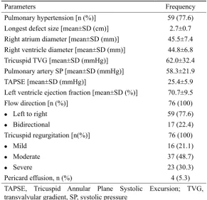
The echocardiography is an important tool to diagnose ASD and estimate pulmonary artery pressure. Based on the transthoracal echocardiography examination, 77.6% of subjects had suffered from PH, indicated by PASP >40 mmHg. Measurement of defects in interatrial septum revealed the mean longest
diameter of ASD were 2.7 cm. Most subjects
had right atrium and right ventricle chamber
across the shunt was mostly left to right side, i.e. 59 subjects (77.6%), whereas bidirectional shunt was observed in 17 subjects (22.4%).
Severe tricuspid regurgitation occurred in 23 subjects (30.3%), moderate regurgitation in
TABLE 3. Echocardiographic indings in ASD patients
Parameters Frequency
Pulmonary hypertension [n (%)] 59 (77.6)
Longest defect size [mean±SD (cm)] 2.7±0.7 Right atrium diameter [mean±SD (mm)] 45.5±7.4 Right ventricle diameter [mean±SD (mm)] 44.8±6.8 Tricuspid TVG [mean±SD (mmHg)] 62.0±32.4 Pulmonary artery SP [mean±SD (mmHg)] 58.3±21.9 TAPSE [mean±SD (mmHg)] 25.4±5.9
Left ventricle ejection fraction [mean±SD (%)] 70.7±9.5
Flow direction [n (%)] 76 (100)
l Left to right 59 (77.6)
l Bidirectional 17 (22.4)
Tricuspid regurgitation [n(%)] 76 (100)
l Mild 16 (21.1)
l Moderate 37 (48.7)
l Severe 23 (30.3)
Pericard effusion, n (%) 4 (5.3) TAPSE, Tricuspid Annular Plane Systolic Excursion; TVG, transvalvular gradient, SP, systolic pressure
Right heart catheterization is an important diagnostic tool to visualize ASD and a gold standard to assess the presence of PAH. It is a crucial step to evaluate the indication of ASD closure, since hemodynamic data can
be obtained. Mean left atrial pressure (LAP) was 11.5 mmHg. It was a surrogate for pulmonary capillary wedge pressure (PCWP), which conirmed the diagnosis of precapillary
or arterial PH in ASD. Mean value of mean
pulmonary artery pressure (mPAP) was 42.0 mmHg. Subjects with mPAP ≥ 25 mmHg and LAP ≤ 15 mmHg were cathegorized as having
PAH. Therefore, 59 subjects (77.6%) had
37 subjects (48.7%) and mild regurgitation in 16 subjects (21.1%). Pericardial effusion
was detected in 4 subjects (5.3%). TABLE 3
depicted the transthoracal echocardiographic
parameters of subjects with secundum ASD.
suffered from PAH, meanwhile 10 subjects (13.2%) had borderline PAH (mPAP 21 - 25
mmHg) and 7 subjects (9.2%) had no PAH. Based on pulmonary artery resistance index (PARI) value (only calculated in 71 subjects), 63.7% subjects had PARI <4 Wood Unit/m2, indicating the feasibility to close the ASD. The PARI value >8 Wood Unit/m2, which is a contraindication to close the defect, occurred
in 24.6% subjects. Mean low ratio was 2.9 and mean Qp/Qs was 1.3, indicating that the
majority of subjects had excess pulmonary
low. TABLE 4 showed the right heart
TABLE 4. Right heart catheterisation results of secundum ASD patients
Parameters Frequency
Left atrial pressure [mean±SD (mmHg)] 11.5±5.3 Mean PA pressure [mean±SD (mmHg)] 42.0±21.8
Flow ratio [mean±SD] 2.9±1.9
PARI [n (%) (Wood Unit/m2)] 71 ( 100)
l < 4 44 (61.9)
l 4-8 8 (11.2)
l > 8 17 (23.9)
Qp/Qs [mean±SD] 1.3±0.8
Systemic oxygen saturation [mean±SD (%)] 91.4±7.5 Pulmonary artery hypertension [n (%)] 76 (100)
l Yes 59 (77.6)
l Borderline 10 (13.2)
l No 7 (9.2)
PARI, pulmonary artery resistance index, PA, pulmonary artery
DISCUSSION
Our research was the irst study describing
the demography, medical and imaging data
of adult patients with uncorrected secundum ASD in Indonesia. Most patients were undiagnosed previously and unaware that
they had congenital heart disease. Indeed, among congenital heart diseases, ASD often asymptomatic or only demonstrates mild atypical symptom during childhood. Its acyanotic nature and functionally tolerable, supplemented by no good screening system during childhood, makes ASD the most common congenital heart disease encountered in adult life in developing countries. Current availability of imaging technique and expertise in our centre, i.e. right heart catheterisation, bring about the accurate diagnosis, supportive data for closure the defect and evaluation of
treatment. However, most adult ASD with
PH could not be treated curatively by defect closure because of its PAH complication. The
closure will be deteriorating for such patients.
Therefore, reliance to medication for reducing
pulmonary artery pressure is encouraged to improve quality of life. The data of the characteristics of such patients is of paramount
important to provide epidemiological proile and burden of disease which affect the local
and national health policy.
Our data showed that the majority of patients were young adult women, which
account for nearly 80%. Epidemiology study
showed that ASD are not uncommon and
can present at any phase of age. Women constitute around 65% until 75% in patients
with secundum ASD.3 Since all of our patients were secundum ASD, it conirmed previous epidemiology data. Most of the patients was in child-bearing ages which pose greater risk
of morbidity and mortality if they become
pregnant. Furthermore, those with PH will
be contraindicated for pregnancy due to increasing mortality rate. Unlike secundum ASD, gender distribution is similar for ostium
primum ASD and sinus venosus defect.3
Most of the patients had been symptomatic
easily fatique and palpitation were the
most common complaints urging patients to visit hospital. Pulmonary hypertension
complicating ASD is encountered in 6-35%
of untreated patients in later adulthood.6 The presence of PH causes progressive dyspneu, presyncope, syncope, edema and functional limitations.7 Furthermore, right ventricular volume overload and right ventricular dysfunction generate more progressive sign and symptom.6 Palpitation is possibly the indication of impending heart failure or arrhytmia. Supraventricular arrhythmia, due to stretching of the conduction system, may be
the irst presenting sign of an ASD.8 In patients
with uncorrected ASD, the clinical parameters tend to worsen over time.6 Nevertheless, a
substantial number of patients with open ASD
have normal parameters, usually the patients
with small defects in whom the hemodynamic
parameters remained stable during several
years of follow up.6 In our cohorts, despite developing PH, around 6% of patients come in later life (sixth decade). The long period of asymptomatic or mild symptom may
be associated with small ASD. Large ASD
(i.e. defect >2cm) are at risk to develop
Eisenmenger syndrome, which prevalence
approximately 10 %.9 It is still unknown as
to whether the PH does not develop in some patients, while in others it progresses rapidly
regardless the defect size.
Based on echocardiogram parameters, most of patients in our study have advanced into
PH. The mean longest defect size was >2cm, which is a signiicantly large defect . This
may partially explain the pathophysiology of PH as a natural history of large ASD defects. Right ventricular volume overload has been
detected in all patients which contribute to
progressing symptom in about 90% patients
with WHO functional class more than II. The
right ventricular dysfunction and right heart
failure have not yet developed in our study patients, indicated by normal tricuspid annular plane systolic excursion (TAPSE) value. Right ventricular noncompliance occurs in fetal life as a result of high pulmonary vascular
resistance allowing the right to left low at the
atrial level. Instantly after delivery, the right ventricular compliance is similar to that of left
ventricle, therefore only little shunting low
through the defect.8 Overtime, pulmonary vascular resistance decreases physiologically
and right ventricular wall becomes thinner,
resulting in reduced compliance and left to
right blood low which develop in children
and young adult period.8
Right heart catheterisation is a tool to
conirm PAH. Pulmonary artery hypertension
related to ASD is included in group I of
PH clinical classiication.5 In addition to diagnose PAH and exclude post capillary PH, right heart catheterisation is required to assess hemodynamic impairment, conduct vasoreactivity test dan decide the operability of defect.5 Our study population indicated that most patients experience PAH (almost 80%). Before performing right heart catheterisation, estimation of PH should be
made by echocardiography. Those with high
probability of PH in echocardiogram need to
be conirmed with right heart catheterisation.5 While 77.6% of subjects had PH based on echocardiography result, the right heart catheterisation consistently detected similar proportion of subjects had PAH. It means, about 20% of subjects could be excellent candidates for ASD closure, because they did not develop PAH. In addition to mPAP level, the decision to close the defect can be reached
by evaluating PARI value. Our data showed the wide range of PARI value, from 0.4 to
33.3 Wood Unit/m.2 The current consensus
suggests that patients with a PARI value <4
with PARI >8 Wood Unit/m2 have inoperable disease.5 Derived from the consensus, 61.9% subjects can be closed and 23.9% subjects
were contraindication to be closed. The rest, 11.2% subjects, were uncertain. However,
some clinicians suggest to give aggresive drugs to reduce PARI value, in the hope that
those with respond to the medication can beneit from closing the ASD.10 While closing
ASD mostly curative, some patients still develop some degree of PAH and therefore
they still need PH lowering medication. The higher proportion of young women in child-bearing age with secundum ASD suffering from signiicant PH raises question as to whether any mechanisms, other than
hemodynamics and anatomy, present that
make PH comes early. Larger data, such as registry, is necessary to answer the question
through investigating the natural history or
novel mechanism associating ASD with PH.
Furthermore, screening for congenital heart disease should be initiated as early as possible, because if it is detected during childhood the
deinitive treatment can be applied and the PH
complication can be avoided. Unfortunately, currently no screening system available in
Indonesia which mandate the simple cardiac
examination such as auscultation to cardiac murmur or electrocardiography.
CONCLUSION
Most adult secundum ASD patients had
developed PAH, mostly young women in
productive ages, mainly visited hospital due to symptom related to PH, the direction of
blood low mostly left to right side and mostly
had reduced functional capacity.
ACKNOWLEDGMENTS
We were grateful to Monika Setiawan,
MD and Aristida Cahyono, MD for the clinical
data preparation. We were indebted to Ms. Sri
Mardilah Wuryani, AMK for her excellent transthoracal echocardiogram acquisition.
REFERENCES
1. Beghetti M, Galiè N. Eisenmenger syndrome
a clinical perspective in a new therapeutic era of pulmonary arterial hypertension. J Am Col Cardiol 2009; 53(9):733-40. http://dx.doi.org/
1016/j.jacc.2008.11.025.
2. Reller MD. Prevalence of secundum atrial
septal defect and associated indings, atrial
septal defect. In: Rao S (ed.) Atrial Septal
Defect. 31-36. Croatia: InTech, ISBN: 978-953-51-0531-2
3. Webb G, Gatzoulis MA. Atrial septal defects
in the adult: recent progress and overview. Circulation 2006; 114(15):1645-53.
4. Fuster V, Alexander RW, O’Rourke RA, et al. Hurst’s the heart, ed. 11. New York: McGraw
Hill. 2005; Vol 2.
5. Galiè N, Humbert M, Vachiery JL, Gibbs S, Lang I, Torbicki A, et al. 2015 ESC/ERS
Guidelines for the diagnosis and treatment of pulmonary hypertension: the joint task force for the diagnosis and treatment of pulmonary hypertension of the European Society of Cardiology (ESC) and the European Respiratory Society (ERS): Endorsed by: Association for European Paediatric and Congenital Cardiology (AEPC), International
Society for Heart and Lung Transplantation (ISHLT). Eur Heart J 2016; 37(1):67-119.
http://dx.doi.org/ 10.1093/eurheartj/ehv317. 6. Engelfriet P, Meijboom F, Boersma E,
Tijssen J, Mulder B. Repaired and open
atrial septal defects type II in adulthood: an epidemiological study of a large European
cohort. Int J Cardiol 2008; 126(3):379-85.
7. Toyono M. Pulmonary arterial hypertension
in adults with atrial septal defect. J Cardiol
8. Sommer RJ, Hijazi ZM, Rhodes JF Jr.
Pathophysiology of congenital heart disease in the adult Part I: shunt lesions. Circulation
2008; 117(8):1090-9. http://dx.doi.org/ 10.1161/CIRCULATIONAHA.107.714402. 9. Huang JB, Liang J, Zhou LY. Eisenmenger
syndrome: not always inoperable. Respir
Care 2012; 57(9):1488-95.http://dx.doi.org/
10.4187/respcare.01418.
10. Nakanishi T. Pulmonary arterial hypertension
associated with congenital heart disease. Personal perspectives. Int Heart J 2015; 56 Suppl:S1-3.