Ventricular Septal Defect Closure with Perforated Patch in Large Ventricular Septal Defect with Severe Pulmonary Hypertension and Non Reactive Oxygen Test
Budi Yuli Setianto, Hariadi Hariawan, Rano Irmawan
Department of Cardiology and Vascular Medicine, Faculty of Medicine Universitas Gadjah Mada – Dr. Sardjito Hospital, Yogyakarta, Indonesia
Abstract
Management of ventricular septal defect (VSD) with severe pulmonary hypertension (PH) had not been extensively studied and is still challenging. The closure of VSD in patients with high pulmonary vascular resistance (PVR) and severe PH is highly risk procedure. If high PVR and severe PH still persist after closure procedure, the patients have poor prognosis. We reported a 24-year-old woman whom was diagnosed with large VSD, bidirectional shunt with L to R dominance, dilatation of left ventricle, and mild to moderate mitral regurgitation, mild tricuspid regurgitation and severe PH. Right heart catheterization showed pre-oxygen test: mean aorta pressure 85 mmHg, mean pulmonary artery pressure 65 mmHg, fl ow ratio 5,4, PVR 2,3 WU and pulmonary vascular resistance index (PVRI) 3,22 WU/m2. The results of post-oxygen test: mean aorta pressure 83 mmHg, mean pulmonary artery pressure 63 mmHg, fl ow Ratio 2,2, PVR 0,3 WU and PVRI 0,42 WU/m2. Patient had been performed VSD closure with perforated patch 3 mm. Three month evaluation by echocardiography showed residual VSD 3 mm, L to R shunt, moderate tricuspid regurgitation and mild PH (TVG 36 mmHg). In Baumgartner criteria of VSD operability, this patient was not operable because the ratio of mean pulmonary and systemic circulation more than 2/3, but in Lopez criteria, patient is operable because PVRI below 6 WU/m2. Patient with high and moderate PH and PVR which is still operable, VSD can be closed partially. Partially VSD closure can be performed by transcatheter procedure after PH decrease and stable.
Keyword: VSD closure- perforated patch – PH severe
Background
The management of ventricular septal defect (VSD) with severe pulmonary hypertension (PH) had not yet extensively investigated.1 The closure procedure in VSD patients with increasing pulmonary vascular resistance (PVR) with severe PH is a risky procedure. The persistence of high PVR and severe PH after the closure procedure is the sign of a bad prognosis.1 In a restropective study, patient with congenital heart disease and severe PH post operatively had worse prognosis than patient with non-operative congenital heart disease.2 In this case we present and discuss a patient with large VSD, severe PH and non reactive oxygen test in whom the VSD closure was performed.
Case
A woman 24 years old came to our hospital with a chief complaint of dyspnea. She felt dyspnea on effort since 10 months before admission, there was neither orthopnea nor paroxysmal nocturnal dyspnea. Patient had been diagnosed as large VSD since April 2014 and has been planned to have VSD closure.
Her physical examination demonstrated a good general condition and compos mentis. Blood pressure was 100/60 mmHg, heart rate was 82 bpm, respiratory rate was 22 x/minute, temperature was 36,5⁰C and peripheral oxygen saturation was 97%. No increased jugular venous pressure. Lung examination was normal. There was cardiomegaly with ictus cordis at intercostal space V left mid clavicula line. In auscultation, S1 was normal and S2 was louder, with holosystolic murmur grade 3/6 with punctum maximum at intercostal space III-IV left parasternal border. Abdominal and extremity examination were normal
Chest X-ray examination in showed cardiomegaly (CTR > 0,5) and a protuding of pulmonary segment indicating the presence of pulmonary hypertension (fi gure 2).
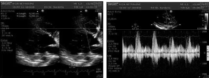
Echocardiography examination results showed a large VSD with diameter 17-20 mm, bidirectional shunt with L-to-R shunt dominance, dilatation of left ventricle with LV diameter 55 mm, ejection fraction 70%, diastolic dysfunction Figure 1. Electrocardiogram showed sinus rhtythm, heart rate 76 bpm and normoaxis
relaxation type, normal right ventricle function, mild to moderate mitral regurgitaion, mild tricuspid regurgitation and severe PH (TVG was 74 mmHg). Right heart catheterization results showed: pre-oxygen test mean aorta pressure was 85 mmHg, mean pulmonary artery pressure was 65 mmHg, fl ow ratio was 5,4, PVR was 2,3 WU and PVRI was 3,22 WU/m2. Post oxygen test: mean aorta pressure was 83 mmHg, mean pulmonary artery pressure was 63 mmHg, fl ow ratio was 22, PVR was 0,3 WU and PVRI was 0,42 WU/m2.
The patient had been done VSD closure in February 2015 with perforated patch 3 mm. The evaluation by echocardiography done in April 2015 showed residual VSD with 3 mm diameter, L to R shunt, normal cardiac chambers, ejection fraction 63%, uncoordinated motion, normal diastolic LV function and normal right ventricle function, moderate tricuspid regurgitation and mild PH (TVG 36 mmHg) (fi gure 3). The patient was hospitalized within 8 days and discharge in stable condition. The peripheral oxigen saturation was 97%. The medication was sildenafi l 3 x 20 mg.
Discussion
Pulmonary hypertension can occur in congenital heart disease because of increasing pulmonary blood fl ow due to L-to-R shunt through the defect. An L-to-R shunt will increase arterial pulmonary pressure and cause a stress to the vessel wall, circumferential dilatation of wall vessel and endotelial dysfunction.3 There is a changing expression of vasoactive mediator such as endothelin-1, prostacyclin and nitrit oxide. Pulmonary vasoconstriction will occur and persist. The increased expression of vascular endothelin-1 and fi broblast growth factors cause remodeling (hypertrophy and proliferation) of vascular smooth
muscle cells and increasing of intracellular matrix deposition. This remodelling will cause increasing of PVR and subsequent right ventricle pressure.3
Patient with moderate to large size congenital heart defect can suffers from PH but not pulmonary vascular disease (PVD) because there is no extensive and permanent remodeling in pulmonary vessel walls. Patient with severe PH and high pulmonary fl ow and normal PVR, may be considered to have VSD closure because the lesion in vessel walls is not yet extensive and may still be reversible. High PVR indicated that such patients have suffered from extensive and irreversible PVD.4 The closure of VSD in patient with moderate to large congenital defect, L to R shunt and moderate to high PVR is very high risk procedure because the extent of lesion in pulmonary vessels is unknown and there is no guarantee for good long term outcome.4 If PVR is still high in post operation, the patient’s prognosis is worse.4 A study in England reported that children whom suffered from severe PH post operatively had worse prognosis than patients with Eisenmenger syndrome.2
When the right heart catheterization was done, a vasoreactive test was usually performed. The gold standard of vasoreactivity test was using inhaled nitric oxide (iNO) not only oxygen.5 In iNOP test 1 (inhaled nitric oxide as a preoperative test 1) study showed that by using oxygen and iNOP (100% oxygen and 80 ppm iNOP) gave 97% and 90% sensitivity and accuracy, respectively, to predict the operability in patient with congenital heart disease and PH, whereas oxygen only (100% oxygen) gave 64% sensitivity and 68% accuracy.6
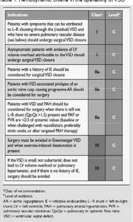
congenital heart disease.7 There is a retrospective study showed that 38 patients with VSD and severe PH and high PVR (>6 WU) was closed, resulting in 8 patients died because of severe PH post operation and 30 patients (79%) had good outcome post operation.8 It showed that PVR treshold in operable patient must be determined precisely, if the treshold too low it will exclude many patients whom actually may still be operable.8 Baumgartner et al. (2009)9 give operability hemodinamic criteria in VSD with PH according to European Society of Cardiology guideline, which is as follows:
Lopes et al. (2009) suggested a hemodynamic criteria for assessment of operability in patients with congenital heart disease and pulmonary artery hypertension. Table 2 showed the criteria suggested by Lopes et al. (2009).10
Patients with high and moderate PH and PVR which is still operable in the criteria by Lopes et al. (2009)10, VSD can be closed partially, so if severe PH still persist post-operatively, right ventricle can be decompressed.11 According to Beghetti (2012) and Myers (2014), partially VSD closure can be closed by transcatheter procedure after PH decrease and stable.4,7
ADO II in baby and Catfi eld et al. (2005)13 who reported transcatheter VSD closure in VSD patient post operatively with patch dehisence.
Conclusion
We reported a 24-year-old woman large VSD and severe PH. The patient had been done VSD closure with perforated patch 3-3,5 mm. In post operative echocardiogram showed residual VSD 3 mm, L to R shunt with improvement of PH from 79 mmHg to 36 mmHg. A VSD closure in this patient was performed by perforated patch, to anticipate if PH persist post operatively, and be closed by transcatheter procedure if PH improve and stable in the follow up.
Reference
Beghetti, M., Tissot, C. 2010. Pulmonary 1.
hypertension in congenital shunts. Rev Esp Cardiol, 63:1179-1193.
Haworth, SG., Hislop, AA. 2009. Treatment 2.
and survival in children with pulmonary arterial hypertension: The UK pulmonary hypertension service for children 2001-2006. Heart, 95:312-317.
D’Alto, M., Romeo, E., Argiento, P., Correra, P., 3.
Santoro, G., Gaio, G., Sarubbi, B, Calabrò, R., Russo, MG. 2013. Hemodynamics of patients developing pulmonary arterial hypertension after shunt closure. Int J Cardiol, 168:3797-3801. Beghetti, M., Galiè, N., Bonnet, D. 2012. Can 4.
“inoperable” congenital heart defects become operable in patients with pulmonary arterial hypertension? Dream or reality? Congenit Heart Dis, 7:3-11.
Galie, N., Hoeper, MM., Humber, M., Torbicki, 5.
A., Vachiery JL., Barbera, JA., Beghetti, M.,
Corris, P., Gaine, S., Gibbs, JS., Gomez-Sanchez, MA., Jondeau, G., Klepetko, W., Opitz, C., Peacock, A., Rubin, L., Zellweger, M., Simonneau, G. 2009. Guidelines for ther diagnosis and treatment of pulmonary hypertension. Eur Heart J, 30:2493-2537 . Balzer, BT., Kort, HW., Day, RW., Corneli, HM., 6.
Kovalchin, JP., Cannon, BC., Kaine, SF., Ivy, DD., Webber, SA., Rothman, A., Ross, RD., Aggarwal, S., Takahashi, M., Waldman, JD. 2002. Inhaled Nitric Oxide as a preoperative test (INOP Test I). Circulation, 106:76-81. Myers, PO., Tissot, C., Beghetti, M. 2014. 7.
Assessment of operability of patients with pulmonary arterial hypertension associated with congenital heart disease. do we have the good tools to predict success? Circ J, 78:5-9.
Kannan BR, Sivasankaran S, Tharakan JA, 8.
Titus T, Ajith Kumar VK, Francis B. 2003. Long-term outcome of patients operated for large ventricular septal defects with increased pulmonary vascular resistance. Indian Heart J, 55:161-166.
Baumgartner, H., Deanfield, JE., Galie, 9.
N., Gatzoulis, MA., Gohlke-Baehwolf, C., Kaemmerer, H., Kilner, P., Meijboom, F., Mulder, BJM., Oechslin, E., Oliver, JM., Serraf, A., Szatmari, A., Thaulow, E., Vouhe, PR., Walma, E. 2010. ESC Guidelines for the management of grown-up congenital heart disease (new version 2010). Eur Heart J, 31: 2915-2957.
Lopes, A ., O’Leary, PW. 2009. Measurement, 10.
interpretation and use of haemodynamic parameters in pulmonary hypertension associated with congenital cardiac disease. Cardiol Young,19:431-435.
Aasim, M., Mohammad, A., Ali, N., Zahidullah, 11.
Rehman, M. 2014. VSD closure following pulmonary arterybanding in congenital vsd with significantpulmonary hypertension. Gomal J Med Sci 2014; 12:23-6
Ramakrishnan, S., Saxena, A., Choudhary, 12.
SK. 2011. Residual VSD closure with an ADO II device in an infant. Congenit Heart Dis, 6:60–63.
Catfield, NJ., Ruygrok, PN., Wilson, NJ., 13.