The journal homepage www.jpacr.ub.ac.id ISSN : 2302 ‐ 4690
72
Effect of D-Alpha Tocopherol Therapy towards Malondialdehyde
1Level and Histology Analysis of Kidney in Rattus norvegicus with
2MLD-STZ Induction
34
Marissa Agnestiansyah,1* Aulanni’am1 and Chanif Mahdi1 5
6 1
Department of Chemistry, Faculty of Science, Brawijaya University, Jl. Veteran, Malang 65145, East Java 7
Indonesia; Corresponding author: [email protected]; Phone: +62341575838 8
9
Received 29 March 2013; Re-submitted 10 April 2013; First revised 28 May 2013; Second revised 3 June 2013; 10
Published online 5 June 2013 for edition May-August 2013 11
12
ABSTRACT 13
Diabetic Nephropathy is a kidney disease which occurs due to complication of diabetes 14
mellitus as a consequence of the damage of the kidney endothelial cells. Hyperglicemia 15
condition in patients with diabetes mellitus that induces an oxidative stress, were related 16
to endothelial cell damage. Oxidative stress as a result of hyperglycemia will activate a 17
number of signal transduction pathways resulting in increase of free radicals. D-alpha 18
tocopherol as one of antioxidant substance, that can act as an inhibitor of free radical 19
chain reactions, play an important role in the reduction of the oxidative stress effect. 20
Effect of D-alpha-tocopherol in reducing oxidative stress is identified by measuring the 21
levels of malondialdehyde (MDA) in kidney and histology of kidney. This study used 22
five groups mice; they were a control group, a diabetic group which was induced with 23
MLD-STZ, and a therapeutic groups with a varieties doses of D-alpha tocopherol (100 24
mg/kgBW, 200 mg/kgBW and 300 mg/kgBW). The results showed that the D-alpha 25
tocopherol was able to reduce the levels of malondialdehyde (MDA) and repair the 26
histology of kidney of mice induced by MLD-STZ. 27
28
Key word: diabetic nephropathy, diabetes mellitus, MLD-STZ, malondialdehyde, D-alpha 29
Complications of diabetes in the kidney is known as diabetic nephropathy. Damage to 33
the kidneys filter or glomerolus occurs in patients with diabetic nephropathy. Glomerolus 34
damage will cause some blood proteins excreted abnormally in urine. This situation is called 35
as glomerolus hyperfiltration1. 36
Glomerular hyperfiltration is due to endothelial kidneys cell damage. One of the causes 37
of endothelial cell damage is oxidative stress that occurs in diabetic people with 38
hyperglycemia2. Hyperglycemia stimulates the release of superoxide in mitochondria, 39
triggering early oxidative stress in patients with diabetes mellitus (DM). Source of oxidative 40
stress in diabetic patients proceed against non-enzimatic pathway, enzymatic and 41
mitochondrial pathways. Enzymatic sources of oxidative stress is derived from the enzymatic 42
glucose. Glucose can undergo autooxidation and generate hydroxyl radicals (•OH). In 43
addition, glucose reacts with non-enzymatic proteins that produce Amadori products 44
followed by the formation of Advanced Glycation End Products (AGEs) which increase the 45
oxidative stress. Polyol pathway in hyperglycemia also produces the radical •O2-. 46
Autooxidation process on hyperglycemia and glycation reactions will trigger the formation of 47
The journal homepage www.jpacr.ub.ac.id ISSN : 2302 ‐ 4690
73 Haber-Weis and Fenton reactions will convert the previous radicals into hydroxyl radicals 49
(•OH). Hydroxyl radicals attack Poly Unsaturated Fatty Acids (PUFAs) in cell membranes, 50
resulting in the formation of hydroperoxide lipids and MDA. The latter compound will cause 51
oxidative damage to kidney cells3. 52
The damage of oxidative stress in people with diabetes mellitus can be resisted by a diet 53
of high levels antioxidant food. One of antioxidant that serves to reduce oxidative damage in 54
diabetics is vitamin E. According to Aggarwal et al.,4 vitamin E has been shown to reduce 55
microalbuminuria and repair kidney damage in patients with diabetic nephropathy. The 56
majority of natural supplements of vitamin E are in the form of D-alpha tocopherol. D-alpha 57
tocopherol can work as a scavenger of oxygen free radicals, lipid peroxyl and singlet oxygen. 58
D-alpha tocopherol is also known as an antioxidant that can maintain the integrity of the cell 59
membrane5. 60
Vitamin E supplementation 100 IU/day significantly increases glutathione and lowers 61
lipid peroxidation and glycosylated hemoglobin (HbA1c) concentrations in the erythrocytes 62
of type 1 diabetic children patients6. Alpha tocopherol supplementation was beneficial in 63
decreasing blood lipid peroxide concentrations without altering antioxidant enzyme activities 64
in Korean patients with type 2 diabetes treated with Continuous subcutaneous insulin infusion 65
(CSII)7. Streptozotocin-induced diabetic rats receiving 200 mg/kgBW alpha tocopherol daily, 66
after 10 days reduced plasma malondialdehyde levels, increased glutathione peroxidase 67
activity and accelerated the rate of wound closure in treated rats8. Erythrocyte 68
malondialdehyde decreased and serum-total antioxidant status increased after alpha 69
tocopherol treatment 800 IU/day during 6 weeks in female type-2 diabetics9. Vitamin E 70
supplementation 1000 IU/day to diabetic type 2 patients for 2 months significantly increased 71
GSH levels and lowered MDA levels which are markers of oxidative stress and this may 72
reduce the risk of microvascular and macrovascular complications associated with diabetes 73
mellitus10. To the best of our knowledge, the use of D-alpha-tocopherol in reducing oxidative 74
stress in diabetic nephropathy has not been studied. Therefore, in this study, we observe 75
MDA levels isolation of kidney organ and histology of kidney tissue in the diabetes mellitus 76
type 1 mice that are treated with D-alpha tocopherol. 77
78
EXPERIMENTAL 79
Animals and experimental design 80
Twenty-five Rattus norvegicus (male, body weight 130-160g) were housed at room 81
temperature in the animal house of Cellular and Molecular Biology Laboratory, Mathematics 82
and Sciences Faculty, Brawijaya University Malang and were exposed to alternate cycles of 83
12 h light and darkness. The mice were divided into five groups as follows : control (non-84
diabetic) group (n = 5), diabetic group (n = 5) which is induced by multiple low dose-85
streptozotocin (MLD-STZ) for five days and incubated for fourteen days until their glucose 86
blood level was more than 300mg/dl. STZ dose used was 20 mg/kg BW for five consecutive 87
days11. Therapeutic groups are treated with variant doses of D-alpha-tocopherol (100; 200, 88
and 300 mg/kg BW) after induced by MLD-STZ. Each D-alpha-tocopherol dose group 89
contained 5 mice. At the end of the experiment, kidneys were collected by cervical 90
dislocation. The kidneys were washed with 0.9% NaCl and the left kidneys were immersed in 91
PBS for five minutes. The right kidneys were immersed in 4% PFA for seven days for further 92
kidney tissues observation. All conditions and handling animals were conducted with 93
protocols approved by Ethical Clearences Committe of Brawijaya University (121-KEP-UB). 94
The journal homepage www.jpacr.ub.ac.id ISSN : 2302 ‐ 4690
74 MDA Measurement using Thiobarbituric acid (TBA) Test
96
A kidney (1.8 gram) was homogenized with 1 mL of NaCl 0.9% in a cold condition by 97
using a block ice for conditioning. The homogenate was centrifuged at a speed of 8000 rpm 98
for 20 minutes and supernatant was taken. Then 100 μL of kidney supernatant was added by 99
550 μL aquadest, 100 μL TCA 100%, 250 μL HCl 1 N, and 100 μL Na-Thio. At each reagent 100
addition was homogenized with a vortex. The mixture was centrifuged at 500 rpm for 10 101
minutes and supernatant was taken. Furthermore, the solution was incubated in the water bath 102
at 100° C for 30 minutes and left until reach to room temperature. The samples were 103
measured at 541 nm for TBA test. 104
105
Histological analysis of kidney tissues 106
Kidneys were fixed in paraformaldehyde solution and were dehydrated with a gradual 107
ethanol series, then were embedded in paraffin to bring out ultrathin sections of kidneys. 108
Furthermore, the ultrathin sections were stained with Hematoxylen-Eosin. First, the ultrathin 109
sections were deparaffinized with xylol and rehydrated with a gradual ethanol series 110
(absolute, 95, 90, 80 and 70%) respectively for 5 minutes. Then those were soaked in 111
aquadest for 5 minutes. Furthermore, the ultrathin sections were dyed with hematoxylen and 112
were incubated for 10 minutes to obtain the best color results. Then the ultrathin sections 113
were washed with flowing water for 30 minutes and rinsed with aquadest. Next, the ultrathin 114
sections were dyed with eosin with alcohol for 5 minutes. The last steps were dehydrated 115
using a gradual series of ethanol (80%, 90%, 95%, and absolute) and cleared with xylol then 116
dried. The dried and stained ultrathin sections were mounted with entellan and were observed 117
under a microscope (Olympus BX53) with a magnification of 600 times. 118
119
RESULTS AND DISCUSSION 120
Therapeutic Effect of D-alpha tocopherol Against MDA Levels of White Rat Kidney 121
Induced MLD-STZ 122
A number of diabetic nephropathy pathogenesis pathway cause of hyperglycemia 123
increases the amount of free radicals in the body. The imbalanced condition between free 124
radicals and cellular antioxidants in the body will induce an oxidative stress and related to 125
oxidative damage. 126
One of these pathogenesis pathway was sorbitol polyol pathway. Sorbitol polyol 127
pathway activation reduces the number of reduced Nicotinamide adenin dinucleotida 128
phosphate (NADPH) which is required to convert Glutathione disulfide (GSSG) into 129
Glutathione (GSH). GSH is an important cellular antioxidant and GSH reduction will lead to 130
oxidative stress. Autooxidation glucose that occurs due to hyperglycemia is also a source of 131
hydrogen peroxide (H2O2) and superoxide (•O2-). Hydrogen peroxide and superoxide via the 132
Habber-Weis reaction include Fenton reaction step will be converted into hidroxyl radicals12. 133
Lipid peroxidation is one cause of oxidative damage which involves the reaction 134
between hydroxyl radical with Poly Unsaturated Fatty Acids (PUFA)13. Lipid peroxidation 135
which happens in the cell membrane of the kidney will cause kidney disfunctioned and it will 136
leading to the end-stage condition that called kidney or renal failure. Levels of oxidative 137
damage caused by lipid peroxidation can be checked through the measurement of MDA14. 138
Unsaturated double bond in PUFA facilitates hydroxyl radical to attack on the acyl 139
chain. PUFA becomes radical lipid through the taking of one hydrogen atom from one 140
methylene group. Lipid radicals react with oxygen in the body forming lipid peroxyl radicals. 141
The journal homepage www.jpacr.ub.ac.id ISSN : 2302 ‐ 4690
75 radicals. This reaction occurs continuously forming a chain reaction. Lipids peroxyl radical 143
have a rearrangement through cyclisation reaction to form MDA13. Lipid hydroperoxide is an 144
unstable compound and its fragmentation will produce a product such as MDA15. 145
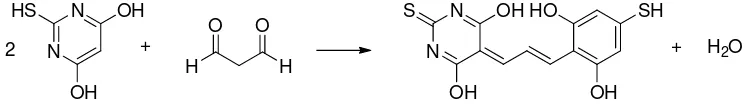
MDA level of kidney tissue was measured by TBA test. TBA test principle is a 146
condensation reaction between one molecules of MDA with two molecules of TBA in acid 147
condition as displayed in Fig. 1. Complex of MDA and TBA produced a pink color that can 148
be measured at a maximum wavelength of 541 nm. MDA levels indicates the number of lipid 149
peroxidation and cell damage that occured. The higher level of MDA was indicate the more 150
severe cell damage that occurs. 151
Figure 1. Reaction between Malondialdehyde and Thiobarbituric Acid 155
156
As shown in Table 1, the levels of MDA in diabetic mice were significantly higher 157
compared with non-diabetic mice. Therapy with D-alpha tocopherol may reduce elevated 158
levels of MDA. MDA levels declined with increasing doses of D-Alpha tocopherol used. 159
Statistical test results showed that there were significant differences (P<0,01) between MDA 160
levels of diabetic mice and therapeutic mice. It suggests that the D-alpha tocopherol able to 161
act as an antioxidant especially as a hydroxyl radical scavenger. The decline in MDA levels 162
related to the decreasing of lipid peroxidation in cell membranes that leads to the reducing of 163
cell membrane damage and inhibition of diabetes mellitus complications. 164
165
Table 1. Profile of MDA level in control, diabetic, and therapeutic mice kidney 166
167
Mice Groups MDA Level
(μg/mL)
Difference in MDA levels to Healthy
Controls (%)
Controls (non-diabetic) 0.438 ± 0.022 0.00
Diabetic 2.242 ± 0.152 412.07
Therapy 100 mg/kg BB 0.636 ± 0.092 45.26
Therapy 200 mg/kg BB 0.549 ± 0.051 25.43
Therapy 300 mg/kg BB 0.509 ± 0.052 16.38
168
Landes16 studied that the inhibition mechanism of lipid peroxidation by D-alpha 169
tocopherol which initiated when lipids (LH) lost an atom hydrogen became lipid radical (L•). 170
Lipid radicals will react with molecular oxygen to produce lipid peroxyl radical (LOO•). 171
Lipid peroxyl radicals can react with other unsaturated lipids and caused a chain radical 172
reaction. At this stage, D-alpha tocopherol will donate one H atom from its hydroxyl (OH) 173
group to lipid peroxyl radical. In the rest, this D-alpha tocopherol become non active alpha 174
tocopherol radical and can be excreted out of the body. 175
The journal homepage www.jpacr.ub.ac.id ISSN : 2302 ‐ 4690
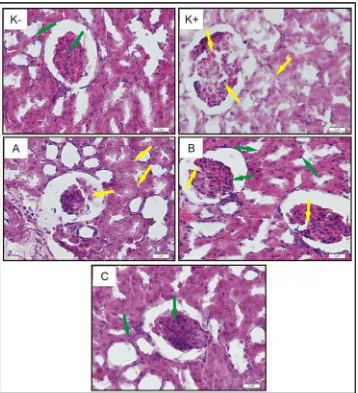
76 Histology of Kidney Tissue from Control Mice, Diabetic Mice, and The Therapeutic Mice 178
Free radicals are the result of normal product of cell metabolism. However, some 179
circumstances may interfere the balance between ROS production and cellular defense 180
mechanisms that lead to cell disfunction and cell damage. Fibrosis and endothelial cell 181
damage due to oxidative stress could cause damage to kidney tissue and kidney disfunction. 182
The histology of kidney tissue was observed to determine both the level of damage and organ 183
repair. 184
185
186 187
Figure 2. Histology of the rat’s kidney magnified 600 times (Control mice (K-), 188
diabetic mice (K+), therapeutic mice treated with doses of D-alpha-tocopherol 100 189
mg/kg BW (A), 200 mg/kg BW (B), and 300 mg/kg BW (C). Glomerolus damage 190
and cell boundaries are not clear ( ), Glomerolus looks intact and boundary 191
between the cells are clearly visible ( ). 192
The journal homepage www.jpacr.ub.ac.id ISSN : 2302 ‐ 4690
77 Comparison of kidney tissue damage between the control mice, diabetic mice and 194
therapeutic mice can be seen in the results of Haematoxylen Eosin staining results as 195
displayed in Fig. 2. Glomerolus cells and tissues in control mice kidney look intact and 196
compact. The boundaries between one cell and another in control mice kidney tissue are 197
clearly visible. The boundary between one cell and another in diabetic mice can not be seen. 198
Glomerolus cells of the diabetic mice not intact. It indicates that MLD-STZ induction has 199
been damage the endothelial cells of diabetic rats kidney. 200
After receiving therapy of D-alpha tocopherol, glomerolus looked better and the 201
boundaries between the cells became clearly visible. The higher dose of D-alpha tocopherol 202
therapeutic bring out a better repair of histology of kidney tissues and the therapeutic dose of 203
D-alpha tocopherol 300 mg/kgBW in diabetic mice can restore kidney tissue structure almost 204
like normal mice kidney. D-alpha tocopherol really can maintain the integrity of cell 205
membranes by inhibited lipid peroxidation reaction. 206
207
CONCLUSION 208
Therapy of D-alpha tocopherol with varieties of doses (100; 200 and 300 mg/kgBW) in 209
diabetic rats which is induced by MLD-STZ showed a decreasing of MDA levels and a repair 210
of histology of kidney tissues in accordance with the increasing dose given. 211
212
ACKNOWLEDGEMENT 213
This study is part of into the development research of Herbal Therapy for DM Diseases. 214
The author would like to thank Dr. Ora Et Labora Immanuel Palandeng, SpTHT-KL as 215
members of research team Herbal Therapies Development for DM Diseases. The authors 216
thank Dr. Sasangka Prasetyawan, MS and Dra. Anna Roosdiana, M.App.Sc. for the 217
discussion and Dr. Sc Akhmad Sabarudin who helped the forming of this manuscript. 218
REFERENCES 219
[1] Pardede, S.O., Sari Pediatri, 2008, 10(1), 8-17. (In Indonesian) 220
[2] Yulianti, E., Jurnal Penelitian Saintek,2009, 14(1), 77-96. (In Indonesian) 221
[3] Wiyono, P., B. I. Ked., 2003, 35(1), 55-60. (In Indonesian) 222
[4] Aggarwal, H.K., D. Jain, S. Sindhu, and R. Yadaf, Dicle Med. J., 2011, 38(2),129-133. 223
[5] Maulana, A.I., The Influence of Mungbean (Phaseolus radiatus) Sprouts Extract to 224
Renal Cell Damaging of Mice (Mus musculus) that be Induced by Paracetamol, 225
Undergraduae Thesis, 2010, Faculty of Medicine, Sebelas Maret University. Surakarta.
226
[8] Musalmah, M., A.H. Fairuz, M.T. Gapor and W.Z.W. Ngah, Asia Pacific J. Clin. Nutr., 230
2002, 11(Suppl), S448-S451. 231
[9] Ble-Castillo J.L., Carmona-Diaz E., Mendez J.D., Larios-Medina F.J., Medina-Santillan 232
R., Cleva-Villanueva G. and Diaz-Zagoya J.C., Biomed. Pharmacother., 2005, 59(6), 233
290-5. 234
[10] Nweke, I.N.,O.C. Ohaeri, C.C. Ezeala, Internet J. Nutr.Wellness, 2009 7, 2. 235
[11] Lukiati, B., Aulanni’am and W. Darmanto, Int. J. Basic App. Sci. (IJBAS-IJENS), 2012, 236
12(02), 22-30. 237
[12] Setiawan, B. and E. Suhartono, Majalah Kedokteran Indonesia.,2005, 55 (2), 86-91. (In 238
The journal homepage www.jpacr.ub.ac.id ISSN : 2302 ‐ 4690
78 [13] Valko, M., C.J. Rhodes, J. Moncol, M. Izakovic, M. Mazur, Chem. Biol. Interact.,2006, 240
160, 1-40. 241
[14] Chang, J.M., C.M Kuo, Y.W. Chiu, H.C. Chen, 2005, J. Lab. Clin.Med., 146 (4), 210-242
215. 243
[15] Gotto, D., L.S.Maria, J.Valentini, C. Paniz, G.Schmitt, S.C Garcia, V.J. Pomblum, 244
J.B.T. Rocha and M.Farina, Quim. Nova, 2009, 32(1), 169-174. 245
[16] Landes, N., Vitamin E Elucidation of The Mechanism of Side Chain Degradation and 246
Gene Regulatory Functions, Disertation, 2005, Mathematisch-Naturwissenschaftlichen 247