SECONDARY METABOLITE FROM ENDOPHYTIC FUNGI Aspergillus sp.
THE LEAVE OF KUNYIT PUTIH (Curcuma zedoaria (BERG) ROSCOE)
METABOLIT SEKUNDER DARI JAMUR ENDOFITIK Aspergillus sp. DAUN TUMBUHAN KUNYIT PUTIH (Curcuma zedoaria (BERG) ROSCOE)
Muharni1*, Fitrya2 , Widia Purwaningrum1, and Ahmad Yogi Nugraha1
1Department of Chemistry, Faculty of Mathematics and Natural Sciences, Sriwijaya University, Palembang, Indonesia
2Department of Pharmacy, Faculty of Mathematics and Natural Sciences, Sriwijaya University, Palembang, Indonesia
*email: [email protected]
Received 9 February 2016; Accepted 2 May 2016; Available online 16 May 2016
ABSTRACT
The compound from endophytic fungi of Aspergillus sp. from leaves of kunyit putih (Curcuma zedoaria (Berg.) Roscoe) has been isolated. Isolation begins with cultivation of Aspergillus sp. in 18 L PDB’s media (Potato Dextrose Broth) for 28 days. The liquid cultivation medium was extracted by partitioning method using ethylacetate and then evaporated. The extract was separated and purified by chromatography techniques. Elucidation stucture of the isolated compound was analysis by spectroscopic method NMR 1D and 2D. Antibacterial activity of isolated compound was tested using the disc diffusion method at concentrations 2500, 1000, 500, and 125 ppm. The isolated compounds obtained in the form of a yellow oil (24.30 mg). The 13C NMR spectrum indicated 24 signals of carbon and base on analysis spectrum DEPT 135 showed 5 signal methynes carbon, 1 signals methylene, 9 signals of methyl and 9 signals quarternary carbon. These signals from 1H and 13C-NMR suggested that this compound contained aromatic group and four carbonyl. The isolated compound show antibacterial activity at concentration 2500 ppm which inhibition zone for E. coli, S. dysenteriae, S. aureus, B. subtilis were 10.3 ; 8.3; 8.4; and 7.8 mm, respectively. Based on the analysis result of NMR 1D and 2D, the compound was methyl 6-(5'-(2"-acetoxy-2” -methylpropanoyl)-3"-methyl-2'-(3"'-methylbutanoyl)phenyl)-3-methylbutanoate and has weak antibacterial activity.
Keyword: Aspergillus sp., Curcuma zedoaria, endophytic fungi
ABSTRAK
kwarterner. Senyawa hasil isolasi menunjukkan aktivitas antibakteri pada konsentrasi 2500 ppm dengan diameter zona bening berturut-turut untuk E.coli, S. dysenteriae, S.aureus, B.subtilis 10,3 ; 8,3; 8,4; dan 7,8 mm. Berdasarkan analisis data spektroskopi NMR 1D dan 2D maka diusulkan senyawa hasil isolasi adalah metil 6-(5'-(2"-asetoksi-2” -metilpropanoil)-3"-metil-2'-(3"'-meilbutanoil)fenil)-3- metilbutanoat dan memiliki sifat aktivitas antibakteri yang lemah.
Kata kunci: Aspergillus sp, Curcuma zedoaria, jamur endofitik
INTRODUCTION
Endophytic fungi are defined as fungi which spend the whole or part of their lifecycle colonizing inter and intracellularly inside the healthy tissue of the host plants, typically causing no apparent symptoms of disease (Zhang, Song, & Tan, 2006; Rodriguez., White, Arnol, & Reman, 2009). Plant endophytic fungi have been recognized as an important and novel resource of natural bioactive products with potential application in agriculture, medicine and food industry (Gunatilaka, 2006; Verma, Kharmar, & Strobel, 2009). Novel antibiotics, antimycotics, immuno-suppressants, and anticancer compounds are rarely founded after the isolation and culturing of individual endophytes which followed by purification and characterization of some of their natural products (Strobel, Daisy, Castillo, & Traditionally Curcuma zedoaria known as herbal medicine which possessing many biological activities. Some bioactive compounds that contained in Curcuma zedoaria have been reported are furanodiene, furanodienone, zedorone, curzerenone, curzeone, germacrone, 13-hydroxy germacrone, dihydrocurdione, curcumenone and zedoaronediole which is of sesquiterpenoid. (Makabe, Maru, Kuwabara, Kamo, & Hirota, 2006). Curcuma zedoaria also contains epikurzerenone and kurzerene compounds (Mau, et.al. 2003) and isocurcumenol
which have antitumour effects (Lakshmi, et. al. 2011) and triterpenoid compounds.
Many endophytic bacterial live in association with their host and may play an important biological roles. Sulistiyani, Lisdiyanti, and Lestari, (2014) have been investigate the endophytic bacterial diversity associated with Curcuma zeodaria and total of 207 bacterial colonies were isolated from rhizomes, stems, and leaves and 73 endophytic bacteria were selected based on morphological characteristics.
Research about secondary metabolite that contained in endophytic fungi of Curcuma zedoaria also has been reported but still limited. Muharni, Fitrya, Ruliza, Susanti and Elfita (2014b) have been reported two compounds bis-(2-ethylhexyl)phtalat and 3-(2,5-diacethyl)-
EXPERIMENTAL SECTION
Materials and Equipment
The leaves of kunyit putih were collected on Mei 2013 from the Indralaya, Ogan Ilir, South Sumatra. Material for isolation and cultivation endophytic fungi: ethanol 70%, NaOCl, chloramphenicol, potato dextrose broth (PDB), potato dextrose agar (PDA). Material for isolation compound: silica gel 60 (70-230 mesh.), thin layer chromatography (TLC) using Merck (Art.5554), silica gel 60 F254, n-hexane, ethylacetate, and methanol. The organic solvents were used from distilled technical grade. Material for antibacterial activity assay: nutrient agar (NA) and nutrient broth (NB), and ampicillin.
The apparatus in the research were counter colony, autoclave, incubator, water bath, microscope, magnetic hotplate, UV lamp, column chromatography and generally apparatus in organic and microbiology laboratory, melting point was determined using Fisher John Apparatus. NMR spectra were recorded at 500 MHz (1H) and 125 MHz (13C) on JEOL JNM ECA-500 spectrometer.
Procedure
Isolation of endophytic fungus
The study begins with the isolation of endophytic fungi from leaves Curcuma zedoaria plant. The procedure refers to Muharni el al. (2014) with slight modifications. The leaves sample were washed and sterilized in 70% ethanol for 5 min and 0.5% NaOCl for 5 min and then washed with sterile distilled water. The segment were placed on petri-plates containing potato dextrose agar medium (PDA). The plates were incubated at 25±2 oC. The plant segment observed every day to see the growth of endophytic fungus. Fungal colony that shows a different characteristic further purified by transferring it in the PDA medium other then some subculture to obtain pure fungal cultures (Aspergillus sp.) (Barik, Tayung,
Jagadev, & Duta, 2010; Kour et al., 2008; Eyberger, Dondapati, & Porter, 2006) Identification of the endophyte
The endophytic fungal strain was identified by the morphological method. The morphological examination was performed by scrutinizing the fungal culture, the mechanism of spore production, and the characteristics of the spores. All experiments and observations were repeated at twice (Guo et al., 2008) . Cultivation of pure fungal strain
The procedure for the cultivation refers to Muharni et al., 2014a with slight modifications. Cultivation of fungus that have been pure (Aspergillus sp), (a small park) were transferred into the medium under sterile conditions to the PDB medium. To isolate the secondary metabolites, the fungal strains were static cultivated in 30 flasks each containing 600 mL of PDB medium and incubated for 28 days at room temperature, so the metabolites sekunderrnya will enter into PDB medium. (Xu et al., 2008). Furthermore filtered to separate filtrate and biomass. The Filtrate containing secondary metabolites, then partitioned with ethylacetate solvent. Then the ethyl acetate phase was further concentrated by rotary vacuum evaporator at 40 oC and obtained ethyl acetate fraction of liquid cultures (5,0 g).
Isolation secondare metabolite from ethylacetate fraction of endophytic fungus
value were combined and treated as a group. Fraction 2nd (0.9 g) was rechromatography using the same method to give pure compound form yellow oil (24.3 mg) (Hundley, 2005). The molecular structure of pure compound were determined on the basis of used: Escherichia coli, Sigella dysentryae, Staphylococcus aureus and Bacillus subtilis. The antibacterial activity were determined by disc diffusion method were described previously for the preliminary of antibacterial activity (Lai, Chyau & Mau, 2004). A sterile paper disc was impregnated with test material and the disc was placed on the nutrient agar medium. Plates were then incubated at 37 oC for 72 h under anaerobic conditions. All disc diffusion tests were performed in three separated experiments and the antibacterial activity was expressed as the mean of inhibition diameters (mm). The test material was
preparated in various concentration and as standard used ampicillin 10 ppm.
RESULT AND DISCUSSION
Isolation and Identification The Secondary Metabolits
The fungus strain was identified as Aspergillus sp. Base on literature study Aspergillus sp species isolated as endophytes were usually obtained from several plant spesies such as, endophytic fungi Aspergillus niger from stem bark of Garcinia griffithii (Elfita, Muharni, Munawar, & Aryani, 2012), Aspergillus niger var taxi from Taxus cuspidata (Zhou et al., 2009), Aspergillus fumigates from fruit of G. griffithii (Elfita, Muharni, & Indah, 2011), and Podocarpus sp (Sun, Rang & Wang, 2008), Aspergillus flavus from sambiloto (Andographis peniculata Nees) (Elfita, Muharni, Munawar, Salni & Oktasari, 2010).
Fungus Aspergillus sp after cultivation in media 18L PDB then extracted with ethyl acetate and obtained 5 g of concentrated ethyl acetate extract. 5.0 g of ethylacetate extract after being separated by column chromatography techniques to yield pure compound in the form of yellow oil (24.3 mg) (Figure 1).
Figure 1. Isolation of the compound from ethyl acetate extract of Aspergillus sp from the leaves of Curcuma zedoaria
O
Curcuma zedoaria Plant segment into PDA Aspergillussp
Culture ofAspergillussp
EtOAc extract
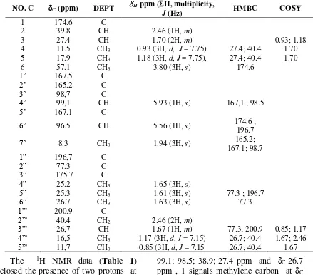
Table 1. The NMR data of isolated compound, recorded at 1H - 500 MHz; 13C-125 MHz, spectral data of recorded in CDCl3
NO. C C (ppm) DEPT H
ppm (H, multiplicity,
J (Hz) HMBC COSY
1 174.6 C
2 39.8 CH 2.46 (1H, m)
3 27.4 CH 1.70 (2H, m) 0.93; 1.18
4 11.5 CH3 0.93 (3H, d, J = 7.75) 27.4; 40.4 1.70 5 17.9 CH3 1.18 (3H, d, J = 7.75), 27.4; 40.4 1.70
6 57.1 CH3 3.80 (3H, s) 174.6
1’ 167.5 C
2’ 165.2 C
3’ 98,7 C
4’ 99,1 CH 5,93 (1H, s) 167,1 ; 98.5
5’ 167.1 C
6’ 96.5 CH 5.56 (1H, s) 174.6 ;
196.7
7’ 8.3 CH3 1.94 (3H, s) 165.2;
167.1; 98.7
1” 196,7 C
2” 77.3 C
3” 175.7 C
4” 25.2 CH3 1.65 (3H, s)
5” 25.3 CH3 1.61 (3H, s) 77.3 ; 196.7
6” 26.7 CH3 1.63 (3H, s) 77.3
1”’ 200.9 C
2”’ 40.4 CH2 2.46 (2H, m)
3”’ 26,7 CH 1.67 (1H, m) 77.3; 200.9 0.85; 1.17
4”’ 16,5 CH3 1.17 (3H, d, J = 7.15) 26.7; 40.4 1.67; 2.46
5”’ 11,7 CH3 0.85 (3H, d, J = 7.15 26.7; 40.4 1.67
The 1H NMR data (Table 1) disclosed the presence of two protons at δH 5.93 and 5.56 ppm (1H, s) were characteristic for vinyl proton. The proton signal at δH 3.80 ppm (3H, s) was assigned to a methoxy group. Furthermore, the presence proton signals at δH 2.46 each (3H, m) for one proton methylene and one proton methine, signals at 1.67 (1H, m), and 1.70 (1H, m) for two methine groups. The 1H NMR data also indicated the presence of eight methylprotons at δH 1.94 ppm, δH 1.65 δH 1.63 ppm and 1.61 ppm, δH 1.18 (3H, d, J = 7.75), 0,93 (3H, d, J = 7.75), 1.17 ppm (3H, d, J = 7.15), and 0.85 ppm (3H, d, J = 7.15.).
The 13C NMR spectrum (Figure 2) indicated 24 signal carbon consist that 10 signals as C sp2 and 14 signals as C sp3. Base on analysis spectrum DEPT 135 showed 5 signal methines carbon at δ
99.1; 98.5; 38.9; 27.4 ppm and δC 26.7 ppm , 1 signals methylene carbon at δC 40.4 ppm, 9 signals methyl carbon at δC 57.1; 26.7; 25.3; 25.2; 17.9; 16.5; 11.7; 11.5 and δC 8.3 ppm and 9 signal quarternary carbon at δC 200.9; 196.7; 175.6; 174.5; 167.3; 167.1; 165.1, 98.7 and δC 77.3 ppm.
HMQC spectrum as summarized in Table 1.
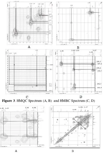
NMR 2D analysis for HMQC spectrum showed the proton signal at δH 5.93 ppm and δH 5.56 ppm attached to carbon signal at δC 99.1 ppm and 98,5 respectively and proton at δH 3.80 attached to carbon at δC 57.1 ppm. HMBC spectrum showed proton at δH 5.93 was correlated to carbon at δc 98.5 and δc 167.1 ppm, while proton at δH 5.56 showed correlation to carbon at δC 174.6 and 196.7 ppm. This data indicated that two proton vinilic was not place at the carbon besides it. Further HMBC spectrum showed correlation proton of δH 3.80 ppm (3H, s) to carbon at δC 174.6 ppm indicated as proton methoxy from ester group.
HMQC spectrum showed that
proton at δH 1.61 attached to carbon at δc 25.3 and HMBC spectrum showed that proton at δH 1.61 was correlated to carbon δc 77.3 dan δc 196.7 ppm. While proton at δH 1.63 ppm at HMQC spectrum showed attached to carbon at δC 26.7 ppm and HMBC spectrum showed correlation to carbon at 77.3 ppm. This data indicated that two proton methyl were bounded at the same carbon that δC 77.3 ppm. The proton at δH 1.94 at HMQC spectrum showed attached to carbon at δC 8.3 ppm and HMBC spectrum showed correlated to carbon at δC 98.7. Proton at δH 2.46 attached to carbon at 39.8 and 40.4 ppm (Figure 3). This data supported that proton at δH 2.46 ppm were one signal methine proton and one as signal methylene proton.
Figure 3. HMQC Spectrum (A, B) and HMBC Spectrum (C, D)
Figure 4. HMBC spectrum (A) and dan COSY spectrum (B)
Furthermore at HMBC spectrum showed correlation proton at δH 1.94 ppm to carbon δc 165.2; δc 167.1; and δc 98.7 ppm indicated the proton attached at aromatic ring. HMQC spectrum also showed that proton at δH 1.18 and 0.93 ppm attached to carbon at δc 17.9 ppm, and δc 11.5 ppm, while HMBC spectrum (Figure 4) showed both of this proton was correlation to carbon δc 27.4 dan 39.8. Proton at δH 1.17 and 0.85 ppm at HMQC
spectrum showed attached to carbon at δC 16.5 and 11.7 ppm at HMBC spectrum showed both of this proton correlation to carbon δc 26.7 ppm 40.4 ppm.
range coupling HMBC experiment.
These spectroscopic data, therefore suggested that compound was identified as methyl 6-(5'-(2"-acetoxy-2” - methylpropanoyl)-3"-methyl-2'-(3"'-
methylbutanoyl)phenyl)-3-methylbutanoate. HMBC correlation and structure this compound showed at Figure 5. Exploration of secondary metabolites research needs to be done in order to get the profile of organic compounds produced by endophytic fungus of Curcuma zedoaria.
Based on the literature study, the biosynthetic pathways of secondary metabolites produced from endophytic fungus has not been found clearly. The substances isolated from endophytic have different biosynthetic pathways: isoprenoid, polyketide, amino acid derivatives, and belonged to diverse structural groups: terpenoids, steroids, xanthones, chinones, phenols, isocumarines, benzopyranones, tetralones, cytochalasines, and enniatines (Barbara, Christine, Anne, & Kristen, 2002). Literature survey also showed these compounds have never found either of endophytic dothiorelon B and dothiorelon C, were isolated from microbial Dothiorella sp who live on the leaves of the species Cynodon dactylon (L) (Poaceae) (Radji, 2005). Other similar compounds ever discovered was 2-{4-methyl-2-[(2-methylpropanoyl)oxy] phenyl}oxiran-2-yl)methyl-3-methyl-butanoic (Yannai, 2004) (Figure 6). Antibacterial Activity
The antibactetrial activity of this compound was evaluated according to the method previously described. The antibacteiral properties of isolated compound was evaluated according to the method described previously (Lai, 2004 ). This compound showed inactive antibacterial for all bacterial test until concentration test 1000 ppm. This compound will show activity by concentration 2500 ppm with the mean of inhibition diameters (mm) for E. coli, S. dysenteriae, S. aureus, B. subtilis 10,3 ; 8.3, 8.4; and 8.8 mm respectively and standard antibacterial ampicillin at concentration 10 ppm showed inhibition diameters 7.5; 8.5; 7.0; and 9.5 mm respectively. Base on this data the compound show weak activity.
Figure 5. Structure, (A), HMBC Correlation (B) and COSY correlation (C) of isolated compound
{4-methyl-2-[(2-methylpropanoyl)oxy]phenyl}oxiran-2-yl)methyl-3-CONCLUSION
A new compound have been isolated from the endophytic fungi Aspergillus sp from the leaves of kunyit putih (Curcuma zedoaria). Based on spectroscopic analysis H-NMR and C-NMR (1D and 2D), was identified as methyl 6-(5'-(2"-acetoxy-2”-methylpropanoyl) -3"-methyl-
2'-(3"'-methylbutanoyl)phenyl)-3-methylbutanoic. Isolated compound showed weak antibacterial activity.
ACKNOWLEDGEMENT
The authors are grateful to the Directorate General of Higher Education which research grant Fundamental 2014 was supported this research.
REFERENCE
Barbara, S., Christine, B., Siegfried, D., Anne, K. R., and Kristen, K. (2002). Endophytic fungi: a source of novel biologically active secondary Fusarium oxysporum isolated from acorus calamus rhizome with antimicrobial activity. European Journal Biological Science. 2(1), 8-16.
Elfita, Muharni, Munawar, Salni, dan A. Oktasari. (2010). Senyawa antimalaria dari jamur endofitik tumbuhan sambiloto (Andographis paniculata Nees. Jurnal Natur Indonesia 13(2) 123-129.
Elfita, Muharni, and T. Indah. (2011). Secondary metabolite of Aspergillus fumigatus, an endophytic fungi of the medicinal plant Garcinia griffithii. Makara Seri Sains, 15(2),124-128.
Elfita, Muharni, Munawar, and S. Aryani. (2012). Secondary metabolite from endophytic Fungi Aspergillus niger of the stem bark of Kandis Gajah
(Garcinia grfiffithii). Indonesian Journal Chemistry, l12(2), 195-200 Eyberger, A. L., Dondapati, R., and
Porter, J.R. (2006). Endophytic fungal isolated from Podophyllum peltatum produce podophyllotoxin. Journal of Natural Products, 69(8), 1121 - 1124.
Gunatilaka A. A. L. (2006). Natural products from plant-associated microorganisms, distribution, structural diversity, bioactivity, and implications of their occurrence. Journal of Natural Products. 69(3), 505-526.
Guo, L., Wu, J. Z., Han, T., Cao, T., Rahman, K., and Qin L.P. (2008). Chemical composition, antifungal and antitumor properties of ether extracts of Scapania verrucosa Heeg. and its endophytic fungus Chaetomium fusiforme. Molecules , 13(9), 2114-2125
Hundley, N.J. (2005). Struktur elucidation of bioactive compounds isolated from endophytes of Alstonia scholaris and Acmena graveolens. Thesis. Department of Chemistry and Biochemistry. Brigham Young University.
Kour, A., Shawl A.S., Rehman, S., Sultan p., Qazi, P. H., and Verma. (2008). Isolation and identification of an endophytic strain of Fikusarium
oxysporum producing
podophylotoxin from Juniperus recurva. World Journal of Microbiology and Biotechnology, 24(7), 1115-1121
Lai, E.Y.C., Chyau, C.C., and Mau, J.L. (2004). Antimicrobial activity and cytotoxicity of the essential oil of Curcuma zedoaria. American Journal of Chinese Medicine. 32(2): 281–290.
International Journal Medicinal Chemistry, 2011, 1-13.
Makabe, H., Maru, N., Kuwabara, A., Kamo, T., and Hirota, M. (2006). Anti-inflammatory Sesquiterpenes from Curcuma zedoaria. Journal Natural Product, 20(7), 680-685. Mau, J. L., Eric, C Lai.,. Nai, P.W, Chien,
C.C., and Charng, C. Y.. (2003). Composition and antioxidant activity of the essential oil from Curcuma zeodaria. Food Chemistry, 82(4), 583–591.
Muharni, Fitrya, Milanti, O.,and Elfita. (2014). Antibacterial and antioxidant activity of pyranon derivated compound from endophytic fungi Penicillium sp of kunyit putih (Curcuma zeodaria), Traditional Medicine journal, 19(3),107-112.
Muharni, Fitrya, M.O. Ruliza, D.A. Susanti, and Elfita. (2014b). Di-2-Ethylhexyl phtalate and pyranon derivated from endophytic fungi Penicillium sp kunyit putih (Curcuma zeodaria) Indonesian Journal Chemistry, 14(3), 290-296. Muharni, Heni, Y., Fitrya, and Roni.
(2015). Evaluation acute toxicity and antibacterial activity of Penicillium sp endophitic fungus extract of kunyit putih (Curcuma zedoaria ) in mice (Mus musculus L.). Journal Chemical and Pharmaceutical Research, 7(9s), 147-151
Radji, M. (2005). Peranan bioteknologi dan mikroba endofit dalam pengembangan obat herbal. Majalah Ilmu Kefarmasian, 2(3), 113 – 126. Rodriguez R.J., White, J. T., Arnol, A. E.,
and Reman, R.S. (2009). Fungal endophytic diversity and funcional
roles. New Phytologist, 182(2), 314–330.
Strobel, G and Daisy , B., Castillo, U., and Harper J. (2004). Natural products from endophytic microorganisms. Journal of Natural products, 67(2), 257 – 268
Sun, D., Ran, X., and Wang, J. (2008). .Isolation and identification of taxol-producing endophytic fungus from Podocarpus. Acta Microbiologica Sinica. 48, 589-595.
Sulistiyani, T. R., Lisdiyanti, P., and Lestari, Y. (2014). Population and diversity of endophytic bacteria associated with medicinal plant Curcuma zedoaria. Microbiology Indonesia, 8(2), 65 - 72
Verma V. C.,Kharmar, R. N., Strobel G. A. (2009). Chemical and functional diversity of natural products from plant associated endophytic fungi. Natural Product Communications, 4(11), 1211-1532
Xu L., Zhou L., Zhao J., Li J., Li X., and Wang J. (2008). Fungal endophytes from Dioscorea zingiberensis and their antibacterial activity. Letters in Applied Microbiology. 46(1), 68-72. Yannai, S. (2004). Dictionary of food
compounds with CD-ROM: Additives, flavors, and ingredients. Boca Raton: Chapman & Hall/CRC. Zhang, H. W., Song, Y. C., and Tan, R. X.
(2006). Biology and chemistry of endophytes. Natural Product Reports, 23(5), 753-771.