Acta Interna - The Journal of Internal Medicine
20
5. Harris,E.D.J., 2007 Rheumatoid Arthritis; W.B Saunders., Philadelphia.
6. Ilse,E.A., Isabelle, P., Hans, P., Ann U., Hulstaert, F., Lydie, M., Filip D.K., 2004 Diagnostic Performance and Predictive Value of Rheumatoid Factor, Anti-citrullinated Peptide Antibodies, and the HLA Shared Epitope for Diagnosis of Rheumatoid Arthritis; Clin. Chemistry 50, No.12.
7. Chernecky,C.C., Berger,B.J, 2008 Laboratory Tests th
and Diagnostic Procedures 5 edition, St louis Saunders. London.
8. Deepa, R., Landon, W.T., Travis L., Amir, R.B., Alan D.K. 2007 Rheumatoid Arthritis: Current p h a r m a c o l o g i c t r e a t m e n t a n d a n e s t h e t i c considerations; Jour. Anesthesia 19 (2).
Puvaneswary Rajendran, et al
21
O R I G I N A L A R T I C L E
THE DIFFERENCE OF SERUM CARBOXY-TERMINAL PROPEPTIDE OF
PROCOLLAGEN TYPE I (PIP) IN STAGE A, B AND C HEART FAILURE
PATIENTS CAUSED BY HYPERTENSION
1 2 2
Nurul Aini , Lucia Kris Dinarti , Hariadi Hariawan
1. Internal Medicine Medical Doctor Specialist Training Program, Medical Faculty of Gadjah Mada University / Dr. Sardjito General Hospital Yogyakarta
2. Cardiology Department, Medical Faculty of Gadjah Mada University / Dr. Sardjito General Hospital Yogyakarta
ABSTRACT
Introduction. Arterial hypertension affects the heart tissue composition which leads to structural remodeling of the myocardium. The imbalance between synthesis and degradation of type I collagen leading to myocardial fibrosis in a form of type I collagen fiber accumulation in the interstitial and perivascular myocardium. Collagen fiber accumulation reduces relaxation stage, diastolic suction, myocardial stiffness and diastolic dysfunction which affect systolic dysfunction leading to heart failure. Concentration of carboxy-terminal pro peptide of pro collagen type I (PIP) in peripheral blood am a synthesis index of type I collagen in HHD. Thus, the measurement of PIP is useful to monitor myocardial fibrosis stage in heart failure and to determine the therapeutic strategy that aims not only to reduce arterial pressure and left ventricular mass but also to prevent myocardial remodeling.
Aim of the study. The aim of the study was to ascertain the difference PIP level in patients with the heart failure stage A, B, and C which are caused by hypertension. The serum concentration of PIP was measured by enzyme immunoassay. This research was a cross sectional research designed for cardiology policlinic's outpatients at Dr. Sardjito General Hospital Yogyakarta from August 2009 until the calculated sample number is fulfilled.
Method. One-way ANOVA was used to analyze the differences between the three groups of heart failure stages after being tested for the normality using Kolmogorov-Smirnov normality test. If the result did not show a normal value, a non-parametric test would be undergone using Kruskal-Wallis test followed by Mann-Whitney U test. The differences considered as significant if p < 0.05 with a confidence interval of 95%.
Result. The research was performed in 64 patients heart failure caused by hypertension consisted of 22 stages A, 19 stage B and 23 stage C.
PIP mean levels of the group stage B 819.78 ± 91,03 ng/ml was higher compared stage A 808.47± 80.8 ng/ml and PIP mean level stage C 852 ± 55.51 ng/ml was higher compared stage B. The PIP mean levels did not differ statistically significantly (p=0. 317).
Conclusion. There were no significant differences in serum level of PIP on the stage heart failure A, B and C.
Keywords: Collagen, fibrosis, hypertension, heart failure, carboxy-terminal pro peptide
INTRODUCTION
Hypertension affects approximately 50 millions of people in the United States and 1 billion of people worldwide. The prevalence of hypertension will increase at population age if the
1
effective preventive measures are not implemented . The data from the Framingham Heart Study mentioned that hypertension contributed as much as 39% of heart failure cases in men and 59% of cases
2
in women .
One complication of hypertension is the development of left ventricular hypertrophy. Left ventricular hypertrophy is characterized by a concentric and circumferential hypertrophy of the myofibrils, increased contractility and thickened ventricular wall, decreased end diastolic volume and
1
relaxation disturbance .
Acta Interna - The Journal of Internal Medicine
22
Nurul Aini, et al
increased synthesis and unchanged or degraded type
3
1 collagen fibers .
Endomyocardial invasive biopsy is one way to assess fibrotic tissues in hypertensive disease but this technique is not widely implemented and managed. Serological determination of collagen derived peptides is used as a fibrillar collagen turnover marker in various conditions leading to
4
cardiac fibrosis .
The increased myocardial fibrosis as a result of the increased type 1 collagen synthesis and its deposit may contribute to the development of heart
3
failure in HHD patients . The hypothesis of this study is a relationship between type I carboxy terminal pro peptide pro collagen level with stage A, B and C heart failure patients caused by hypertension.
METHOD
This study used a cross sectional study, comparing the average serum level of type I carboxy terminal pro peptide procollagen (PIP) in 3 groups of heart failure patients, consisting of hypertension group (stage A heart failure), hypertension with left ventricular hypertrophy (Stage B heart failure), and hypertension with left ventricular hypertrophy and heart failure symptoms (stage C heart failure). This study began in August 2009 to complete.
The Course of the Study
Research subjects were the outpatients in cardiology polyclinic of Dr. Sardjito Yogyakarta General Hospital diagnosed to be having heart failure based on ACC/AHA criteria and willing to follow the course of this study.
The inclusion criteria of this study were hypertensive heart failure diagnosed based on ACC/AHA criteria, heart failure patients on treatment and those who were willing to sign the informed consent form. The exclusion criteria were acute coronary syndrome, IHD, primary valvular heart diseases, permanent arrhythmia, diabetes mellitus, severe chronic kidney diseases, hyperthyroid, alcoholic liver diseases, metabolic bone diseases (osteomalacia, Paget's disease, rheumatism, bone metastasis), stroke and malignancies.
Research Finding and Discussion Studied variables were:
1. The independent variable was the type I carboxy terminal propeptide procollagen (PIP).
2. Dependent variables were hypertension (stage A heart failure), hypertension with left ventricular hypertrophy (stage B heart failure), hypertension with left ventricular hypertrophy along with the symptoms of heart failure (stage C heart failure). 3. Other variables measured were age, sex, body
stature, body weight, BMI, blood profile, random blood glucose test, ALT, AST, BUN, creatinine, anti-hypertensive drugs, stage of hypertension, duration of hypertension, heart rate, left ventricular mass, left ventricular mass index, and left ventricular hypertrophy.
Measurement
1. The Auscultatory method of blood pressure measurement with Sphygmomanometer, subjects should be seated quietly for 5 minutes with the arm supported at heart level. Spine manometer which pumped up beats disappears then increase 30 mm Hg. The stethoscope bell was placed in position then lowered slowly (2-3 mmHg/Sec). Systolic blood pressure is the point at which the first sound is the heart (korotkoff 1) and diastolic blood pressure is the point before the disappearance of sound (korotkoff 5).
2. The BMI was calculated by dividing body
5
weight (kg) and body height (m2).
3. Left ventricular mass (LVM) measured by using a recommended formula by American Society of Echocardiography, of which LVM = 0.8 [1.04 x {(LVIDd + IVSd + PWd) 3- LVIDd3}] + 0.6
6-7
grams.
4. Left ventricular Mass Index (LVMI) = LVM/BSA. The BSA stands for body surface area, and was calculated by using Dubois formula: 0.007184 x W0.425 x H0.725 (W is for body weight in kg and H is for body height in cm)
6-7
5. Type I carboxy terminal propeptide procollagen (PIP) serum was tested using Enzyme immunoassay human Procollagen Type I-C-peptide (EIA human PICP) Takara bio INC method with mg/ml as the applied unit.
Acta Interna - The Journal of Internal Medicine
24
Study subjects consisted of 22 (34, 37%) patients in stage A heart failure group, 19 (29,68%) patients in stage B heart failure group, and 23 (35,93%) patients in stage C heart failure group. Overall, there were 21 (32, 81%) male subjects and 43 (67, 18%) female subjects.
Hypertension stage
Pre hypertension n(%) 4(18.8) 3(15.78) 6(26.08)
Stage 1 n(%) 12(54.54) 11(57.89) 7(30.43)
Stage II n (%) 6(27.27) 5(26.31) 10(43.47)
Duration HT Years ( mean±SD)
7.6±6.5 7.4±4.1 11.6±8.0
Treatment
ACEI n(%) 13(59) 12(63.15) 18(78.26)
ARB n(% 9(40.9) 11(57.89) 10(43.47)
â-blockern (%) 5(22.72) 4(21.05) 11(47.82)
CCB n(%) 10(45.45) 14(73.68) 9(39.13)
Furosemidn(%) 4(18.18) 6(31.57) 16(69.56)
Spironolactonn(%) 0(0) 0(0) 4(17.39)
Thiazides n (%) 5(22.72) 7(36.84) 1(4.34)
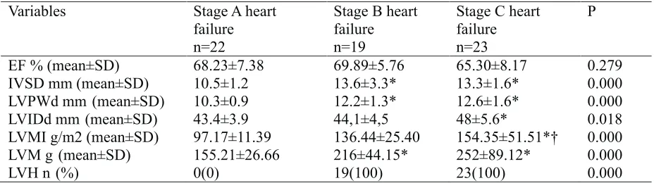
Echocardiography was used as a parameter to measure the magnitude of left ventricular hypertrophy. Obtained the power of good inter-observer agreement in interpretation of the ECG with a kappa value of 0.69, echocardiography parameters are indicated in table 2.
Table 2. Comparison of three groups of variable echocardiography parameters of heart failure
Kruskal-Wallis followed Mann-Whitney U test, statistically significant p<0.05, *p<0.05vs stage A; †p<0.05 vs stage B. 11.39 g/m2, stage B 136.44 ±25.40 g/m2 and stage C 154.35 ± 51.51 g/m2)), significant difference of
EF % (mean±SD) 68.23±7.38 69.89±5.76 65.30±8.17 0.279
IVSD mm(mean±SD) 10.5±1.2 13.6±3.3* 13.3±1.6* 0.000
LVPWd mm (mean±SD) 10.3±0.9 12.2±1.3* 12.6±1.6* 0.000
LVIDd mm (mean±SD) 43.4±3.9 44,1±4,5 48±5.6* 0.018
LVMI g/m2 (mean±SD) 97.17±11.39 136.44±25.40 154.35±51.51*† 0.000 LVM g (mean±SD) 155.21±26.66 216±44.15* 252±89.12* 0.000
LVH n (%) 0(0) 19(100) 23(100) 0.000
found about 100% in stage B and C group of heart failure.
The PIP level examinations (table 3) of the three heart failure groups in this study showed results as the following: 808.47 ± 80.8 ng/ml for stage A group, 819.78 ± 91.03 ng/ml for stage B group and 852 ± 55.51 ng/ml for the stage C group. The mean PIP level of the stage B group was higher than stage A group and the mean PIP level of the stage C group was higher than stage B group, but these differences were not statistically significant (p=0. 317).
Nurul Aini, et al
25 Table 3. The difference in mean levels of PIP on the three groups of heart failure
Variable Stage A heart
PIP ng/ml 808.47±80.8 819.78±91.03 852±55.51 0.317
Statistically significant p < 0.05
DISCUSSIONS
This study was conducted in order to obtain the PIP level of the three groups of heart failure patients. This study resulted in a higher proportion of female subjects compared to male subjects.
The mean age of onset of the heart failure in stage A, B, and C group respectively were as follow 57.68 ± 11.44 years old, 56.89 ± 13.09 years old, and 62.91 ± 7.59 years old.
The mean BMI for stage A group was 23.04 ± 3.39 kg/m2, 24.17 ± 3.12kg/m2 for stage B group and 26,88 ± 3,35 kg/m2 for the stage C group. The prevalence of hypertension will increase as the increased of BMI, central obesity degree, and
9
fasting blood glucose level .
The mean systolic blood pressure for stage A, B, and C group respectively were as follows (145 ± 15.35 mmHg, 143, 1 ± 15.3 mmHg, 148 ± 18.5 mm Hg), stage I hypertension had its highest distribution in stage A heart failure group as many as 12 patients (54.54%), stage II had its highest distribution in stage C heart failure group as many as 10 patients (43.47%).
Measurement of LVMI based on BSA resulted in significant differences among stage A mean LVMI (g/m2) of 97.17±11.39, stage B of 136.44±25.40 and stage C of 154.35±51.51 with p value of p= 0.000. A LVH frequency of 100% was found in stage B and C heart failure groups. A study conducted by Chukwuka et al., 2008 found a significant difference of LVPWd, LVM and LVMI parameters between normotensive and hypertensive
10
subjects .
This study also found the increasing thickness of the interventricular septum during diastole, the thickening of the left ventricular posterior wall during diastole in stage B and C heart failure groups, while the mean internal diameter during diastole was still within normal limits in stage A and B groups. Accumulation of collagen fibers decrease of the level of relaxation, diastolic
suction and passive stiffness because impaired diastolic function further reduces diastolic filling and lower transduction Cardio myosit contraction in the power of thus affecting the development of
11
myocardial systolic performance .
The mean PIP level found in this study were 819.78 ± 91.03 ng/ml in stage B group which was higher than what was found in stage A group of 808.47 ± 80.8ng/ml and the mean PIP level in stage C group was 852 ± 55.51 ng/ml which was higher than stage B group, even though those scores were not statistically significant among the three groups (p=0. 317).
The results found in this study were in accordance with the results of a survey conducted by Richard et al, in 2007 which comparing PIP level 115 hypertensive patients with LVH, 38 hypertensive patients without LVH and 38 normotensive patients. The study showed an increased PIP level in patients with hypertension compared to normotensive patients, and there were only slight differences between patients with and without LVH. Hypertension with LVH correlated with cardiomyocytes hypertrophy and the amount of myocardial collagen, myocardial fibrosis could cause diastolic dysfunction and heart failure. PIP level in the circulation acted as type I collagen synthesis index and had some correlation with the
12
amount of myocardial fibrosis .
These study results were also consistent with the reports made by Liang et al., In 2004 who studied about the PIP level in 61 CHF patients and 20 control subjects, and the study showed that PIP level increased significantly compared to normal subjects and increased in parallel upon NYHA classifications and also no correlation was found with any underlying diseases. PIP serum level examinations showed synthesis and metabolism of cardiac extracellular collagen matrix during left
13
ventricle reconstruction in CHF .
Acta Interna - The Journal of Internal Medicine
A study conducted by Querejeta et al. (2000), found PIP serum and collagen volume fraction (CVF) concentration to be higher (p<0.001) in hypertensive patients compared to normotensive subjects. PIP level found in hypertensive patients with severe fibrosis was higher (p<0.05) compared to hypertensive patients with non-severe fibrosis
4
(108±6 ìg/L) and normotensive patients. A study by Querejeta et al, in 2004 found PIP level was higher in patients with heart failure compared to hypertensive patients without heart failure (p<0.05), there was direct correlations between CVF and
3
peripheral PIP .
The results of this study showed that there was no significant difference on PIP level among the three heart failure groups (P=0.317), this was caused by no treadmill and coronary angiography undergone to exclude other heart diseases which correlated to fibrotic process, which was ischemic heart disease (IHD). In order to exclude IHD, researchers only performed electrocardiography and echocardiography. In classifying stage B and C heart failure, researchers examined whether there were any symptoms of heart failure before or prior heart failure symptoms are subjective.
Several weaknesses of this study were on the research design employed, a cross sectional design which had a weakness in determining the causal relationship of the diseases. This study did not perform treadmill test and coronary angiography to exclude other heart diseases which correlated to fibrotic process, which was Ischemic Heart Disease (IHD). In classifying stage B and C heart failure, researchers examined whether there were any symptoms of heart failure before or at the moment of examination, prior heart failure symptoms are subjective.
Most of heart failure was treated with an ACEI, ARB, â-blockers, CCB, furosemide, spironolactone and thiazides could be a confounding factor, but it was still the therapeutic standard for heart failure patients.
CONCLUSION
There were no significant differences in the level of type I carboxy terminal propeptide procollagen (PIP) in the serum of heart failure
groups of stage A, B and C. Researchers have not yet been able to give any suggestions for this study. Further research needs to be done by doing treadmill test and coronary angiography to exclude IHD and to classify stage B and C heart failure groups in a more objective way.
REFERENCES
1. Chobanian, A.V., Bakris, G.L., Black, H.R., Cushman, W.C.,Green, L.A., Izzo, J.L., et al. The seventh Report of the Joint National Committee on Prevention, Detection, Evaluation, and Treatment of High Blood Pressure the JNC 7 Report. JAMA: 2003. 289:2560-2572.
2. IzzoJr, J.L., Gradman, A.H. Mechanisms and management of hypertensive heart disease: from left ventricular hypertrophy to heart failure. MED Clin N Am. 2004. 88:1257–1271.
3. Querejeta, R., López, B., González, A., Sánchez, E., Larman, M.,Ubago, J.M., Díez, J. Increased Collagen Type I Synthesis in Patients with Heart Failure of Hypertensive Origin Relation to Myocardial Fibrosis Circulation. 2004. 110:1263-1268.
4. Querejeta, R., Varo, N., ˜a Lo´pez, B., Larman, M., Artin˜ano, E.,Etayo, J.C., et al. Serum Carboxy-Terminal Pro peptide of Pro collagen Type I Is a Marker of Myocardial Fibrosis in Hypertensive Heart Disease. Circulation. 2000. 101:1729-1735. 5. Soegondo, S. Obesitas. dalam: A.W. Sudoyo., B.
Setiyohadi., I. Alwi., M. Simadibrata, K., S. Setiati (editor). Buku Ajar Ilmu Penyakit Dalam, Jilid III, edisi V, Pusat Penerbitan Departemen Ilmu Penyakit Dalam FK-UI, Jakarta. 2009. 1973-1983.
6. Dangri, P., Agarwal, S., Kaira, O., Rajpal, S. Echocardiographic assessment of left ventricular hypertrophy in patients with chronic renal failure. Indian Journal of Nephrology.2003. 13:92-3.
7. Selvetella, G., Notte, A., Maffei A., Calistri, V., Scamardella, V. Left ventricular Hypertrophy is Associated With asymptomatic Cerebral Damage in Hypertensive Patients. Stroke.2003; 34:1766-7.8. Hunt, S.A., Abraham, W.T., Chin, M.H., Feldman, A.M.,Francis, G.S., Ganiats, T.G., et al. ACC/AHA 2005 Guideline Update for the Diagnosis and Management of Chronic Heart Failure in the Adult.Circulation.2005.112:e154-e235.
26
Nurul Aini, et al
8. Vikrant, S., Tiwari, S.C. Essential Hypertension Pathogenesis and Pathophysiology. Journal, Indian Academy of Clinical Medicine. 2001. 2:141- 61. 9. Chukwuka, U.A., Anayo, O.C., Amaechi, A.J.
Evaluation of left ventricular structures in normotensive and hypertensive subjects by two dimensional echocardiography: Anthropometric correlates in hypertension. Internet Journal of Medical Update. 2008. 3:3-7
10. Díez, J., Frohlich, E.D. A translational Approach to Hypertensive Heart Disease. Hypertension. 2010. 55:1-8.
11. Richard, M.B., Thomas, K., Begona, L., Magnus, E., Arantxa, G., Javier, D., Et al. Myocardial fibrosis and diastolic dysfunction in patients with hypertension: results from the Swedish Irbesartan Left Ventricular Hypertrophy Investigation versus Atenolol (SIVHIA). (Abstract) Journal of Hypertension. 2007. 25:1958-1966.
12. Liang, S., Jinghan, W., Luosha, Z., Taixing, W. Carboxy terminal pro peptide of type I pro collagen and amino terminal pro peptide of type III pro collagen in patients with chronic heart failure. (abstract). Journal of Clinical Cardiology 2004;1