Isolation of a
Brassica napus
L. cDNA encoding a putative
high-mobility-group HMG I
/
Y protein
Tomas Masek
a, Petr Smykal
a, Iveta Janotova
a, David Honys
b, Vera Capkova
b,
Paul M. Pechan
a,c,*
aDepartment of Plant Physiology,Faculty of Science,Charles Uni6ersity,Vinicna5,Prague,CZ128 44,Czech Republic bInstitute of Experimental Botany,Czech Academy of Science,Roz6ojo6a135,Prague6,CZ165 02,Czech Republic cTechnical Uni6ersity Munich,Lehrstuhl fu¨r Pflanzenbau und Pflanzenzu¨chtung,85350Freising-Weihenstephan,Germany
Received 22 February 2000; received in revised form 20 June 2000; accepted 26 June 2000
Abstract
A cDNA encoding a high-mobility-group protein has been isolated from a microspore-specific library of Brassica napus. The 930 bp cDNA contains a 612 bp open reading frame encoding a protein of 203 amino acids residues exhibiting significant homology to HMG-I/Y protein from Arabidopsis thaliana (62%). The predicted protein contains four copies of the ‘AT-hook’ motif which is involved in binding A/T-rich DNA. Southern blotting indicates that the HMG-I/Y gene is a single-copy gene in
B.napus. Transcription of the HMG-I/Y gene was detected in all tissues examined, with the highest expression in pollen-derived embryos. In situ localization studies of flower organs indicate the transcript to be preferentially located in petals and sepals. Subcellular localization analysis performed during pollen development showed that the transcript of the HMG-I/Y gene is predominantly associated with polysomes. © 2000 Elsevier Science Ireland Ltd. All rights reserved.
Keywords:AT-hook;Brassica napus; HMG-I/Y protein; Pollen; Rape
www.elsevier.com/locate/plantsci
1. Introduction
High-mobility-group (HMG) proteins represent a family of the most abundant and ubiquitous non-histone proteins common to eukaryotic or-ganisms. HMG proteins were originally defined as low molecular weight (up to 30 kDa) non-histone proteins, that can be extracted from chromatin with high salt buffers and are soluble in 2%
trichloracetic acid or 2 – 5% perchloric acid [1]. These proteins are rich in basic amino acids (25%), acidic (25%) and proline (7%) and display high electrophoretic mobility [2]. HMG proteins have been more thoroughly studied in vertebrates. They have been classified in three families: HMG-1/2, HMG-14/17 and HMG-I/Y proteins [1]. HMG-1/ 2 proteins bind to single- and double-stranded DNA, HWG14/17 proteins are preferentially asso-ciated with nucleosomes, while HMG-I/Y proteins display preferential binding to A/T-rich tracts of DNA.
The different isoforms of the mammalian protein, HMG-I and HMG-Y, arise by differential splicing of the transcript of a single gene [3]. Their DNA-binding domain is termed the ‘AT hook’ that consists of a repeating PRGRP amino acid sequence. In the case of mammalian HMG-I/Y, there are three copies of this domain. These proteins bind directly to the A/T rich domain [2].
Abbre6iations: BAP, 6-benzylaminopurine; BSA, bovine serum al-bumin; cDNA, complementary DNA; 2,4-D, 2,4-dichlorphenoxy-acetic acid; DAPI, 4,6-diaminophenyl indol; DDPCR, differential display polymerase chain reaction; DMPC, dimethylpyrocarbonate; dscDNA, double stranded complementary DNA; EtBr, ethidium bromide; GuTC, guanidium thiocyanate; HMG, high mobility group; NAA,a-naphthylacetic acid.
The nucleotide sequence data reported are in the EMBL, GeneBank and DDBJ Sequence Databases under the accession num-ber AF 127919.
* Corresponding author. Tel.: +49-8024-93333; fax: + 49-8024-93150.
E-mail address:[email protected] (P.M. Pechan).
Animal HMG-I/Y proteins have been shown to participate in the assembly of protein complexes of several inducible genes [4,5]. It has also been shown that HMG-I/Y protein is able to relieve the repression of transcription caused by histone H1 and may be able to modulate DNA-binding to the nuclear scaffold and influence local chromatin structure [6,7].
The existence of plant HMG-I/Y proteins was first implied by the isolation of soybean SB 16A cDNA [8], which encodes a protein with four AT-hook motifs. cDNAs, encoding related HMG-I/Y proteins, have been subsequently isolated from a variety of plants, including for example rice [9,10], oat [11], Japanese jack bean [12] and Ara -bidopsis [13].
Plant HMG-I/Y show low sequence homology to mammalian HMG-I/Y. They are fully ho-mologous only in their DNA-binding domains. The plant HMG-I/Y proteins have molecular masses of approximately 21 kDa, which is twice as big as their animal counterparts, and display three or four copies of the AT-hook motif [14]. The N-terminal region shows sequence similarity to the N-terminal region of histone H1 [8,14] while the AT-hook motifs in the C-terminal part of the protein are arranged with similar spacing to that found in animal HMG-I/Y proteins [1,14,15].
A protein combining an HMG-I-like DNA-binding domain with a putative transcription do-main was isolated from tobacco [16]. Further implication of plant HMG-I/Y proteins in the transactivation of gene expression has been sup-ported by a study of rice and oat HMG-I/Y proteins binding to the oat phytochrome A gene promoter [10]. The binding to soybean nodulin N23 promoter [17], pea plastocyanin gene pro-moter [2,18,19] and the pea PetF propro-moter [20] has been also attributed to HMG-I/Y-like proteins.
Expression of the HMG-I/Y gene has been studied in several plants such as Arabidopsis thaliana [13], pea [21] and Japanese jack bean [12]. Transcripts were found in all tissues examined, with the highest expression levels in actively prolif-erating tissues.
Although the biological function of the HMG proteins is still unknown, some already mentioned likely roles point to participation in transcriptional activation events.
Here we report on the isolation and characteri-zation of an HMG-I/Y cDNA from Brassica na-pus, specifically in relation to its expression regulation in the reproductive organs.
2. Material and methods
2.1. Plant material
Plants ofB. napusL. cv. Topas were grown in a growth chamber (Heraeus 1500): 15/10°C day/ night temperature, 16 h light duration, 300 mmol m−2s−1photon influence irradiation and 50 – 70%
relative humidity. Flower buds containing mi-crospores and pollen grains at an appropriate developmental stage were collected. Correlation of developmental stages, as assessed by nuclear DAPI staining and the size of buds, and subse-quent pollen in vitro culture at 32°C were done according to Ref. [22]. Microspores and pollen grains were either cultured to divert pollen devel-opment to the embryogenic pathway or used di-rectly for RNA isolation. Other tissues of interest were frozen to −70°C until used. Callus used for Northern analyses was cultured in darkness on B5 medium supplemented by 30 g l−1 sucrose, 1 mg
l−1NAA, 1 mg l−1BAP and 1 mg l−12,4-D [23].
2.2. RNA isolation
RNA was extracted according to the protocol in Ref. [24]. Briefly, samples were homogenized un-der liquid nitrogen in 500 ml grinding buffer (6 M GuTC, 0.5% lauryl sarcosine, 25 mM Na – citrate, 0.1 M mercaptoethanol, pH 7.0) per 100 mg of sample, 900 ml of phenol/chloroform/ isoamylalco-hol (25:24:1) was added and the samples vortexed. After a 15 min incubation, the samples were cen-trifuged and aqueous phase precipitated with one volume of 100% iso-propanol. The RNA pellet was washed twice with 75% ethanol, dissolved in formamide and stored at −70°C.
2.3. Isolation of polysomes
Tris – HCl, pH 9, 50 mM KCl, 50 mM MgOAc, 1 mM cycloheximide, 2 mM DTT, 2% Tween and 250 mM sucrose. The postmitochondrial superna-tant was obtained by centrifugation of lysate at 20 000×g (twice, 15 min) at 40°C in a Beckman ultracentrifuge (Beckman Instruments, Palo Alto, CA) using a 50 Ti rotor. The postmitochondrial supernatant was subsequently loaded onto a 60% sucrose cushion (containing 50 mM Tris – HCl, pH 8,5, 50 mM KCl, 5 mM MgOAc, 1 mM cyclohex-imide, 2 mM DTT) and centrifuged at 226 000×g for 3 h at 4°C. The pellet and the supernatant were considered as polysomal and non-polysomal (in-cluding RNPs) fractions, respectively.
2.4. Northern blot analysis
Samples of denatured total RNA (10 – 15 mg) were loaded onto 1.0% agarose-formaldehyde gel and after electrophoresis [26] transferred to Quiabond nylon membrane (Quiagen) by capillary blotting and fixed by UV cross-linking. The rRNA bands were used to verify equal loading and as size markers. Hybridization was carried out for 16 – 20 h at 65°C in roller hybridization tubes containing the probe in hybridization buffer (50% formamide, 10% SDS, 50 mM Na – phosphate, pH 7.0, 0.5% lauryl sarcosine, 1% blocking solution (Boehringer)). Anti-sense RNA probes were pre-pared using T7 primer, [32P]UTP (Amersham) and
Riboprobe in vitro transcription system (Promega). The membranes were washed twice in 2×SSC, 0.1% SDS at room temperature followed by washing twice in 0.1×SSC, 0.1% SDS at 65°C to remove non-bound and non-specifically bound probes. The filters were exposed to FOMA X-ray films with an intensifying screen at −70°C.
2.5. Construction and screening of cDNA library
mRNA was isolated from late-unicellular to early-bicellular microspores and pollen grains, re-spectively, and reversely transcribed to cDNA. The dscDNA was ligated into l ZAP II EcoRI and XhoI pre-digested vector according to the manufacturer instructions (Stratagene). The result-ing cDNA library was amplified once prior to screening to yield 109pfuml−1. Fifty thousand pfu
was plated on XL-I Blue MRF-Cells (Stratagene)
per 200 mm Petri dish. Duplicate plaque filters were screened with a 650 bp long PCR digoxigenin
labelled DNA fragment probe (Boehringer) ob-tained from a differential display PCR [27]. Posi-tive clones were recovered as pBluescript SK plasmid using in vivo excision (Stratagene). Re-striction and sequencing analysis was carried out and the longest cDNA clone completely sequenced by labelled terminator kit (Perkin-Elmer) on a ABI Prism 310 sequencing machine.
2.6. In situ hybridization (ISH)
Plant material was fixed overnight (o/n) in His-tochoice (Amresco) at 4°C. Tissue was afterwards dehydrated through successive baths of EtOH (30, 50, 70, 96 and 100%) and Histochoice clearing agent 1X and embedded in three successive baths of Paraplast Plus at 56°C, (Sigma). Paraffin sec-tions (7 mm) were cut and mounted on Vectabond (Vector) treated slides, dried at 50°C, de-paraffinized (1X Histochoice Clearing Agent) and stored at 4°C until used for ISH experiments. Sections were rehydrated through successive baths of EtOH and incubated 2×15 min in PBS con-taining 0.1% active DMPC (Sigma) and after-wards dehydrated through successive baths of ROH. The digoxigenine-labelled probes (500 ng ml−1) were added to the hybridization mix (50%
formamide, 300 mM NaCl, 10 mM Tris – HCl, pH
7.5, 1 mM EDTA, 0.02% Ficoll, 0.02%
polyvinylpyrrolidone, 0.02% BSA, 10% dextran sulphate, 60 mM DTT, 150mg ml−1yeast tRNA)
and denatured (at 100°C for 10 min). The hy-bridization reaction was carried out with 20 ml hybridization mix on each section, covered by cover slides. Hybridization was performed o/n at 55°C in a box saturated with 5×SSC to avoid evaporation. After incubation the sections were washed for 30 min in 2×SSC (room temperature), 1 h in 2×SSC (60°C), 1 h in 0. 1×SSC (60°C) and equilibrated for 5 min in washing buffer (100 mM Tris – HCl, 150 mM NaCl, pH 7.5). Signal colorimetric detection followed the Boehringer protocol. Sections were mounted in glycerol and analyzed using AX70 microscope (Olympus).
2.7. Southern blot analysis
Genomic DNA was extracted from liquid
nitro-gen frozen young leaf samples using a
EcoRI HindIII and XbaI restriction endonucle-ases and, after electrophoresis on a 0.8% agarose gel, blotted onto a neutral nylon membrane (Appligene). The probe was prepared by a Klenow [32P]ATP labelling system of the entire PCR
HMG-I/Y fragment. The hybridization and washes were carried out according to Ref. [26].
3. Results
3.1. Characterization of a cDNA encoding HMG-I/Y protein homologue
We obtained the cDNA clone 5/96-4 after screening a B. napus cDNA library with a probe from a DNA fragment previously isolated in our laboratory by a DD-PCR technique as part of our search for pollen-specific genes. After screening, we have isolated five cDNA clones, among them 5/96-4. Sequencing of this clone and subsequent search of the EMBL nucleotide and protein se-quence database revealed significant homology to a HMG-I/Y gene from A. thaliana [13].
The 930 bp cDNA contains an open reading frame of 612 bp along with 58 bp of the 5% -un-translated region and 260 bp of the 3%-untranslated
region. The translational start site of the HMG-I/ Y coding region (AAAATCATGGC) is very simi-lar to the dicot consensus sequence (aaAa(A/ C)-ATGGC) [29]. On the basis of high similarity to other plant HMG-I/Y genes, and especially to closely related A. thaliana, where a 73 bp intron was identified [30], it is likely that a single intron
in B. napus HMG-I/Y gene also exists. Corre-sponding conservative splicing sites Ag/gT (5% -exon/intron) and Ag/AT (3%-intron/exon), are
predicted at position 147 – 150 (AgAT). Thus a single intron may be located in exactly the same position as in Japanese jack bean [12], Arabidopsis [30] and pea [21].
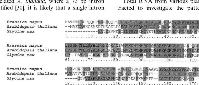
The sequenced cDNA encodes a protein of 203 amino acid residues with ATG initiation codon in position 59 and putative termination codon (TAA) in position 668. The putative B. napus HMG-I/Y protein has a calculated molecular mass of 2 169 967 Da and pI of 10.44. Four copies of the AT-hook (GRPP/R) motif are distributed throughout the C-terminus part (Fig. 1). The protein is very similar to HMG-I/Y proteins from other plant species, with the highest similarity to A.thaliana (62% identity at the protein level). Like other plant HMG-I/Y proteins the N-terminal se-quence is similar to the N-terminal sese-quence of histone H1 [8,14,31]. The B. napus HMG-I/Y pre-dicted protein has two nuclear localization se-quences, at position 97 (PKRGRRGR) and 160 (PKKQKTE), targeting the protein into the nu-cleus (PSORT database) and several putative phosphorylation sites at positions 100 – 104, 133 – 137, 160 – 164 and 193 – 197 (NetPhos 2.0-protein phosphorylation prediction server, http:// www.cbs.dtu.dk/services/NetPhos/).
3.2. Expression of the HMG-I/V gene
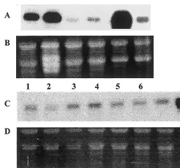
Total RNA from various plant tissues was ex-tracted to investigate the pattern of HMG-I/Y
Fig. 1. Sequence alignment of B. napus HMG-I/Y putative encoded protein using the AntherProt program to A. thaliana
Fig. 2. Expression of HMG-I/Y gene in various plant tissues and during pollen development. Total RNA (15 mg) was separated by electrophoresis, blotted and probed with anti-sense RNA HMG-VY probe. (A) Line 1, root; 2, leaf tissue; 3, seed; 4, callus; 5, pollen derived embryos in torpedo stage of development; 6, petals. (B and D) EtBr stained gel was used to verify RNA integrity and equal loading. (C) Line 7, early-unicellular microspores; 8, mid-unicellular microspores; 9, late-unicellular microspores; 10, early-bicellular mi-crospores; 11, mid-bicellular pollen grains; 12, late bicellular pollen grains; 13, mature tricellular pollen grains; 14, leaf tissue as a estimation of expression.
HMG transcript was detected predominantly in anthers and petals. A strong signal was also de-tected in root epidermis. These experiments confi-rmed the results obtained by Northern blot analysis.
Subcellular localization of the HMG-I/Y tran-script was investigated by Northern analysis using separated polysomal and non-polysomal fractions. Total RNA was extracted from samples corre-sponding to various B. napus microspores and pollen developmental stages, with subsequent polysomal and non-polysomal fraction separation. The non-polysomal fraction contained predomi-nantly RNPs and dissociated monosomes and ri-bosomal subunits. Northern blot hybridizations with the HMG-I/Y probe indicated predominantly polysomal localization of the transcript, suggesting possible translational competence immediately upon transcription (Fig. 4).
Fig. 3. Localization of the HMG-I/V transcript by in situ hybridization. In situ hybridization experiments were carried out as described in Section 2 with 500 ng ml−1 HMG-I/Y
anti-sense probe (panels A and C) and with a blank hy-bridization mix as a control (panels B and D). Panels: A and B, heart-shaped embryo; C and D, flower bud-developmental stage tricellular pollen grains. Bar: 40 mm. Arrows indicate areas of highest transcript concentration.
expression. The samples included microspores and pollen grains at various developmental stages, starting from early-unicellular, mid-unicellular and late-unicellular microspores, early to mid-bicellu-lar, late-bicellular and mature pollen grains. Also included were several sporophytic tissues such as roots, leaves, seeds, callus tissue, torpedo stage pollen-derived embryos and petals (Fig. 2). The HMG-I/Y transcript signal was detected in all tissues examined, with the highest expression in 3-week-old pollen-derived embryos. Estimated ex-pression in roots and leaves of 2-week-old plantlets was lower. The HMG-I/Y transcript was hardly detectable in mature seeds ofB.napus. Low levels of HMG-I/Y mRNA were detected in all pollen developmental stages examined. The esti-mated size of the detected transcript was about 900 bp. Northern blot analysis was also carried out with pollen grains from other plant species, including several Brassica species and Nicotiana tabacum: results indicate the presence of ho-mologous transcripts in related Brassica species, but not in tobacco (results not shown).
Fig. 4. HMG-I/Y transcript is localized on polysomal com-plexes. The polysomal and non-polysomal complexes (con-taining RNPs) were separated by ultracentrifugation, total RNA isolated and, after electrophoresis, probed with anti-sense RNA HMG-I/Y probe. RNA (10 mg) isolated from polysomal fraction and equal amount of non-polysomal frac-tion were loaded. (A) Line 1+6, RNA isolated from early-unicellular microspores, polysomal and nonpolysomal fractions, respectively. Line 2+7, RNA from late-unicellular microspores, polysomal and non-polysomal fractions, respec-tively. Line 3+8, RNA from mid-bicellular pollen grains, polysomal and non-polysomal fractions, respectively. Line 4+9, RNA isolated from late bicellular pollen grains, polyso-mal and non-polysopolyso-mal fraction, respectively. Line 5+10. RNA from mature pollen grains, polysomal and non-polyso-mal fractions, respectively. (B) EtBr stained gel was used to verify RNA integrity and equal loading.
clone). This result was confirmed by Southern hybridization of B. napus DNA samples cut by EcoRI,BamHI andXbaI, where only one band of estimated size of 1 kbp was detected (result not shown).
The results show that the B. napus HMG-I/Y gene is a single-copy gene with high homology to all Brassica species examined. The estimated size of the detected signal was about 1000 bp. The lack of a detected signal in distant species such as tobacco and maize suggests lower homology of corresponding genes, at least at nucleotide level (data not shown).
4. Discussion
HMG genes were originally cloned from verte-brates and additionally from yeast and lower eu-karyotes. HMG genes have been subsequently identified in plants. Best characterised is the HMG-I/Y subgroup that binds to DNA A/T-rich promoter regions. Here we describe the first isola-tion of a B. napus cDNA encoding a putative HMG-I/Y protein.
The HMG-I/Y transcript was detected in all tissues examined, with the highest level of expres-sion in microspore-derived embryos. This is a novel observation. The level of expression in leaves and roots was lower. A weaker signal was detected in samples derived from mature seeds and callus. The surprisingly low level of expression in callus was probably due to older tissue used for Northern blot analyses. Discrepancies between re-ported high level of expression in pea andB. napus seeds could be explained by use of younger, and thus transcriptionally more active, seeds for peas [21]. In agreement with previous data [13], the highest expression is found in rapidly dividing tissues, pointing to HMG-I/Y regulation of gene expression in actively dividing tissues.
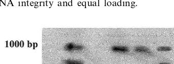
As the isolated HMG-I/Y cDNA originated from a pollen cDNA library, we were also inter-ested to analyze the activity of the gene during pollen development. The level of detected tran-script remains constant, with no significant alter-nation even during transcriptionally very active stages around the first pollen mitosis. These results confirm a ubiquitous pattern of expression of HMG-I/Y genes. Interestingly, the HMG-I/Y transcript is predominantly localized on the Fig. 5. Southern blot analysis — HMG-I/V is a single copy
gene. Genomic DNA (10 mg) was digested with HindIII, separated on 0. 8% agarose gel and, after blotting, hybridized with HMG-I/Y cDNA probe. The position of the 1000 bp DNA marker is indicated. Line 1,B.napus; 2,B.oleracea; 3,
B.carinata; 4,B.campestris 6ar.chinensis; 5, B.rapa.
3.3. Genomic DNA analysis
Southern blot analysis of genomic DNA was done to estimate the copy number of the gene encoding the HMG-I/Y protein. High molecular weight genomic DNA from young leaves of B. napus and closely related B. oleracea, B. carinata, B. campestris 6ar. Chinenis and B. rapa was
di-gested with restriction nuclease HindIII. (Fig. 5). The radioactively labelled cDNA clone was used as a probe for the hybridization experiments.
polysomal fraction, indicating that the gene is available for transcription throughout the pollen development. This observation may point to one of the few published instances where genes may remain transcriptionally active in tricellular pollen [32].
Southern blot analysis of genomic DNA re-vealed the presence of only a single copy of a corresponding gene in the B. napus genome and also in genomes of closely related Brassica species a situation similar toArahidopsis[13] and pea [21]. Detection of only one band in B. campestris and B. carinata was possibly due to alternations in the 3% non-coding region of HMG-I/Y corresponding genes. In contrast, loss of detectable signal in the case of B. oleracea is very surprising because B. oleraceais one of theB. napusparents. Two copies of the HMG-I/Y gene have been isolated from Japanese jack been [12].
In contrast to Arabidopsis HMG-I/Y protein, the putative B. napus protein does not contain an additional alanine-rich stretch, which is suggested to play a role as a dimerization domain for the formation of homo- or heterodimers [33]. This domain, which is found also in mammalian HMG-I/Y proteins could be involved in protein – protein interactions. Insertion of nine amino acid residues (preferentially alanine) at position 114 – 122 of Arabidopsis protein is in the same position as an insertion of 11 amino acid residues which distin-guishes mammalian HMG-I from HMG-Y [13]. Using the PSORT database several putative phosphorylation sites were found in the putative HMG-I/Y protein. Maize HMG protein had been shown to be phosphorylated by a protein kinase of casein type II isolated from maize endosperm nu-clei [34].
Acknowledgements
We are grateful to Dr B. Allore (Canada), Dr I. Tachezy for assisting in sequencing and J. Novotny for construction of the microspore cDNA library. The work was supported by EC COST grant 822.60/97, Grant Agency of Charles University 246/97B and Czech Ministry of Educa-tion grant 980145. The assistance of AvH Stiftung is also gratefully acknowledged. The work was carried out at a joint laboratory of Charles Uni-versity and PIAS.
References
[1] M. Bustin, D.A. Lehn, D. Landsman, Structural fea-tures of HMG chromosomal protein and their genes, Biochim. Biophys. Acta 1049 (1990) 231 – 243.
[2] C.I. Webster, L.C. Packman, K.H. Pwee, J.C. Gray, High mobility group proteins HMG-1 and HMG-I/Y bind to a positive regulatory region of pea plastocyanin gene promoter, Plant J. 11 (1997) 703 – 715.
[3] M. Friedmann, L.T. Holth, Y. Zoghbi, R. Reeves, Orga-nization, inducible-expression and chromosome localiza-tion of the human HMG I(Y) nonhistone protein gene, Nucleic Acids Res. 21 (1993) 4259 – 4267.
[4] D. Thanos, T. Maniatis, The high mobility group protein HMG I/Y is required for NF-kB dependent virus induction of the human IFN-B gene, Cell 71 (1992) 777 – 789.
[5] W. Du, D. Thanos, T. Maniatis, Mechanisms of tran-scriptional synergism between distinct virus-inducible en-hancer elements, Cell 74 (1993) 887 – 898.
[6] K. Zhao, E. Ka¨s, E. Gonzales, U.K. Laemmli, SAR-de-pendent mobilization of histone H1 by HMG-I/Y in vitro: HMG-I/Y is enriched in H1-depleted chromatin, EMBO J. 8 (1993) 3237 – 3247.
[7] D.A. Hill, R. Reeves, Competition between HMG-I/Y, HMG-I and histone H1 on four-way junction DNA, Nucleic Acids Res. 25 (1997) 3523 – 3531.
[8] T. Laux, J. Seurinck, R.B. Goldberg, A soybean embryo cDNA encodes a DNA-binding protein with histone and HMG protein like domains, Nucleic Acid Res. 19 (1991) 4768.
[9] A.H. Meijer, E.L. van Dijk, J.H:C. Hoge, Novel mem-bers of a family of AT-hook-containing DNA-binding proteins from rice are identified through their in vitro interaction with consensus target sites of plant and animal homeodomain proteins, Plant Mol. Biol. 31 (1996) 607 – 618.
[10] J. Nietelo-Sotelo, A. Ichida, P.H. Quail, PF1: an A-T hook-containing DNA binding protein from rice that interacts with a functionally defined d(AT)-rich element in oat phytochrome A3 gene promoter, Plant Cell 6 (1994) 287 – 301.
[11] J. Nietelo-Sotelo, A. Ichida, P.H. Quail, Positive factor 1 (PF 1) from oat is an HMG-I/Y and histone 1-like protein that binds a functionally defined (AT)-rich ele-ment in oat phytochrome A gene (PHYA3) promoter, Nucleic Acid Res. 22 (1994) 1115 – 1116.
[12] S. Yamamoto, T. Minamikawa, Two genes for high mobility group protein HMG-Y are present in genome of Cana6alia gladiata D.C, Plant Mol. Biol. 33 (1997) 537 – 544.
[13] R. Gupta, C.I. Webster, A.R. Walker, J.C. Gray, Chro-mosomal location and expression of the single-copy gene encoding high-mobility-group protein HMG-I/Y inAra
-bidopsis thaliana, Plant Mol. Biol. 34 (1997) 529 – 536. [14] K.D. Grasser, Plant chromosomal high mobility group
(HMG) proteins, Plant J. 7 (1995) 185 – 192.
[16] G. Tjaden, G.M. Coruzzi, A novel AT-rich DNA bind-ing protein that combines an HMG I-like DNA bindbind-ing domain with a putative transcription domain, Plant J. 6 (1994) 107118.
[17] K. Jacobsen, N.B. Laursen, E.O. Jensen, A. Marker, C. Poulsen, K.A. Marker, HMG-I like proteins from leaf and nodule nuclei interact with different motifs in soy-bean nodulin promoters, Plant Cell 1 (1990) 85 – 94. [18] K.H. Pwee, C.I. Webster, J.C. Gray, HMG protein
binding to an A/T-rich positive regulatory region of the pea plastocyanin gene promoter, Plant Mol. Biol. 26 (1994) 1907 – 1920.
[19] K.H. Pwee, J.C. Gray, The pea plastocyanin promoter directs cell-specific but not full light-regulated expression in transgenic tobacco plants, Plant J. 3 (1993) 437 – 449. [20] T.J. Pedersen, L.J. Arwood, S. Spiker, M.J. Guiltinan, F. Thompson, High mobility group chromosomal proteins bind to AT-rich tracts flanking plant genes, Plant Mol. Biol. 16 (1991) 95 – 104.
[21] R. Gupta, C.I. Webster, J.C. Gray, The single-copy gene encoding high-mobility-group protein HMG-I/Y from pea contains a single intron and is expressed in all organs, Plant Mol. Biol. 35 (1997) 987 – 992.
[22] P.M. Pechan, W.A. Keller, Indentification of potentially embryogenic microspores in Brassica napus, Physiol. Plant. 74 (1988) 377 – 384.
[23] Z.-H. Xu, M.R. Davey, E.C. Cocking, Plant regenera-tion from root protoplasts of Brassica, Plant Sci. Lett. 24 (1982) 117 – 121.
[24] P. Chomczynski, N. Sacchi, Single-step method of RNA isolation by acid guanidiurn thiocyanate – phenol – chlo-roform extraction, Anal. Biochem. 162 (1987) 156 – 159. [25] S. deVries, H. Hoge, T. Bisseling, Isolation of total and polysomal RNA from plant tissues, in: S..B. Gelvin
(Ed.), Plant Molecular Biology Manual, Kluwer, Dor-drecht, 1988, pp. 1 – 13/B6.
[26] J. Sambrook, E.F. Fritsch, T. Maniatis, Molecular Cloning: A Laboratory Manual, 2nd edn, Cold Spring Harbor, New York, 1989.
[27] B.P. Sokolov, D.J. Prockop, A rapid and simple PCR-based method for isolation of cDNAs from differentially expressed genes, Nucleic Acids Res. 22 (1994) 4009 – 4015.
[28] L. Rogers, G. Bendich, in: S.B. Gelvin, R.A. Schilper-oort, D.P. Verma (Eds.), Plant Molecular Biology Man-ual, Kluwer, Dordrecht, 1990, pp. A6/1 – 11.
[29] D.R. Cavener, S.C. Ray, Eukaryotic start and stop translation sites, Nucleic Acids Res. 19 (1991) 3185 – 3192.
[30] R. Gupta, C.I. Webster, J.C. Gray, Characterisation and promoter analysis of the Arabidopsis gene encoding high-mobility-group protein HMG-I/Y, Plant Mol. Biol. 36 (1998) 897 – 907.
[31] P. Razafimabatratra, N. Chaubet, G. Phillipps, C. Gigot, Nucleotide sequence and expression of a maize H1 histone cDNA, Nucleic Acids Res. 19 (1991) 1491 – 1496.
[32] J.P. Mascarenhas, Gene activity during pollen develop-ment, Annu. Rev. Plant Physiol. Plant Mol. Biol. 41 (1990) 317 – 338.
[33] R. Eckner, M.L. Bimstiel, Cloning of cDNAs encoding for human HMG I and HMG Y proteins: both are capable of binding to the octamer sequence motif, Nu-cleic Acids Res. 17 (1989) 5947 – 5959.
[34] K.D. Grasser, U.G. Maier, G. Feix, A nuclear casein type 11 kinase from maize endosperm phosphorylating HMG proteins, Biochem. Biophys. Res. Commun. 162 (1989) 456 – 463.