PLANTS ARE EXPLOITED as a source of food and shelter by a wide range of parasites, including viruses, bacteria, fungi, nematodes, insects and even other plants. Plants have responded to this pressure by evolving mechanisms to recognize and counterattack prospec-tive colonists. If a plant detects an in-vasion, then a set of inducible defense responses is deployed; these include programmed cell death (referred to as the hypersensitive response or HR), tis-sue reinforcement at the infection site, production of anti-microbial metab-olites and induction of ‘defense-associ-ated’ gene expression1. Activation of
‘local’ responses at the point of infec-tion can be followed by establishment of secondary immunity throughout the plant (‘systemic’ acquired resistance or
SAR), which is long lasting and effective against a broad spectrum of pathogens2.
Activation of inducible defenses is contingent upon recognition of an in-vasion. Surveillance in the plant is the collective duty of a complex array of constitutively expressed Rgenes (for re-sistance). Individual Rgenes have nar-row recognition capabilities and they trigger resistance only when the invad-ing pathogen expresses a correspondinvad-ing ‘Avr gene’ (for avirulence). These gen-etic attributes have inspired a molecular
model in which Avr-encoded proteins
are delivered to the plant cell to facili-tate invasion and there, they are recog-nized by the corresponding R protein as a signal that invaders have arrived. Studies arising from molecular cloning
of numerous Avr and R genes have at
least partially validated this ‘receptor-elicitor’ model. These efforts have been summarized in several excellent re-views, including a recent article in TiBS3.
This review focuses on the molecular events that occur ‘downstream’ of pathogen recognition. For inducible fenses to be effective, they must be de-ployed rapidly; the ability of pathogens to outpace a tardy counterattack is well
documented. On the other hand, these defenses cannot be unleashed with im-punity, as they are resource-intensive and can inflict substantial collateral damage on host tissues. Thus, deploy-ment must be confined to the proper place and time. These requirements sug-gest that a complex, highly integrated regulatory network controls defense re-sponses. With these considerations in mind, we highlight recent insights into the mediatory signals between the ini-tial recognition event and ultimate acti-vation of defense responses.
Ion fluxes and reactive oxygen intermediates are the first messengers
Much effort has been devoted to iden-tification of the earliest responses to pathogen invasion. These studies typi-cally employ biochemical and physio-logical analysis of cultured cells or in-tact plants that have been challenged with pathogens or purified elicitors. The earliest detectable cellular events are ion fluxes across the plasma membrane and a burst of oxygen metabolism that produces reactive oxygen intermediates (ROIs), such as superoxide (O22) and
hydrogen peroxide (H2O2)4. A clear
causal link between these events and defense induction was demonstrated in cultured cells of parsley, in which elici-tor-induced ion fluxes are required for induction of the oxidative burst, but not vice versa5(Fig. 1). The oxidative burst
is in turn required for activation of de-fense gene induction and production of antimicrobial metabolites.
Thus, receptor-dependent pathogen recognition triggers ion channel fluxes, which subsequently induce ROI produc-tion (Fig. 1). The molecules that connect these events have yet to be defined. Inhibitor studies suggest that receptor ac-tivation is linked to ion channel stimu-lation and ROI production by mechanisms involving protein phosphorylation and
GTP-binding proteins4, but genetic
requirements for these steps have not been demonstrated. A recently identified pathogen-responsive calmodulin isoform6 TIBS 25 –FEBRUARY 2000
79
0968 – 0004/00/$ – See front matter © 2000, Elsevier Science Ltd. All rights reserved. PII: SO986-0004(99)01532-725 Bork, P. et al. (1997) A superfamily of conserved domains in DNA damage-responsive cell cycle checkpoint proteins. FASEB J. 11, 68–76
26 Opperman, T. et al. (1999) A model for a
umuDC-dependent prokaryotic DNA damage checkpoint. Proc. Natl. Acad. Sci. U. S. A. 96, 9218–9223 27 Bridges, B.A. (1998) DNA repair: getting past a lesion –
at a cost. Curr. Biol. 8, R886–R888
28 Maenhaut-Michel, G. et al. (1992) A umuDC-independent SOS pathway for frameshift mutagenesis. Mol. Gen. Genet. 373, 373–380
29 Gibbs, P.E.M. et al. (1998) A human homolog of the Saccharomyces cerevisiae REV3 gene, which encodes the catalytic subunit of DNA polymerase z. Proc. Natl. Acad. Sci. U. S. A. 95, 6876–6880
30 Cordonnier, A.M. et al. (1999) Impaired translesion
synthesis in xeroderma pigmentosum variant extracts. Mol. Cell. Biol. 19, 2206–2211
31 Masutani, C. et al. (1999) The XPV (xeroderma pigmentosum variant) gene encodes human DNA polymerase h. Nature 399, 700–704
32 Johnson, R.E. et al. (1999) hRAD30 mutations in the variant form of xeroderma pigmentosum. Science 285, 263–265
REVIEWS
Signal transduction in the plant
immune response
John M. McDowell and Jeffery L. Dangl
Complementary biochemical and genetic approaches are being used to dissect the signaling network that regulates the innate immune response in plants. Receptor-mediated recognition of invading pathogens triggers a signal amplification loop that is based on synergistic interactions between nitric oxide, reactive oxygen intermediates and salicylic acid. Alternative re-sistance mechanisms in Arabidopsis are deployed against different types of pathogens; these mechanisms are mediated by either salicylic acid or the growth regulators jasmonic acid and ethylene.
J.M. McDowellis at the Dept of Plant Pathology, Plant Physiology, and Weed Science, Virginia Polytechnic Institute, Blacksburg, VA 24061-0331, USA; and
J.L. Danglis at the University of North Carolina, Dept of Biology and Curriculum in Genetics, C.B. 3280 Coker Hall,Chapel Hill, NC 27599-3280, USA.
REVIEWS
TIBS 25 –FEBRUARY 200080
might represent a downstream target of ion fluxes. Furthermore, several pathogen-responsive MAP kinase iso-forms have been identified recently. In one case, the MAP kinase translocates to the nucleus after pathogen challenge, and ac-tivation and translocation occur indepen-dently, or upstream, of the oxidative burst7. Multiple pathogens as well as
wounding and tissue damage activate a second MAP kinase8. Again, this activation
is independent or upstream of the oxida-tive burst, suggesting a mechanism for pathogen-induced, but ROI-independent, transcriptional activation.
The role of ROIs in the defense re-sponse is particularly interesting and controversial at the moment. ROIs have been associated with apoptosis of mam-malian cells, indicating a role in cell death during the HR in plants9. This
analogy is supported by identification of a plant equivalent to the mammalian NADPH oxidase complex that produces ROIs in neutrophils. Plant homologs of two NADPH oxidase components
(gp91phox and Rac) have been cloned.
The phenotypes of transgenic plants ex-pressing constitutively active or domi-nant negative variants of Rac indicate that it is a regulator of cell death in plants10. The plant gp91phox homologs
contain Ca21-binding motifs that are not
found in the mammalian homolog and could provide a regulatory connection
to pathogen-induced ion fluxes11.
However, no direct evidence exists cur-rently to associate this protein with
defense induction. Plant gp91phox
homologs are encoded by a multi-gene family and might thus be functionally specialized or redundant, or both12.
A requirement for NADPH oxidase in plant cell death is further supported by observations that pharmacological in-hibitors of the NADPH oxidase complex can interfere with induction of the HR9.
However, alternative sources of ROIs have been postulated in several studies, and the relative contributions of various oxidases must be clarified13. A more
vex-ing question has arisen from reports that exogenously supplied ROIs are in-sufficient to activate HR cell death in some cell systems5. Although necessary,
ROIs are not sufficient to trigger cell death and must therefore require accomplices.
Salicylic acid and nitric oxide potentiate ROI-dependent responses
Two putative accomplices have been identified in recent investigations of interactions between salicylic acid (SA), nitric oxide (NO) and ROIs. SA has long been associated with defense induction in plants: exogenous SA is sufficient to induce plant defense gene expression and systemic acquired resistance (but not cell death), whereas transgenic plants expressing an SA-degrading en-zyme from bacteria (NahG) are unable
to activate local or systemic defense re-sponses2. NO collaborates with ROIs to
trigger cell death in the mammalian im-mune response14, and three recent
stud-ies provide evidence that NO also inter-acts with ROIs and SA in plants to induce the HR and defense gene expres-sion. First, Shirasu et al. demonstrated that addition of exogenous SA ‘primes’-cultured soybean cells to initiate cell death in response to pathogen
chal-lenge15. This potentiation occurs
through an undefined, phosphorylation-dependent agonist that does not require translation. In this system, SA in combi-nation with cantharidin (a protein phos-photase inhibitor) is sufficient to trigger ROI production in the absence of pathogens, but this oxidative burst is not sufficient to trigger cell death. However, a subsequent study demon-strated that exogenous NO in combi-nation with SA plus cantharidin (or ex-ogenous ROIs) is sufficient to induce cell death without pathogen challenge16.
An independent study in intact tobacco plants demonstrated that exogenous NO triggers expression of pathogen-respon-sive genes, and that this gene induction is dependent on SA accumulation17.
The physiological relevance of re-sponses to exogenous NO was sup-ported by indirect evidence in both studies for accumulation of endogenous NO in response to pathogen challenge. The effects of both exogenous and en-dogenous NO were counteracted by simultaneous addition of NO scavengers or pharmacological inhibitors of mam-malian nitric oxide synthase (NOS). More significantly, induction of endogenous NO was dependent upon recognition of the challenging pathogen: NO accumulation does not occur in dis-ease-susceptible tobacco lines17 or in
soybean cells that are challenged with an isogenic pathogen that does not express the appropriate avrgene16. This
correlation provides strong, although still circumstantial, evidence for a link between NO and defense activation, and genetic confirmation of NOS involvement is awaited eagerly.
A model for signal amplification and propagation
How might these three small molecules interact to promote defense induction? An important observation in the above studies is that the contributions of NO, SA and H2O2appear to be synergistic rather than additive, implying that they interact directly and cooperatively in a signal- amplification mechanism. Furthermore,
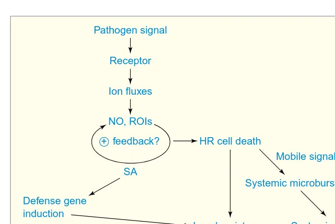
Ti BS Pathogen signal
Receptor
HR cell death
Defense gene induction
Mobile signal?
Local resistance Ion fluxes
NO, ROIs
SA + feedback?
Systemic microbursts
Systemic resistance
Figure 1
TIBS 25 –FEBRUARY 2000
81
ROIs and NO stimulate SA biosynthesis, and SA in turn potentiates ROI–NO-de-pendent responses, as described above. These features suggest that receptor-dependent pathogen perception triggers a positive feedback loop of ROI–NO production and SA accumulation, which rapidly amplifies the initial signal and guarantees timely defense activation (Fig. 1). Culmination of this cycle in HR cell death could release ROIs, NO and SA into intercellular spaces, and these com-pounds could directly inhibit pathogen growth or ‘warn’ neighboring cells of an imminent invasion, or both.
A spatial extension of this scenario was provided in a report claiming that localized production of ROIs is sufficient to trigger SAR, and that establishment of SAR was correlated with oxidative ‘mi-crobursts’ in distal tissues18. The
impli-cation is that systemic resistance can be induced by subsequent reiterations of the ROI–SA–cell-death cycle at lower amplitude throughout the plant. However, it is currently unclear whether ROIs themselves are the ‘mobile signal’ that induces distal microbursts and SAR. Other candidates for the mobile signal include SA (although the weight of available evidence argues against this) and nitrosylated glutathione19.
The above model makes no predic-tions about the biochemical roles of SA, ROIs or NO. SA can bind to catalase (an
H2O2-degrading enzyme) and other
heme-containing enzymes in vitro, but the relevance of this capability in planta
has not been demonstrated conclu-sively20. The literature on mammalian
species provides an almost overwhelm-ing number of candidate targets for NO and ROIs14. Perhaps the most intuitive
scenario is that ROIs and NO interact to form cytotoxic radicals that could di-rectly kill host or pathogen cells, or both. Alternatively, but not exclusively, ROIs and NO could serve as second messengers through redox-mediated al-terations of downstream regulators. For example, an SA- and pathogen-inducible plant homolog of cyclooxygenase has been reported21. This enzyme is a
promi-nent target of NO in mammalian cells and catalyses the production of lipid sig-nal intermediates. Guanylate cyclase is a second target of NO, and Durner et al.
provide evidence that cyclic GMP is an intermediate in defense-gene induc-tion17. Durner et al. also demonstrate
that defense-gene expression is induced by cyclic ADP ribose, which transduces NO-dependent signals in mammalian systems through modulation of ion
channel activity. The obvious multiplic-ity of NO targets implies that dissection of NO signaling will be a challenging task requiring physiological and genetic approaches.
Another question not addressed in this model is the repression of defense responses. The apparent reiterative am-plification of input signals that can kill plant cells implies the existence of key regulators that limit the propagation of the HR. SA can inhibit NOS in mam-malian cells, suggesting a potential mechanism for self-limitation of the HR (Ref. 19). Similarly, H2O2induces the ex-pression of antioxidant enzymes in cells adjacent to the HR site9. Defense
re-sponses could also be under negative genetic control, as suggested by reces-sive mutants in which cell death or other defense responses are activated
spontaneously. For example, the lsd1
mutation renders the plant hypersensi-tive to cell-death-inducing stimuli, such as pathogens, SA or exogenous superox-ide22. A particularly interesting aspect of
this mutant is that cell death lesions, once initiated, can spread to engulf the entire leaf. This suggests that the wild-type gene somehow delimits the area of the HR. The LSD1gene encodes a zinc-finger protein, which raises the possi-bility that cell death is a default pathway under negative transcriptional regu-lation23. If true, this regulatory
configu-ration might have arisen to meet a need for rapid defense activation. However, a critical need for future exploration clearly exists in this area.
SA-independent defense responses are triggered by distinct pathogens
The ROI–SA–cell-death response, de-scribed above, has received the major-ity of recent experimental attention. However, this response is not germane to every plant–pathogen interaction. For example, recent genetic studies in
Arabidopsishave revealed resistance re-sponses that operate independently of SA accumulation and are mediated by jasmonic acid (JA) and the gaseous hor-mone ethylene (ET)24. JA and ET are also
plant growth regulators, suggesting overlap between the regulatory compo-nents of development and defense.
Arabidopsis mutants, compromised in their ability to respond to JA or to pro-duce SA, have been used elegantly to demonstrate that the SA-dependent and ET–JA-dependent responses are utilized differentially against pathogens with contrasting modes of attack25 (Fig. 2).
The ET–JA-dependent defense response
is activated by pathogens that kill plant cells to obtain nutrients. In contrast, the SA-dependent response is triggered by a pathogen that obtains nutrients from living plant tissue. This observation raises the intriguing possibility (yet to be generalized) that plants can activate distinct defense responses tailored to specific types of parasites. Several stud-ies have also suggested that the ET–JA and SA responses are mutually in-hibitory24. Such potential cross talk is
again suggestive of a capacity for selec-tive defense deployment. It will be of great interest to define and compare the executioners in SA- and ET–JA-dependent responses, and to determine whether both responses are triggered by the same perception or early-response components.
Why such complexity?
A clear theme in this review is that simple linear pathways are insufficient to explain the regulation of inducible de-fenses in plants. Indeed, the branched models that we have presented in this review will soon be obsolete, as more regulatory components are defined. This apparent complexity probably re-flects a requirement for tight regulation of an important, but costly response. Regulatory complexity might also have
REVIEWS
Ti BS Biotrophic
pathogen
Resistance SA
NahG
Necrotrophic pathogen
Resistance ET, JA
coi1
Figure 2
REVIEWS
TIBS 25 –FEBRUARY 200082
arisen, in part, as a consequence of co-evolution between plants and pathogens. It is tempting to speculate that pathogens have evolved mecha-nisms to suppress defense responses by interfering with key downstream regula-tors, thereby forcing the plant in turn to evolve bypass mechanisms. In fact, some pathogens might have learned to turn defense responses to their own ne-farious ends. For example, induction of the ROI–SA–cell-death response could work to the advantage of pathogens that live by killing host cells and perhaps, the plant has evolved the ET–JA re-sponse (with a concomitant repression of the SA response) to avoid turning the gun on itself unnecessarily. If these sce-narios are valid, then a thorough com-parison of defense response circuitry in several plant species could provide valuable insight into the molecular mechanisms of response regulation and the evolutionary mechanisms by which alternate regulatory configurations could have arisen.
Agricultural applications
A final and perhaps most significant payoff from endeavors in this area could come in the form of strategies for engi-neering resistance in crops. Several promising approaches have already been initiated from our relatively limited knowledge base. For example, an analog of SA is being marketed as an alternative to costly and environmentally harmful fungicides26. Cross-species transgenic
approaches might also prove useful, as overexpression of the SAR regulatory gene NPR1in Arabidopsisconfers resis-tance to two different pathogens without
a discernible growth penalty to the plant27. Finally, Molina and
co-workers demonstrate that one commonly used fungicide induces host defense responses28, suggesting that already are
we unwittingly manipulating plant defense responses to our own ends.
Acknowledgements
We apologize for the omission of many relevant citations owing to space limitations. We thank Dave Mackey, Daniel Aviv, Petra Epple and Mats Ellerstrom for comments on the manuscript. The authors are supported by grants from the NIH, DOE, USDA and NSF (J.L.D.), and postdoctoral awards from the NIH and USDA (J.M.M.).
References
1 Hammond-Kosack, K.E. and Jones, J.D.G. (1996) Inducible plant defense mechanisms and resistance gene function. Plant Cell 8, 1773–1791
2 Ryals, J.L. et al. (1996) Systemic acquired resistance. Plant Cell 8, 1809–1819
3 Van der Biezen, E.A. and Jones, J.D.G. (1998) Plant disease-resistance proteins and the gene-for-gene concept. Trends Biochem. Sci. 12, 454–456 4 Scheel, D. (1998) Resistance response physiology and
signal transduction. Curr. Opin. Plant Biol. 1, 305–310 5 Jabs, T. et al. (1997) Elicitor-stimulated ion fluxes
and reactive oxygen species from the oxidative burst signal defense gene activation and phytoalexin synthesis in parsley. Proc. Natl. Acad. Sci. U. S. A. 94, 4800–4805
6 Heo, W.D. et al. (1999) Involvement of specific calmodulin isoforms in salicylic acid-independent activation of plant disease resistance responses. Proc. Natl. Acad. Sci. U. S. A. 96, 766–771
7 Ligterink, W. et al. (1997) Receptor-mediated activation of a MAP kinase in pathogen defense of plants. Science 276, 2054–2057
8 Romeis, T. et al. (1999) Rapid Avr9- and Cf-9-dependent activation of MAP kinases in tobacco cell cultures and leaves: convergence of resistance gene, elicitor, wound, and salicylate responses. Plant Cell 11, 273–287
9 Lamb, C. and Dixon, R.A. (1997) The oxidative burst in plant disease resistance. Annu. Rev. Plant Physiol. Plant Mol. Biol. 48, 251–275
10 Kawasaki, T. et al. (1999) The small GTP-binding
protein rac is a regulator of cell death in plants. Proc. Natl. Acad. Sci. U. S. A. 96, 10922–10926 11 Keller, T. et al. (1998) A plant homolog of the
neutrophil NADPH oxidase gp91phox subunit gene encodes a plasma membrane protein with Ca21binding
motifs. Plant Cell 10, 255–266
12 Torres, M.A. et al. (1998) Six Arabidopsis thaliana homologues of the human respiratory burst oxidase (gp91phox). Plant J. 14, 365–370
13 Bolwell, G.P. (1999) Role of active oxygen species and NO in plant defence responses. Curr. Opin. Plant Biol. 2, 287–294
14 Hausladen, A. and Stamler, J.S. (1998) Nitric oxide in plant immunity. Proc. Natl Acad. Sci. U. S. A. 95, 10345–10347
15 Shirasu, K. et al. (1997) Salicylic acid potentiates an agonist-dependent gain control that amplifies pathogen signals in the activation of defense mechanisms. Plant Cell 9, 261–270
16 Delledonne, M. et al. (1998) Nitric oxide functions as a signal in plant disease resistance. Nature 394, 585–588 17 Durner, J. et al. (1998) Defense gene induction in
tobacco by nitric oxide, cyclic GMP, and cyclic ADP-ribose. Proc. Natl. Acad. Sci. U. S. A. 95, 10328–10333
18 Alvarez, M.E. et al. (1998) Reactive oxygen
intermediates mediate a systemic signal network in the establishment of plant immunity. Cell 92, 773–784 19 Durner, J. and Klessig, D.F. (1999) Nitric oxide as a signal in plants. Curr. Opin. Plant Biol. 2, 369–374 20 Durner, J. et al. (1997) Salicylic acid and disease
resistance in plants. Trends Plant Sci. 2, 266–274 21 Sanz, A. et al. (1998) PIOX, a new pathogen-induced
oxygenase with homology to animal cyclooxygenase. Plant Cell 10, 1523–1537
22 Jabs, T. et al. (1996) Initiation of runaway cell death in an Arabidopsis mutant by extracellular superoxide. Science 273, 1853–1856
23 Dietrich, R.A. et al. (1997) A novel zinc-finger protein is encoded by the Arabidopsis lsd1 gene and functions as a negative regulator of plant cell death. Cell 88, 685–694
24 Dong, X. (1998) SA, JA, ethylene, and disease resistance in plants. Curr. Opin. Plant Biol. 1, 316–323 25 Thomma, B. et al. (1998) Separate
jasmonate-dependent and salicylate-jasmonate-dependent defense-response pathways in Arabidopsis are essential for resistance to distinct microbial pathogens. Proc. Natl. Acad. Sci. U. S. A. 95, 15107–15111
26 Görlach, J. et al. (1996) Benzothiabiazole, a novel class of inducers of systemic acquired resistance, activates gene expression and disease resistance in barley. Plant Cell 8, 629–643
27 Cao, H. et al. (1998) Generation of broad-spectrum disease resistance by overexpression of an essential regulatory gene in systemic acquired resistance. Proc. Natl. Acad. Sci. U. S. A. 95, 6531–6536
28 Molina, A. et al. (1998) Impaired fungicide activity in plants blocked in disease resistance signal transduction. Plant Cell 10, 1903–1914
http://tto.trends.com
Editor: Adrian Bird
Institute for Cell and Molecular Biology at the University of Edinburgh, UK
New articles published recently in Elsevier Trends Journals Technical Tips Onlineinclude:
• Tanaka, K.J. and Nishikata, T. (1999) A non-radioactive gel shift protocol enables recovery of RNA-binding proteins (http://tto.trends.com) t01794
• Goldberg, M. (1999) The use of green fluorescent fusion proteins for efficient and reliable in vitro binding assays (http://tto.trends.com) t01801