I . I nt roduc t ion t o t he Ce ll
1 . T he Evolut ion of t he Ce ll
I nt roduc t ion
From M ole c ule s t o t he First Ce ll
From Proc a ryot e s t o Euc a ryot e s
From Single Ce lls t o M ult ic e llula r Orga nism s
Re fe re nc e s
Ge ne ra l
Cit e d
2 . Sm a ll M ole c ule s, Ene rgy, a nd Biosynt he sis
I nt roduc t ion
T he Che m ic a l Com pone nt s of a Ce ll
Biologic a l Orde r a nd Ene rgy
Food a nd t he De riva t ion of Ce llula r Ene rgy
Biosynt he sis a nd t he Cre a t ion of Orde r
T he Coordina t ion of Ca t a bolism a nd Biosynt he sis
Re fe re nc e s
Ge ne ra l
Cit e d
3 . M a c rom ole c ule s: St ruc t ure , Sha pe , a nd I nform a t ion
I nt roduc t ion
M ole c ula r Re c ognit ion Proc e sse s
N uc le ic Ac ids
Prot e in St ruc t ure
Prot e ins a s Ca t a lyst s
Re fe re nc e s
I nt roduc t ion
Look ing a t t he St ruc t ure of Ce lls in t he M ic rosc ope
I sola t ing Ce lls a nd Grow ing T he m in Cult ure
Fra c t iona t ion of Ce lls a nd Ana lysis of T he ir M ole c ule s
T ra c ing a nd Assa ying M ole c ule s I nside Ce lls
Re fe re nc e s
Ge ne ra l
Cit e d
I I . M ole c ula r Ge ne t ic s
5 . Prot e in Func t ion
I nt roduc t ion
M a k ing M a c hine s Out of Prot e ins
T he Birt h, Asse m bly, a nd De a t h of Prot e ins
Re fe re nc e s
Ge ne ra l
Cit e d
6 . Ba sic Ge ne t ic M e c ha nism s
I nt roduc t ion
RN A a nd Prot e in Synt he sis
DN A Re pa ir
DN A Re plic a t ion
Ge ne t ic Re c om bina t ion
V iruse s, Pla sm ids, a nd T ra nsposa ble Ge ne t ic Ele m e nt s
Re fe re nc e s
Ge ne ra l
Cit e d
7 . Re c om bina nt DN A T e c hnology
I nt roduc t ion
T he Fra gm e nt a t ion, Se pa ra t ion, a nd Se que nc ing of DN A M ole c ule s
N uc le ic Ac id H ybridiza t ion
DN A Cloning
DN A Engine e ring
Re fe re nc e s
8 . T he Ce ll N uc le us
I nt roduc t ion
Chrom osom a l DN A a nd I t s Pa c k a ging
T he Globa l St ruc t ure of Chrom osom e s
Chrom osom e Re plic a t ion
RN A Synt he sis a nd RN A Proc e ssing
T he Orga niza t ion a nd Evolut ion of t he N uc le a r Ge nom e
Re fe re nc e s
Cit e d
9 . Cont rol of Ge ne Ex pre ssion
I nt roduc t ion
An Ove rvie w of Ge ne Cont rol
DN A-binding M ot ifs in Ge ne Re gula t ory Prot e ins
H ow Ge ne t ic Sw it c he s Work
Chrom a t in St ruc t ure a nd t he Cont rol of Ge ne Ex pre ssion
T he M ole c ula r Ge ne t ic M e c ha nism s T ha t Cre a t e Spe c ia lize d Ce ll
T ype s
Post t ra nsc ript iona l Cont rols
Re fe re nc e s
Ge ne ra l
Cit e d
I I I . I nt e rna l Orga niza t ion of t he Ce ll
1 0 . M e m bra ne St ruc t ure
I nt roduc t ion
T he Lipid Bila ye r
M e m bra ne Prot e ins
Re fe re nc e s
Ge ne ra l
Cit e d
1 1 . M e m bra ne T ra nsport of Sm a ll M ole c ule s a nd t he I onic Ba sis of
M e m bra ne Ex c it a bilit y
I nt roduc t ion
Princ iple s of M e m bra ne T ra nsport
1 2 . I nt ra c e llula r Com pa rt m e nt s a nd Prot e in Sort ing
I nt roduc t ion
T he Com pa rt m e nt a liza t ion of H ighe r Ce lls
T he T ra nsport of M ole c ule s int o a nd out of t he N uc le us
T he T ra nsport of Prot e ins int o M it oc hondria a nd Chloropla st s
Pe rox isom e s
T he Endopla sm ic Re t ic ulum
Re fe re nc e s
Ge ne ra l
Cit e d
1 3 . V e sic ula r T ra ffic in t he Se c re t ory a nd Endoc yt ic Pa t hw a ys
I nt roduc t ion
T ra nsport from t he ER T hrough t he Golgi Appa ra t us
T ra nsport from t he
T ra ns
Golgi N e t w ork t o Lysosom e s
T ra nsport from t he Pla sm a M e m bra ne via Endosom e s: Endoc yt osis
T ra nsport from t he
T ra ns
Golgi N e t w ork t o t he Ce ll Surfa c e :
Ex oc yt osis
T he M ole c ula r M e c ha nism s of V e sic ula r T ra nsport a nd t he
M a int e na nc e of Com pa rt m e nt a l Dive rsit y
Re fe re nc e s
Ge ne ra l
Cit e d
1 4 . Ene rgy Conve rsion: M it oc hondria a nd Chloropla st s
I nt roduc t ion
T he M it oc hondrion
T he Re spira t ory Cha in a nd AT P Synt ha se
Chloropla st s a nd Phot osynt he sis
T he Evolut ion of Ele c t ron-T ra nsport Cha ins
T he Ge nom e s of M it oc hondria a nd Chloropla st s
Re fe re nc e s
Ge ne ra l
Cit e d
1 5 . Ce ll Signa ling
I nt roduc t ion
Ge ne ra l Princ iple s of Ce ll Signa ling
Signa ling via G-Prot e in-link e d Ce ll-Surfa c e Re c e pt ors
Signa ling via Enzym e -link e d Ce ll-Surfa c e Re c e pt ors
T a rge t -Ce ll Ada pt a t ion
Re fe re nc e s
Ge ne ra l
Cit e d
1 6 . T he Cyt osk e le t on
I nt roduc t ion
T he N a t ure of t he Cyt osk e le t on
I nt e rm e dia t e Fila m e nt s
M ic rot ubule s
Cilia a nd Ce nt riole s
Ac t in Fila m e nt s
Ac t in-binding Prot e ins
M usc le
Re fe re nc e s
Ge ne ra l
Cit e d
1 7 . T he Ce ll-Division Cyc le
I nt roduc t ion
T he Ge ne ra l St ra t e gy of t he Ce ll Cyc le
T he Ea rly Em bryonic Ce ll Cyc le a nd t he Role of M PF
Y e a st s a nd t he M ole c ula r Ge ne t ic s of Ce ll-Cyc le Cont rol
Ce ll-Division Cont rols in M ult ic e llula r Anim a ls
Re fe re nc e s
Ge ne ra l
Cit e d
1 8 . T he M e c ha nic s of Ce ll Division
I nt roduc t ion
An Ove rvie w of M Pha se
M it osis
Cyt ok ine sis
Re fe re nc e s
Ge ne ra l
Cit e d
I V . Ce lls in T he ir Soc ia l Cont e x t
Ce ll J unc t ions
Ce ll-Ce ll Adhe sion
T he Ex t ra c e llula r M a t rix of Anim a ls
Ex t ra c e llula r M a t rix Re c e pt ors on Anim a l Ce lls: T he I nt e grins
T he Pla nt Ce ll Wa ll
Re fe re nc e s
Cit e d
2 0 . Ge rm Ce lls a nd Fe rt iliza t ion
I nt roduc t ion
T he Be ne fit s of Se x
M e iosis
Eggs
Spe rm
Fe rt iliza t ion
Re fe re nc e s
Ge ne ra l
Cit e d
2 1 . Ce llula r M e c ha nism s of De ve lopm e nt
I nt roduc t ion
M orphoge ne t ic M ove m e nt s a nd t he Sha ping of t he Body Pla n
Ce ll Dive rsific a t ion in t he Ea rly Anim a l Em bryo
,
Ce ll M e m ory, Ce ll De t e rm ina t ion, a nd t he Conc e pt of Posit iona l
V a lue s
T he N e m a t ode Worm : De ve lopm e nt a l Cont rol Ge ne s a nd t he Rule s of
Ce ll Be ha vior
Drosophila
a nd t he M ole c ula r Ge ne t ic s of Pa t t e rn Form a t ion. I .
Ge ne sis of t he Body Pla n
Drosophila
a nd t he M ole c ula r Ge ne t ic s of Pa t t e rn Form a t ion. I I .
H om e ot ic Se le c t or Ge ne s a nd t he Pa t t e rning of Body Pa rt s
,
Pla nt De ve lopm e nt
N e ura l De ve lopm e nt
Re fe re nc e s
Ge ne ra l
Cit e d
2 2 . Diffe re nt ia t e d Ce lls a nd t he M a int e na nc e of T issue s
I nt roduc t ion
M a int e na nc e of t he Diffe re nt ia t e d St a t e
T issue s w it h Pe rm a ne nt Ce lls
Re ne w a l by St e m Ce lls: Epide rm is
,
Re ne w a l by Pluripot e nt St e m Ce lls: Blood Ce ll Form a t ion
,
Ge ne sis, M odula t ion, a nd Re ge ne ra t ion of Sk e le t a l M usc le
Fibrobla st s a nd T he ir T ra nsform a t ions: T he Conne c t ive -T issue Ce ll
Fa m ily
Appe ndix
Re fe re nc e s
Ge ne ra l
Cit e d
2 3 . T he I m m une Syst e m
I nt roduc t ion
T he Ce llula r Ba sis of I m m unit y
T he Func t iona l Prope rt ie s of Ant ibodie s
T he Fine St ruc t ure of Ant ibodie s
T he Ge ne ra t ion of Ant ibody Dive rsit y
T Ce ll Re c e pt ors a nd Subc la sse s
M H C M ole c ule s a nd Ant ige n Pre se nt a t ion t o T Ce lls
Cyt ot ox ic T Ce lls
H e lpe r T Ce lls a nd T Ce ll Ac t iva t ion
Se le c t ion of t he T Ce ll Re pe rt oire
Re fe re nc e s
Ge ne ra l
Cit e d
2 4 . Ca nc e r
I nt roduc t ion
Ca nc e r a s a M ic roe volut iona ry Proc e ss
T he M ole c ula r Ge ne t ic s of Ca nc e r
Re fe re nc e s
I: Introduction to the Cell
1. The Evolution of the Cell Introduction
From Molecules to the First Cell
From Procaryotes to Eucaryotes
From Single Cells to Multicellular Organisms
References
Introducción
From Molecules to the First Cell
Complex Chemical Systems Can Develop in an Environment That Is Far from Chemical Equilibrium
Polynucleotides Are Capable of Directing Their Own Synthesis
Self-replicating Molecules Undergo Natural Selection
Specialized RNA Molecules Can Catalyze Biochemical Reactions
Information Flows from Polynucleotides to Polypeptides
Membranes Defined the First Cell
All Present-Day Cells Use DNA as Their Hereditary Material
Summary
Referencias
key terms
I n t ro d u c t io n
All living creatures are made of cells - small membrane-bounded compartments filled with a concentrated aqueous solution of chemicals. The simplest forms of life are solitary cells that propagate by dividing in two. Higher organisms, such as ourselves, are like cellular cities in which groups of cells perform specialized functions and are linked by intricate systems of communication. Cells occupy a halfway point in the scale of biological complexity. We study them to learn, on the one hand, how they are made from molecules and, on the other, how they cooperate to make an organism as complex as a human being.
All organisms, and all of the cells that constitute them, are believed to have descended from a common ancestor cell through evolution by natural selection. This involves two essential
processes: (1) the occurrence of random variation in the genetic information passed from an
individual to its descendants and (2) selection in favor of genetic information that helps its
possessors to survive and propagate. Evolution is the central principle of biology, helping us to make sense of the bewildering variety in the living world.
This chapter, like the book as a whole, is concerned with the progression from molecules to multicellular organisms. It discusses the evolution of the cell, first as a living unit constructed from smaller parts and then as a building block for larger structures. Through evolution, we introduce the cell components and activities that are to be treated in detail, in broadly similar sequence, in the chapters that follow. Beginning with the origins of the first cell on earth, we consider how the properties of certain types of large molecules allow hereditary information to be transmitted and expressed and permit evolution to occur. Enclosed in a membrane, these
molecules provide the essentials of a self-replicating cell. Following this, we describe the major transition that occurred in the course of evolution, from small bacteriumlike cells to much larger and more complex cells such as are found in present-day plants and animals. Lastly, we suggest ways in which single free-living cells might have given rise to large multicellular organisms,
becoming specialized and cooperating in the formation of such intricate organs as the brain.
Clearly, there are dangers in introducing the cell through its evolution: the large gaps in our knowledge can be filled only by speculations that are liable to be wrong in many details. We cannot go back in time to witness the unique molecular events that took place billions of years ago. But those ancient events have left many traces for us to analyze. Ancestral plants, animals, and even bacteria are preserved as fossils. Even more important, every modern organism
provides evidence of the character of living organisms in the past. Present-day biological
manuscripts that have been corrupted through repeated copying and editing. The task is hard, and the evidence is incomplete, but it is possible at least to make intelligent guesses about the major stages in the evolution of living cells.
Fro m Mo le c u le s t o t h e Firs t Ce ll
1
S im p le B io lo g ic a l Mo le c u le s Ca n Fo rm Un d e r
P re b io t ic Co n d it io n s
1 , 2
The conditions that existed on the earth in its first billion years are still a matter of dispute. Was the surface initially molten? Did the atmosphere contain ammonia, or methane? Everyone seems to agree, however, that the earth was a violent place with volcanic eruptions, lightning, and
torrential rains. There was little if any free oxygen and no layer of ozone to absorb the ultraviolet radiation from the sun. The radiation, by its photochemical action, may have helped to keep the atmosphere rich in reactive molecules and far from chemical equilibrium.
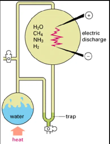
Simple organic molecules (that is, molecules containing carbon) are likely to have been
produced under such conditions. The best evidence for this comes from laboratory experiments. If mixtures of gases such as CO2, CH4, NH3, and H2 are heated with water and energized by electrical discharge or by ultraviolet radiation, they react to form small organic molecules - usually a rather small selection, each made in large amounts (Figure 1-1). Among these products are compounds, such as hydrogen cyanide (HCN) and formaldehyde (HCHO), that readily undergo further reactions in aqueous solution (Figure 1-2). Most important,
representatives of most of the major classes of small organic molecules found in cells are generated, including amino acids, sugars, and the purines and pyrimidines required to make nucleotides.
Although such experiments cannot reproduce the early conditions on the earth exactly, they make it plain that the formation of organic molecules is surprisingly easy. And the developing earth had immense advantages over any human experimenter; it was very large and could produce a wide spectrum of conditions. But above all, it had much more time - tens to hundreds of millions of years. In such circumstances it seems very likely that, at some time and place, many of the simple organic molecules found in present-day cells accumulated in high
concentrations.
Co m p le x Ch e m ic a l S y s t e m s Ca n D e v e lo p in a n En v iro n m e n t
Th a t I s Fa r fro m Ch e m ic a l Eq u ilib riu m
One amino acid can join with another by forming a peptide bond, and two nucleotides can join together by a phosphodiester bond. The repetition of these reactions leads to linear polymers known as polypeptides and polynucleotides, respectively. In present-day living cells, large polypeptides - known as proteins - and polynucleotides - in the form of both ribonucleic acids (RNA) and deoxyribonucleic acids (DNA)are commonly viewed as the most important
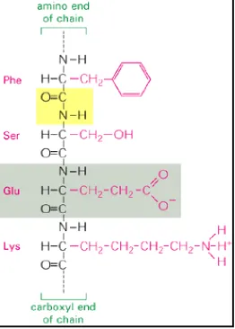
constituents. A restricted set of 20 amino acids constitute the universal building blocks of the proteins, while RNA and DNA molecules are constructed from just four types of nucleotides each. Although it is uncertain why these particular sets of monomers were selected for
biosynthesis in preference to others that are chemically similar, we shall see that the chemical properties of the corresponding polymers suit them especially well for their specific roles in the cell.
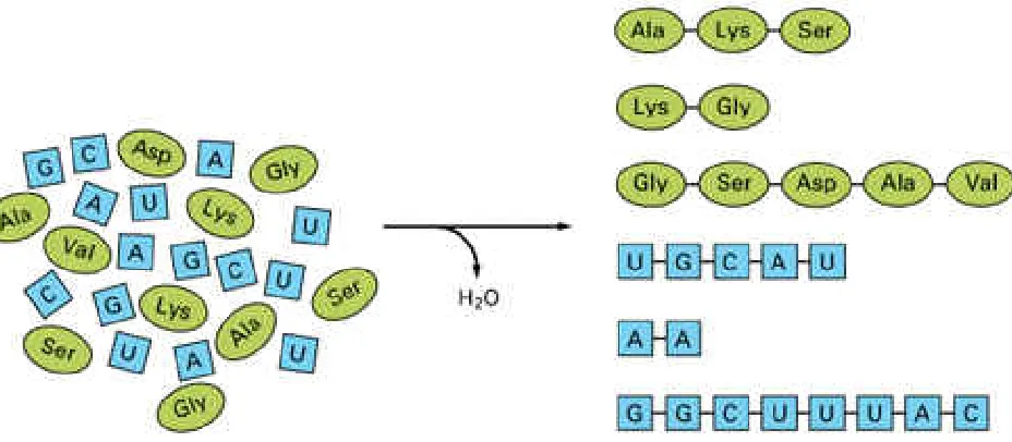
The earliest polymers may have formed in any of several ways - for example, by the heating of dry organic compounds or by the catalytic activity of high concentrations of inorganic
polyphosphates or other crude mineral catalysts. Under laboratory conditions the products of similar reactions are polymers of variable length and random sequence in which the particular amino acid or nucleotide added at any point depends mainly on chance (Figure 1-3). Once a polymer has formed, however, it can itself influence subsequent chemical reactions by acting as a catalyst.
The origin of life requires that in an assortment of such molecules there must have been some possessing, if only to a small extent, a crucial property: the ability to catalyze reactions that lead, directly or indirectly, to production of more molecules of the catalyst itself. Production of catalysts with this special self-promoting property would be favored, and the molecules most efficient in aiding their own production would divert raw materials from the production of other substances. In this way one can envisage the gradual development of an increasingly complex chemical system of organic monomers and polymers that function together to generate more molecules of the same types, fueled by a supply of simple raw materials in the environment. Such an
autocatalytic system would have some of the properties we think of as characteristic of living matter: it would comprise a far from random selection of interacting molecules; it would tend to reproduce itself; it would compete with other systems dependent on the same feedstocks; and if deprived of its feedstocks or maintained at a wrong temperature that upsets the balance of reaction rates, it would decay toward chemical equilibrium and "die."
But what molecules could have had such autocatalytic properties? In present-day living cells the most versatile catalysts are polypeptides, composed of many different amino acids with
chemically diverse side chains and, consequently, able to adopt diverse three-dimensional forms that bristle with reactive sites. But although polypeptides are versatile as catalysts, there is no known way in which one such molecule can reproduce itself by directly specifying the formation of another of precisely the same sequence.
P o ly n u c le o t id e s Are Ca p a b le o f D ire c t in g Th e ir Ow n
S y n t h e s is
3sequence. This capacity depends on complementary pairing of nucleotide subunits, which
enables one polynucleotide to act as a template for the formation of another. In the simplest case a polymer composed of one nucleotide (for example, polycytidylic acid, or poly C) can line up the subunits required to make another polynucleotide (in this example, polyguanylic acid, or poly G) along its surface, thereby promoting their polymerization into poly G (Figure 1-4). Because C subunits preferentially bind G subunits, and vice versa, the poly-G molecule in turn can promote synthesis of more poly C.
Consider now a polynucleotide with a more complex sequence of subunits - specifically, a
molecule of RNA strung together from four types of nucleotides, containing the bases uracil (U), adenine (A), cytosine (C), and guanine (G), arranged in some particular sequence. Because of complementary pairing between the bases A and U and between the bases G and C, this molecule, when added to a mixture of activated nucleotides under suitable conditions, will line them up for polymerization in a sequence complementary to its own. The resulting new RNA molecule will be rather like a mold of the original, with each A in the original corresponding to a U in the copy and so on. The sequence of nucleotides in the original RNA strand contains
information that is, in essence, preserved in the newly formed complementary strands: a second round of copying, with the complementary strand as a template, restores the original sequence (Figure 1-5).
Such complementary templating mechanisms are elegantly simple, and they lie at the heart of information transfer processes in biological systems. Genetic information contained in every cell is encoded in the sequences of nucleotides in its polynucleotide molecules, and this information is passed on (inherited) from generation to generation by means of complementary base-pairing interactions.
Templating mechanisms, however, require additional catalysts to promote polymerization; without these the process is slow and inefficient and other, competing reactions prevent the formation of accurate replicas. Today, the catalytic functions that polymerize nucleotides are provided by highly specialized catalytic proteinsthat is, by enzymes. In the "prebiotic soup"
primitive polypeptides might perhaps have provided some catalytic help. But molecules with the appropriate catalytic specificity would have remained rare unless the RNA itself were able
somehow to reciprocate and favor their production. We shall come back to the reciprocal
relationship between RNA synthesis and protein synthesis, which is crucially important in all living cells. But let us first consider what could be done with RNA itself, for RNA molecules can have a variety of catalytic properties, besides serving as templates for their own replication. In particular, an RNA molecule with an appropriate nucleotide sequence can act as catalyst for the accurate replication of another RNA molecule - the template - whose sequence can be arbitrary. The special versatility of RNA molecules is thought to have enabled them to play a central role in the origin of life.
S e lf- re p lic a t in g Mo le c u le s Un d e rg o N a t u ra l S e le c t io n
3 , 4polynucleotide can pair with free complementary nucleotides in their environment to form a new polymer, so they can pair with complementary nucleotide residues within the polymer itself. A sequence GGGG in one part of a polynucleotide chain can form a relatively strong association with a CCCC sequence in another region of the same molecule. Such associations produce complex three-dimensional patterns of folding, and the molecule as a whole takes on a specific shape that depends entirely on the sequence of its nucleotides (Figure 1-6).
The three-dimensional folded structure of a polynucleotide affects its stability, its actions on other molecules, and its ability to replicate, so that not all polynucleotide shapes will be equally
successful in a replicating mixture. Moreover, errors inevitably occur in any copying process, and imperfect copies of the originals will be propagated. With repeated replication, therefore, new variant sequences of nucleotides will be continually generated. Thus, in laboratory studies,
replicating systems of RNA molecules have been shown to undergo a form of natural selection in which different favorable sequences eventually predominate, depending on the exact conditions. Most important, RNA molecules can be selected for the ability to bind almost any other molecule specifically. This too has been shown, in experiments in vitro that begin with a preparation of
short RNA molecules with random nucleotide sequences manufactured artificially. These are passed down a column packed with beads to which some chosen substance is bonded. RNA molecules that fail to bind to the chosen substance are washed through the column and
discarded; those few that bind are retained and used as templates to direct production of multiple copies of their own sequences. This new RNA preparation, enriched in sequences that bind the chosen substance, is then used as the starting material for a repetition of the procedure. After several such cycles of selection and reproduction, the RNA is found to consist of multiple copies of a relatively small number of sequences, each of which binds the test substance quite
specifically.
An RNA molecule therefore has two special characteristics: it carries information encoded in its nucleotide sequence that it can pass on by the process of replication, and it has a specific folded structure that enables it to interact selectively with other molecules and determines how it will respond to the ambient conditions. These two features - one informational, the other functional - are the two properties essential for evolution. The nucleotide sequence of an RNA molecule is analogous to the genotype - the hereditary information - of an organism. The folded
three-dimensional structure is analogous to the phenotype - the expression of the genetic information on which natural selection operates.
S p e c ia liz e d RN A Mo le c u le s Ca n Ca t a ly z e B io c h e m ic a l
Re a c t io n s
5ribozyme), causing particular chemical groups on one or more of its nucleotides to become
highly reactive.
Certain catalytic activities would have had a cardinal importance in the primordial soup. Consider in particular an RNA molecule that helps to catalyze the process of templated polymerization, taking any given RNA molecule as template. (This ribozyme activity has been directly
demonstrated in vitro, albeit in a rudimentary form.) Such a molecule, by acting on copies of
itself, can replicate with heightened speed and efficiency (Figure 1-7A). At the same time, it can promote the replication of any other type of RNA molecules in its neighborhood (Figure 1-7B). Some of these may have catalytic actions that help or hinder the survival or replication of RNA in other ways. If beneficial effects are reciprocated, the different types of RNA molecules,
specialized for different activities, may evolve into a cooperative system that replicates with unusually great efficiency.
I n fo rm a t io n Flo w s fro m P o ly n u c le o t id e s t o P o ly p e p t id e s
6There are strong suggestions, therefore, that between 3.5 and 4 billion years ago, somewhere on earth, self-replicating systems of RNA molecules, mixed with other organic molecules including simple polypeptides, began the process of evolution. Systems with different sets of polymers competed for the available precursor materials to construct copies of themselves, just as organisms now compete; success depended on the accuracy and the speed with which the copies were made and on the stability of those copies.
However, as we emphasized earlier, while the structure of polynucleotides is well suited for information storage and replication, their catalytic abilities are limited by comparison with those of polypeptides, and efficient replication of polynucleotides in modern cells is absolutely
dependent on proteins. At the origin of life any polynucleotide that helped guide the synthesis of a useful polypeptide in its environment would have had a great advantage in the evolutionary struggle for survival.
But how could the information encoded in a polynucleotide specify the sequence of a polymer of a different type? Clearly, the polynucleotides must act as catalysts to join selected amino acids together. In present-day organisms a collaborative system of RNA molecules plays a central part in directing the synthesis of polypeptides - that is, protein synthesis - but the process is aided by other proteins synthesized previously. The biochemical machinery for protein synthesis is
remarkably elaborate. One RNA molecule carries the genetic information for a particular polypeptide in the form of a code, while other RNA molecules act as adaptors, each binding a specific amino acid. These two types of RNA molecules form complementary base pairs with one another to enable sequences of nucleotides in the coding RNA molecule to direct the
incorporation of specific amino acids held on the adaptor RNAs into a growing polypeptide chain. Precursors to these two types of RNA molecules presumably directed the first protein synthesis without the aid of proteins (Figure 1-7C).
Today, these events in the assembly of new proteins take place on the surface of ribosomes -
these particles plays a central catalytic role in the process of protein synthesis and forms more than 60% of the ribosome's mass. At least in evolutionary terms, it appears to be the
fundamental component of the ribosome.
It seems likely, then, that RNA guided the primordial synthesis of proteins, perhaps in a clumsy and primitive fashion. In this way RNA was able to create tools - in the form of proteins - for more efficient biosynthesis, and some of these could have been put to use in the replication of RNA and in the process of tool production itself.
The synthesis of specific proteins under the guidance of RNA required the evolution of a code by which the polynucleotide sequence specifies the amino acid sequence that makes up the
protein. This code - the genetic code - is spelled out in a "dictionary" of three-letter words:
different triplets of nucleotides encode specific amino acids. The code seems to have been selected arbitrarily (subject to some constraints, perhaps); yet it is virtually the same in all living organisms. This strongly suggests that all present-day cells have descended from a single line of primitive cells that evolved the mechanism of protein synthesis.
Me m b ra n e s D e fin e d t h e Firs t Ce ll
7One of the crucial events leading to the formation of the first cell must have been the
development of an outer membrane. For example, the proteins synthesized under the control of a certain species of RNA would not facilitate reproduction of that species of RNA unless they remained in the neighborhood of the RNA; moreover, as long as these proteins were free to diffuse among the population of replicating RNA molecules, they could benefit equally any competing species of RNA that might be present. If a variant RNA arose that made a superior type of enzyme, the new enzyme could not contribute selectively to the survival of the variant
RNA in its competition with its fellows. Selection of RNA molecules according to the quality of the proteins they generated could not occur efficiently until some form of compartment evolved to contain the proteins made by an RNA molecule and thereby make them available only to the RNA that had generated them (Figure 1-8).
The need for containment is easily fulfilled by another class of molecules that has the simple physicochemical property of being amphipathic, that is, consisting of one part that is hydrophobic
(water insoluble) and another part that is hydrophilic (water soluble). When such molecules are placed in water, they aggregate, arranging their hydrophobic portions as much in contact with one another as possible and their hydrophilic portions in contact with the water. Amphipathic molecules of appropriate shape spontaneously aggregate to form bilayers, creating small closed
vesicles whose aqueous contents are isolated from the external medium (Figure 1-9). The phenomenon can be demonstrated in a test tube by simply mixing phospholipids and water together: under appropriate conditions, small vesicles will form. All present-day cells are
phospholipid molecules from the prebiotic soup, enclosing a self-replicating mixture of RNA and other molecules. It is not clear at what point in the evolution of biological catalysts and protein synthesis this first occurred. In any case, once RNA molecules were sealed within a closed membrane, they could begin to evolve in earnest as carriers of genetic instructions: they could be selected not merely on the basis of their own structure, but also according to their effect on the other molecules in the same compartment. The nucleotide sequences of the RNA molecules could now be expressed in the character of a unitary living cell.
All P re s e n t - D a y Ce lls Us e D N A a s Th e ir He re d it a ry
Ma t e ria l
3 , 6 , 8The picture we have presented is, of course, speculative: there are no fossil records that trace the origins of the first cell. Nevertheless, there is persuasive evidence from present-day
organisms and from experiments that the broad features of this evolutionary story are correct. The prebiotic synthesis of small molecules, the self-replication of catalytic RNA molecules, the translation of RNA sequences into amino acid sequences, and the assembly of lipid molecules to form membrane-bounded compartments - all presumably occurred to generate primitive cells 3.5 to 4 billion years ago.
It is useful to compare these early cells with the simplest and smallest present-day cells, the mycoplasmas. Mycoplasmas are small bacteria of a degenerate type that normally lead a parasitic existence in close association with animal or plant cells (Figure 1-10). Some have a diameter of about 0.3 mm and contain only enough nucleic acid to direct the synthesis of about 400 different proteins. Some of these proteins are enzymes, some are structural; some lie in the cell's interior, others are embedded in its membrane. Together they synthesize essential small molecules that are not available in the environment, redistribute the energy needed to drive biosynthetic reactions, and maintain appropriate conditions inside the cell.
The first cells on the earth were presumably less sophisticated than a mycoplasma and less efficient in reproducing themselves. There was, however, a more fundamental difference between these primitive cells and a mycoplasma, or indeed any other present-day cell: the hereditary information in all cells alive today is stored in DNA rather than in the RNA that is thought to have stored the hereditary information during the earliest stages of evolution. Both types of polynucleotides are found in present-day cells, but they function in a collaborative
manner, each having evolved to perform specialized tasks. Small chemical differences fit the two kinds of molecules for distinct functions. DNA acts as the permanent repository of genetic
information, and, unlike RNA, it is found in cells principally in a double-stranded form, composed of a pair of complementary polynucleotide molecules. This double-stranded structure makes DNA in cells more robust and stable than RNA; it also makes DNA relatively easy to replicate (as will be explained in Chapter 3) and permits a repair mechanism to operate that uses the intact strand as a template for the correction or repair of the associated damaged strand. DNA guides the synthesis of specific RNA molecules, again by the principle of complementary base-pairing, though now this pairing is between slightly different types of nucleotides. The resulting single-stranded RNA molecules then perform two primeval functions: they direct protein synthesis both as coding RNA molecules (messenger RNAs) and as RNA catalysts (ribosomal and other
The suggestion, in short, is that RNA preceded DNA in evolution, having both genetic and
catalytic properties; eventually, DNA took over the primary genetic function and proteins became the major catalysts, while RNA remained primarily as the intermediary connecting the two
(Figure 1-11). With the advent of DNA cells were enabled to become more complex, for they could then carry and transmit an amount of genetic information greater than that which could be stably maintained in RNA molecules.
S u m m a ry
Living cells probably arose on earth about 3.5 billion years ago by spontaneous reactions between molecules in an environment that was far from chemical equilibrium. From our knowledge of present-day organisms and the molecules they contain, it seems likely that the development of the directly autocatalytic mechanisms fundamental to living systems began with the evolution of families of RNA molecules that could catalyze their own replication. With time, one of these families of cooperating RNA catalysts developed the ability to direct synthesis of polypeptides. Finally, as the accumulation of additional protein catalysts allowed more efficient and complex cells to evolve, the DNA double helix replaced RNA as a more stable molecule for storing the increased amounts of genetic information required by such cells.
Figure 1-2. A few of the compounds that might form in the experiment described in Figure 1-1.
Figure 1-4. Polynucleotides as templates. Preferential binding occurs between pairs of
nucleotides (G with C and U with A) by relatively weak chemical bonds (above). This pairing
enables one polynucleotide to act as a template for the synthesis of another (left).
Figure 1-5. Replication of a polynucleotide sequence (here an RNA molecule). In step 1 the original RNA molecule acts as a template to form an RNA molecule of complementary sequence. In step 2 this complementary RNA molecule itself acts as a template, forming RNA molecules of the original sequence. Since each templating molecule can produce many copies of the
Figure 1-6. Conformation of an RNA molecule. Nucleotide pairing between different regions of the same polynucleotide (RNA) chain causes the molecule to adopt a distinctive shape.
Figure 1-8. Evolutionary significance of cell-like compartments. In a mixed population of self-replicating RNA molecules capable of influencing protein synthesis (as illustrated in Figure 1-7), any improved form of RNA that is able to promote formation of a more useful protein must share this protein with its neighboring competitors. However, if the RNA is enclosed within a
compartment, such as a lipid membrane, then any protein the RNA causes to be made is
retained for its own use; the RNA can therefore be selected on the basis of its guiding production of a better protein.
Figure 1-9. Formation of membranes by phospholipids. Because these molecules have
Figure 1-10. Spiroplasma citrii, a mycoplasma that grows in plant cells. (Courtesy of Jeremy
Burgess.)
Figure 1-11. Suggested stages of evolution from simple self-replicating systems of RNA molecules to present-day cells. Today, DNA is the repository of genetic information and RNA acts largely as a go-between to direct protein synthesis.
References
Bendall, D.S., ed. Evolution from Molecules to Men. Cambridge, UK: Cambridge University Press, 1983.
Evolution of Catalytic Function. Cold Spring Harbor Symp. Quant. Biol.52,: 1987..
Curtis, H.; Barnes, N.S. Biology, 5th ed. New York: Worth, 1989.
Darnell, J.E.; Lodish, H.F.; Baltimore, D. Molecular Cell Biology, 2nd ed., Chapter 26. New York: Scientific American Books, 1990.
Darwin, C. On the Origin of Species. London: Murray, 1859. Reprinted, New York: Penguin, 1984.
Dawkins, R. The Blind Watchmaker. New York: Viking Penguin, 1988.
Margulis, L.M.; Schwartz, K.V. Five Kingdoms: An Illustrated Guide to the Phyla of Life on Earth, 2nd ed. New York: W.H. Freeman, 1988.
Watson, J.D.; Hopkins, N.H.; Roberts, J.W.; Steitz, J.A.; Weiner, A.M. Molecular Biology of the Gene, 4th ed., Chapter 28. Menlo Park, CA: Benjamin-Cummings, 1987.
Cit e d
1. Lazcano, A.; Fox, G.E.; Oró, J.F. Life before DNA: the origin and evolution of early archaean cells. In The Evolution of Metabolic Function (R.P. Mortlock, ed.), pp. 237-295. Boca Raton, FL: CRC Press, 1992.
Schopf, J.W.; Hayes, J.M.; Walter, M.R. Evolution of earth's earliest ecosystems: recent progress and unsolved problems. In Earth's Earliest Biosphere: Its Origin and Evolution (J.W. Schopf, ed.), pp. 361-384. Princeton, NJ: Princeton University Press, 1983.
2. Miller, S.L. Which organic compounds could have occurred on the prebiotic earth? Cold
Spring Harbor Symp. Quant. Biol. 52: 17-27. 1987. (PubMed)
3. Orgel, L.E. Molecular replication. Nature 358: 203-209. 1992. (PubMed)
4. Ellington, A.D.; Szostak, J.W. In vitro selection of RNA molecules that bind specific ligands.
Nature 346: 818-822. 1990. (PubMed)
Joyce, G.F. Directed molecular evolution. Sci. Am. 267(6): 90-97. 1992. (PubMed)
sequences. Science261: 1411-1418. 1993. (PubMed)
Cech, T.R. RNA as an enzyme. Sci. Am. 255(5): 64-75. 1986. (PubMed)
6. Alberts, B.M. The function of the hereditary materials: biological catalyses reflect the cell's evolutionary history. Am. Zool. 26: 781-796. 1986.
Maizels, N.; Weiner, A.M. Peptide-specific ribosomes, genomic tags, and the origin of the genetic code. Cold Spring Harbor Symp. Quant. Biol. 52: 743-749. 1987. (PubMed)
7. Cavalier-Smith, T. The origin of cells: a symbiosis between genes, catalysts, and membranes.
Cold Spring Harbor Symp. Quant. Biol. 52: 805-824. 1987. (PubMed)
8. Muto, A.; Andachi, Y.; Yuzawa, H.; Yamao, F.; Osawa, S. The organization and evolution of transfer RNA genes in Mycoplasma capricolum. Nucl. Acid Res.18: 5037-5043. 1990.
9. Sogin, M.L. Early evolution and the origin of eukaryotes. Curr. Opin. Genet. Devel. 1: 457-463.
1991.
Vidal, G. The oldest eukaryotic cells. Sci. Am. 250(2): 48-57. 1984. (PubMed)
10. Woese, C.R. Bacterial evolution. Microbiol. Rev. 51: 221-271. 1987. (PubMed)
Zillig, W. Comparative biochemistry of Archaea and Bacteria. Curr. Opin. Genet. Devel. 1:
544-551. 1991.
11. Clarke, P.H. Enzymes in bacterial populations. In Biochemical Evolution (H. Gutfreund, ed.), pp. 116-149. Cambridge, UK: Cambridge University Press, 1981.
De Duve, C. Blueprint for a Cell: The Nature and Origin of Life. Burlington, NC: Neil Patterson Publishers, 1991.
12. Li, W.-H.; Graur, D. Fundamentals of Molecular Evolution. Sunderland, MA: Sinauer Associates, 1991.
Sidow, A.; Bowman, B.H. Molecular phylogeny. Curr. Opin. Genet. Devel. 1: 451-456. 1991.
13. Dickerson, R.E. Cytochrome c and the evolution of energy metabolism. Sci. Am. 242(3):
136-153. 1980.
14. Cavalier-Smith, T. The origin of eukaryote and archaebacterial cells. Ann. N.Y. Acad. Sci.
503: 17-54. 1987. (PubMed)
14. Gray, M.W. The evolutionary origins of organelles. Trends Genet.5: 294-299. 1989.
(PubMed)
Margulis, L. Symbiosis in Cell Evolution. New York: W.H. Freeman, 1981.
15. Gray, M.W. Origin and evolution of mitochondrial DNA. Annu. Rev. Cell Biol.5: 25-50. 1989.
Sogin, M.L.; Gunderson, J.H.; Elwood, H.J.; Alonso, R.A.; Peattie, D.A. Phylogenetic meaning of the kingdom concept: an unusual ribosomal RNA from Giardia lamblia. Science 243: 75-77.
1989. (PubMed)
Vossbrinck, C.R.; Maddox, J.V.; Friedman, S.; Debrunner-Vossbrinck, B.A.; Woese, C.R. Ribosomal RNA sequence suggests microsporidia are extremely ancient eukaryotes. Nature
326: 411-414. 1987. (PubMed)
16. Bryant, D.A. Puzzles of chloroplast ancestry. Curr. Biol.2: 240-242. 1992.
17. Sleigh, M.A. Protozoa and Other Protists. London: Edward Arnold, 1989.
18. Buchsbaum, R. Animals Without Backbones, 3rd ed. Chicago: University of Chicago Press, 1987.
Field, K.G. Molecular phylogeny of the animal kingdom. Science239: 748-753. 1988. (PubMed)
Knoll, A.H. The end of the proterozoic eon. Sci. Am. 265(4): 64-73. 1991. (PubMed)
Levinton, J.S. The big bang of animal evolution. Sci. Am.267(5): 84-91. 1992. (PubMed)
Shapiro, J.A. Bacteria as multicellular organisms. Sci. Am. 258(6): 82-89. 1988.
Valentine, J.W. The evolution of multicellular plants and animals. Sci. Am.239(3): 140-158.
1978. (PubMed)
19. Bode, P.M.; Bode, H.R. Patterning in Hydra. In Pattern Formation (G.M. Malacinski, S.V. Bryant, eds.), pp. 213-244. New York: Macmillan, 1984.
20. Raff, R.A.; Kaufman, T.C. Embryos, Genes, and Evolution. New York: Macmillan, 1983.
Slack, J.M.W.; Holland, P.W.H.; Graham, C.F. The zootype and the phylotypic stage. Nature
361: 490-492. 1993. (PubMed)
21. Chothia, C. One thousand families for the molecular biologist. Nature 357: 543-544. 1992.
(PubMed)
Miklos, G.L.; Campbell, H.D. The evolution of protein domains and the organizational complexities of metazoans. Curr. Opin. Genet. Devel. 2: 902-906. 1992.
Key terms
•
Amino Acids•
Catalysis•
Purines•
Pyrimidines•
Radiation Effects•
Support, U.S. Gov't, Non-P.H.S.•
Thermodynamics•
Base Sequence•
Biogenesis*•
Biopolymers*•
DNA/genetics*•
DNA Replication*•
DNA-Directed DNA Polymerase/metabolism•
DNA-Directed RNA Polymerase/metabolism•
Models, Genetic*•
Models, Molecular•
Molecular Conformation•
Molecular Sequence Data•
Nucleic Acid Conformation•
Oligonucleotides/chemistry*•
Support, U.S. Gov't, Non-P.H.S.•
Base Sequence•
Binding Sites•
Chromatography, Affinity•
Cloning, Molecular•
DNA/chemical synthesis•
Dyes/metabolism*•
Ligands•
Molecular Sequence Data•
Molecular Structure•
Nucleic Acid Conformation•
Polymerase Chain Reaction•
RNA/metabolism•
RNA/isolation & purification*•
RNA/biosynthesis•
Support, Non-U.S. Gov't•
Templates•
Transcription, Genetic•
Triazines/metabolism•
DNA/metabolism•
DNA/chemistry*•
Mutagenesis•
Polymerase Chain Reaction•
RNA/metabolism•
RNA/chemistry*•
Base Sequence•
Binding Sites•
Chromatography, Affinity•
Cloning, Molecular•
DNA/chemical synthesis•
Dyes/metabolism*•
Ligands•
Molecular Sequence Data•
Molecular Structure•
Nucleic Acid Conformation•
Polymerase Chain Reaction•
RNA/metabolism•
RNA/isolation & purification*•
RNA/biosynthesis•
Support, Non-U.S. Gov't•
Templates•
Triazines/metabolism•
Catalysis•
Enzymes/metabolism*•
Introns•
RNA/metabolism*•
RNA Splicing•
RNA, Messenger/metabolism•
RNA, Ribosomal/metabolism•
Tetrahymena/metabolism•
Animal•
Bacterial Physiology•
Catalysis•
Cell Physiology*•
Chromosomes/physiology•
DNA Replication•
Eukaryotic Cells/physiology•
Evolution*•
Genes*•
Membranes/physiology*•
Models, Biological•
Viruses/genetics•
Cells*•
Eukaryotic Cells*•
Evolution•
Fossils•
Microscopy, Electron, Scanning•
Bacteria*/genetics•
Bacteria*/classification•
Base Sequence•
Evolution*•
Phylogeny•
RNA, Bacterial•
RNA, Ribosomal•
Support, U.S. Gov't, Non-P.H.S.•
Chloroplasts•
DNA, Mitochondrial•
Evolution*•
Mitochondria•
Organelles*•
RNA, Ribosomal/analysis•
Support, Non-U.S. Gov't•
Animal•
Base Sequence•
Databases, Factual•
Evolution•
Human•
Proteins/genetics*•
Proteins/classification*•
Sequence Homology, Nucleic Acid•
Substances:
•
Pyrimidines•
Purines•
Amino Acids•
DNA-Directed DNA Polymerase•
DNA-Directed RNA Polymerase•
DNA•
Oligonucleotides•
Biopolymers•
procion blue MX-R•
Cibacron Blue F 3GA•
Triazines•
Ligands•
Dyes•
RNA, Ribosomal•
RNA, Messenger•
Enzymes•
RNA, Bacterial•
DNA, Mitochondrial•
Proteins•
I: Introduction to the Cell 1. The Evolution of the Cell
Introduction
From Molecules to the First Cell From Procaryotes to Eucaryotes
From Single Cells to Multicellular Organisms References
Fro m P ro c a ry o t e s t o Eu c a ry o t e s
9
Introduction
Procaryotic Cells Are Structurally Simple but Biochemically Diverse
Metabolic Reactions Evolve
Evolutionary Relationships Can Be Deduced by Comparing DNA
Sequences
Cyanobacteria Can Fix CO2 and N2
Bacteria Can Carry Out the Aerobic Oxidation of Food Molecules
Eucaryotic Cells Contain Several Distinctive Organelles
Eucaryotic Cells Depend on Mitochondria for Their Oxidative Metabolism
Chloroplasts Are the Descendants of an Engulfed Procaryotic Cell
Eucaryotic Cells Contain a Rich Array of Internal Membranes
Protozoa Include the Most Complex Cells Known
In Eucaryotic Cells the Genetic Material Is Packaged in Complex Ways
Summary
I n t ro d u c t io n
It is thought that all organisms living now on earth derive from a single primordial cell born more than 3 billion years ago. This cell, out-reproducing its competitors, took the lead in the process of cell division and evolution that eventually covered the earth with green, changed the composition of its atmosphere, and made it the home of intelligent life. The family resemblances among all organisms seem too strong to be explained in any other way. One important landmark along this evolutionary road occurred about 1.5 billion years ago, when there was a transition from small cells with relatively simple internal structures - the so-called procaryotic cells, which include the various types of bacteria - to a flourishing of larger and radically more complex eucaryotic cells such as are found in higher animals and plants.
P ro c a ry o t ic Ce lls Are S t ru c t u ra lly S im p le b u t B io c h e m ic a lly
D iv e rs e
1 0Bacteria are the simplest organisms found in most natural environments. They are spherical or rod-shaped cells, commonly several micrometers in linear dimension (Figure 1-12). They often possess a tough protective coat, called a cell wall, beneath which a plasma membrane encloses a single cytoplasmic compartment containing DNA, RNA, proteins, and small molecules. In the electron microscope this cell interior appears as a matrix of varying texture without any obvious organized internal structure (see Figure 1-12B).
Bacteria are small and can replicate quickly, simply dividing in two by binary fission. When food is plentiful, "survival of the fittest" generally means survival of those that can divide the fastest. Under optimal conditions a single procaryotic cell can divide every 20 minutes and thereby give rise to 5 billion cells (approximately equal to the present human population on earth) in less than 11 hours. The ability to divide quickly enables populations of bacteria to adapt rapidly to changes in their environment. Under laboratory conditions, for example, a population of bacteria
maintained in a large vat will evolve within a few weeks by spontaneous mutation and natural selection to utilize new types of sugar molecules as carbon sources.
richness in their underlying biochemical composition. Two distantly related groups can be
recognized: the eubacteria, which are the commonly encountered forms that inhabit soil, water, and larger living organisms; and the archaebacteria, which are found in such incommodious environments as bogs, ocean depths, salt brines, and hot acid springs (Figure 1-13).
There are species of bacteria that can utilize virtually any type of organic molecule as food, including sugars, amino acids, fats, hydrocarbons, polypeptides, and polysaccharides. Some are even able to obtain their carbon atoms from CO2 and their nitrogen atoms from N2. Despite their relative simplicity, bacteria have existed for longer than any other organisms and still are the most abundant type of cell on earth.
Me t a b o lic Re a c t io n s Ev o lv e
1 0 , 1 1A bacterium growing in a salt solution containing a single type of carbon source, such as glucose, must carry out a large number of chemical reactions. Not only must it derive from the glucose the chemical energy needed for many vital processes, it must also use the carbon atoms of glucose to synthesize every type of organic molecule that the cell requires. These reactions are catalyzed by hundreds of enzymes working in reaction "chains" so that the product of one reaction is the
substrate for the next; such enzymatic chains, called metabolic pathways, will be discussed in the following chapter.
Originally, when life began on earth, there was probably little need for such elaborate metabolic reactions. Cells with relatively simple chemistry could survive and grow on the molecules in their surroundings. But as evolution proceeded, competition for these limited natural resources would have become more intense. Organisms that had developed enzymes to manufacture useful
organic molecules more efficiently and in new ways would have had a strong selective advantage. In this way the complement of enzymes possessed by cells is thought to have gradually
increased, generating the metabolic pathways of present organisms. Two plausible ways in which a metabolic pathway could arise in evolution are illustrated in Figure 1-14.
If metabolic pathways evolved by the sequential addition of new enzymatic reactions to existing ones, the most ancient reactions should, like the oldest rings in a tree trunk, be closest to the center of the "metabolic tree," where the most fundamental of the basic molecular building blocks are synthesized. This position in metabolism is firmly occupied by the chemical processes that involve sugar phosphates, among which the most central of all is probably the sequence of
reactions known as glycolysis, by which glucose can be degraded in the absence of oxygen (that is, anaerobically). The oldest metabolic pathways would have had to be anaerobic because there was no free oxygen in the atmosphere of the primitive earth. Glycolysis occurs in virtually every living cell and drives the formation of the compound adenosine triphosphate, or ATP, which is used by all cells as a versatile source of chemical energy. Certain thioester compounds play a fundamental role in the energy-transfer reactions of glycolysis and in a host of other basic
biochemical processes in which two organic molecules (a thiol and a carboxylic acid) are joined by a high-energy bond involving sulfur (Figure 1-15). It has been argued that this simple but powerful chemical device is a relic of prebiotic processes, reflecting the reactions that occurred in the
Linked to the core reactions of glycolysis are hundreds of other chemical processes. Some of these are responsible for the synthesis of small molecules, many of which in turn are utilized in further reactions to make the large polymers specific to the organism. Other reactions are used to degrade complex molecules, taken in as food, into simpler chemical units. One of the most
striking features of these metabolic reactions is that they take place similarly in all kinds of organisms, suggesting an extremely ancient origin.
Ev o lu t io n a ry Re la t io n s h ip s Ca n B e D e d u c e d b y Co m p a rin g
D N A S e q u e n c e s
1 2The enzymes that catalyze the fundamental metabolic reactions, while continuing to serve the same essential functions, have undergone progressive modifications as organisms have evolved into divergent forms. For this reason the amino acid sequence of the same type of enzyme in different living species provides a valuable indication of the evolutionary relationship between these species. The evidence obtained closely parallels that from other sources, such as the fossil record. An even richer source of information is locked in the living cell in the sequences of
nucleotides in DNA, and modern methods of analysis allow these DNA sequences to be
determined in large numbers and compared between species. Comparisons of highly conserved sequences, which have a central function and therefore change only slowly during evolution, can reveal relationships between organisms that diverged long ago (Figure 1-16), while very rapidly evolving sequences can be used to determine how more closely related species evolved. It is expected that continued application of these methods will enable the course of evolution to be followed with unprecedented accuracy.
Cy a n o b a c t e ria Ca n Fix CO2 a n d N 2
1 3As competition for the raw materials for organic syntheses intensified, a strong selective
advantage would have been gained by any organisms able to utilize carbon and nitrogen atoms (in the form of CO2 and N2) directly from the atmosphere. But while they are abundantly available, CO2 and N2 are also very stable. It therefore requires a large amount of energy as well as a
number of complicated chemical reactions to convert them to a usable form - that is, into organic molecules such as simple sugars.
In the case of CO2 the major mechanism that evolved to achieve this transformation was
photosynthesis, in which radiant energy captured from the sun drives the conversion of CO2 into organic compounds. The interaction of sunlight with a pigment molecule, chlorophyll, excites an electron to a more highly energized state. As the electron drops back to a lower energy level, the energy it gives up drives chemical reactions that are facilitated and directed by protein molecules.
donor by chlorophyll in a reaction that is driven by sunlight. The strong electron donor is then used to reduce CO2 or N2. Comparison of the mechanisms of photosynthesis in various present-day bacteria suggests that one of the first sources of electrons was H2S, from which the primary waste product would have been elemental sulfur. Later the more difficult but ultimately more rewarding process of obtaining electrons from H2O was accomplished, and O2 was released in large
amounts as a waste product.
Cyanobacteria (also known as blue-green algae) are today a major route by which both carbon and nitrogen are converted into organic molecules and thus enter the biosphere. They include the most self-sufficient organisms that now exist. Able to "fix" both CO2 and N2 into organic
molecules, they are, to a first approximation, able to live on water, air, and sunlight alone; the mechanisms by which they do this have probably remained essentially constant for several billion years. Together with other bacteria that have some of these capabilities, they created the
conditions in which more complex types of organisms could evolve: once one set of organisms had succeeded in synthesizing the whole gamut of organic cell components from inorganic raw materials, other organisms could subsist by feeding on the primary synthesizers and on their products.
B a c t e ria Ca n Ca rry Ou t t h e Ae ro b ic Ox id a t io n o f Fo o d
Mo le c u le s
1 3Many people today are justly concerned about the environmental consequences of human activities. But in the past other organisms have caused revolutionary changes in the earth's environment (although very much more slowly). Nowhere is this more apparent than in the composition of the earth's atmosphere, which through oxygen-releasing photosynthesis was transformed from a mixture containing practically no molecular oxygen to one in which oxygen constitutes 21% of the total (Figure 1-17).
Since oxygen is an extremely reactive chemical that can interact with most cytoplasmic
constituents, it must have been toxic to many early organisms, just as it is to many present-day anaerobic bacteria. However, this reactivity also provides a source of chemical energy, and, not surprisingly, this has been exploited by organisms during the course of evolution. By using
oxygen, organisms are able to oxidize more completely the molecules they ingest. For example, in the absence of oxygen glucose can be broken down only to lactic acid or ethanol, the end
products of anaerobic glycolysis. But in the presence of oxygen glucose can be completely degraded to CO2 and H2O. In this way much more energy can be derived from each gram of glucose. The energy released in respiration - the aerobic oxidation of food molecules - is used to drive the synthesis of ATP in much the same way that photosynthetic organisms produce ATP from the energy of sunlight. In both processes there is a series of electron-transfer reactions that generates an H+ gradient between the outside and inside of a membrane-bounded compartment;
the H+ gradient then serves to drive the synthesis of the ATP. Today, respiration is used by the
great majority of organisms, including most procaryotes.
organisms with which life had begun? In a world that was rich in oxygen, which they could not use, they were at a severe disadvantage. Some, no doubt, became extinct. Others either
developed a capacity for respiration or found niches in which oxygen was largely absent, where they could continue an anaerobic way of life. Others became predators or parasites on aerobic cells. And some, it seems, hit upon a strategy for survival more cunning and vastly richer in implications for the future: they are believed to have formed an intimate association with an aerobic type of cell, living with it in symbiosis. This is the most plausible explanation for the metabolic organization of present-day cells of the eucaryotic type (Panel 1-1, pp. 18-19) with which this book will be chiefly concerned.
Eucaryotic cells, by definition and in contrast to procaryotic cells, have a nucleus (caryon in
Greek), which contains most of the cell's DNA, enclosed by a double layer of membrane (Figure 1-18). The DNA is thereby kept in a compartment separate from the rest of the contents of the cell, the cytoplasm, where most of the cell's metabolic reactions occur. In the cytoplasm, moreover, many distinctive organelles can be recognized. Prominent among these are two types of small bodies, the chloroplasts and mitochondria (Figures 1-19 and 1-20). Each of these is enclosed in its own double layer of membrane, which is chemically different from the membranes surrounding the nucleus. Mitochondria are an almost universal feature of eucaryotic cells, whereas
chloroplasts are found only in those eucaryotic cells that are capable of photosynthesis - that is, in plants but not in animals or fungi. Both organelles almost certainly have a symbiotic origin.
Eu c a ry o t ic Ce lls D e p e n d o n Mit o c h o n d ria fo r Th e ir
Ox id a t iv e Me t a b o lis m
1 5Mitochondria show many similarities to free-living procaryotic organisms: for example, they often resemble bacteria in size and shape, they contain DNA, they make protein, and they reproduce by dividing in two. By breaking up eucaryotic cells and separating their component parts, it is
possible to show that mitochondria are responsible for respiration and that this process occurs nowhere else in the eucaryotic cell. Without mitochondria the cells of animals and fungi would be anaerobic organisms, depending on the relatively inefficient and antique process of glycolysis for their energy. Many present-day bacteria respire like mitochondria, and it seems probable that eucaryotic cells are descendants of primitive anaerobic organisms that survived, in a world that had become rich in oxygen, by engulfing aerobic bacteria - keeping them in symbiosis for the sake of their capacity to consume atmospheric oxygen and produce energy. Certain present-day
microorganisms offer strong evidence of the feasibility of such an evolutionary sequence. There are several hundred species of single-celled eucaryotes that resemble the hypothetical ancestral eucaryote in that they live in oxygen-poor conditions (in the guts of animals, for example) and lack mitochondria altogether. Comparative nucleotide sequence analyses have revealed that at least two groups of these organisms, the diplomonads and the microsporidia, diverged very early from the line leading to other eucaryotic cells (Figure 1-21). There is another eucaryote, the amoeba Pelomyxa palustris, that, while lacking mitochondria, nevertheless carries out oxidative
metabolism by harboring aerobic bacteria in its cytoplasm in a permanent symbiotic relationship. Diplomonads and microsporidia, on the one hand, and Pelomyxa, on the other, therefore
resemble two proposed stages in the evolution of eucaryotes such as ourselves.
separation of functions left the eucaryotic plasma membrane free to evolve important new
features. In particular, because eucaryotic cells need not maintain a large H+ gradient across their
plasma membrane, as required for ATP production in procaryotes, it became possible to use controlled changes in the ion permeability of the plasma membrane for cell-signaling purposes. Thus, a variety of ion channels appeared in the eucaryotic plasma membrane. Today, these
channels mediate the elaborate electrical signaling processes in higher organisms - notably in the nervous system -and they control much of the behavior of single-celled free-living eucaryotes such as protozoa (see below).
Ch lo ro p la s t s Are t h e D e s c e n d a n t s o f a n En g u lfe d
P ro c a ry o t ic Ce ll
1 6Chloroplasts carry out photosynthesis in much the same way as procaryotic cyanobacteria, absorbing sunlight in the chlorophyll attached to their membranes. Some bear a close structural resemblance to the cyanobacteria, being similar in size and in the way that their
chlorophyll-bearing membranes are stacked in layers (see Figure 1-20). Moreover, chloroplasts reproduce by dividing, and they contain DNA that is nearly indistinguishable in nucleotide sequence from
portions of a bacterial chromosome. All this strongly suggests that chloroplasts share a common ancestry with cyanobacteria and evolved from procaryotes that made their home inside eucaryotic cells. These procaryotes performed photosynthesis for their hosts, who sheltered and nourished them. Symbiosis of photosynthetic cells with other cell types is, in fact, a common phenomenon, and some present-day eucaryotic cells contain authentic cyanobacteria (Figure 1-22).
Figure 1-23 shows the evolutionary origins of the eucaryotes according to the symbiotic theory. It must be stressed, however, that mitochondria and chloroplasts show important differences from, as well as similarities to, present-day aerobic bacteria and cyanobacteria. Their quantity of DNA is very small, for example, and most of the molecules from which they are constructed are
synthesized elsewhere in the eucaryotic cell and imported into the organelle. Although there is good evidence that they originated as symbiotic bacteria, they have undergone large evolutionary changes and have become greatly dependent on - and subject to control by - their host cells.
The major existing eucaryotes have in common both mitochondria and a whole constellation of other features that distinguish them from procaryotes (Table 1-1). These function together to give eucaryotic cells a wealth of different capabilities, and it is debatable which of them evolved first. But the acquisition of mitochondria by an anaerobic eucaryotic cell must have been a crucial step in the success of the eucaryotes, providing them with the means to tap an abundant source of energy to drive all their complex activities.
Eu c a ry o t ic Ce lls Co n t a in a Ric h Arra y o f I n t e rn a l
Me m b ra n e s
Eucaryotic cells are usually much larger in volume than procaryotic cells, commonly by a factor of 1000 or more, and they carry a proportionately larger quantity of most cellular materials; for
increase in cell volume requires an increase in cell surface. But it is a fact of geometry that simply scaling up a structure increases the volume as the cube of the linear dimension while the surface area increases only as the square. Therefore, if the large eucaryotic cell is to keep as high a ratio of surface to volume as the procaryotic cell, it must supplement its surface area by means of convolutions, infoldings, and other elaborations of its membrane.
This probably explains in part the complex profusion of internal membranes that is a basic feature of all eucaryotic cells. Membranes surround the nucleus, the mitochondria, and (in plant cells) the chloroplasts. They form a labyrinthine compartment called the endoplasmic reticulum (Figure 1-24), where lipids and proteins of cell membranes, as well as materials destined for export from the cell, are synthesized. They also form stacks of flattened sacs constituting the Golgi apparatus (Figure 1-25), which is involved in the modification and transport of the molecules made in the endoplasmic reticulum. Membranes surround lysosomes, which contain stores of enzymes required for intracellular digestion and so prevent them from attacking the proteins and nucleic acids elsewhere in t