Lipoprotein (a) and the risk of ischemic stroke in young women
Robert J. Wityk
a,* , Steven J. Kittner
b,c, Jennifer L. Jenner
d, J. Richard Hebel
c,
Anne Epstein
b, Marcella A. Wozniak
b, Paul D. Stolley
c, Barney J. Stern
e,
Michael A. Sloan
b,c, Thomas R. Price
b,c, Robert J. McCarter
c, Richard F. Macko
b,
Constance J. Johnson
a, Christopher J. Earley
a, David W. Buchholz
a,
Ernst J. Schaefer
daDepartment of Neurology,Johns Hopkins Uni
6ersity School of Medicine,Baltimore,MD,USA
bDepartment of Neurology,Uni6ersity of Maryland School of Medicine,Baltimore,MD,USA
cDepartment of Epidemiology and Pre6enti6e Medicine,Uni6ersity of Maryland School of Medicine,Baltimore,MD,USA dLipid Metabolism Laboratory,JM USDA Human Nutrition Research Center on Aging,Tufts Uni6ersity,Boston,MA,USA
eDepartment of Neurology,Emory Uni6ersity School of Medicine,Atlanta,GA,USA
Received 16 December 1998; received in revised form 4 August 1999; accepted 3 September 1999
Abstract
Background and purpose: lipoprotein (a) (lp (a)) is a lipid-containing particle similar to LDL which has been found in atherosclerotic plaque. The role of lp (a) in ischemic stroke remains controversial, but some studies suggest lp (a) is particularly important as a risk factor for stroke in young adults. We investigated the role of lp (a) as a risk factor for stroke in young women enrolled in the Stroke Prevention in Young Women Study. Methods: subjects were participants in a population-based, case-control study of risk factors for ischemic stroke in young women. Cases were derived from surveillance of 59 regional hospitals in the central Maryland, Washington DC, Pennsylvania and Delaware area. Lp (a) was measured in 110 cases and 216 age-matched controls. Demographics, risk factors, and stroke subtype were determined by interview and review of medical records.Results: lp (a) values were higher in blacks than whites, but within racial groups, the distribution of lp (a) values was similar between cases and controls. After adjustment for age, race, hypertension, diabetes, cigarette smoking, coronary artery disease, total cholesterol and HDL cholesterol, the odds ratio for an association of lp (a) and stroke was 1.36 (95% CI 0.80 – 2.29). There was no dose-response relationship between lp (a) quintile and stroke risk. Among stroke subtypes, only lacunar stroke patients had significantly elevated lp (a) values compared to controls.Conclusions: we found no association of lp (a) with stroke in a population of young women with ischemic stroke. Small numbers of patients limit conclusions regarding risk in ischemic stroke subtypes, but we could not confirm previous suggestions of an association of lp (a) with atherosclerotic stroke in young adults. © 2000 Elsevier Science Ireland Ltd. All rights reserved.
Keywords:Ischemic stroke; Lipoprotein (a); Stroke in the young
www.elsevier.com/locate/atherosclerosis
1. Introduction
The role of lipoprotein (a) (lp (a)) as a risk factor for ischemic stroke remains controversial [1]. Lp (a) is a particle structurally similar to low-density lipoprotein (LDL), but has the covalently-linked glycoprotein apo
(a) as its distinguishing characteristic. Serum concentra-tions of lp (a) are primarily genetically determined and are minimally influenced by environmental factors, such as diet and most lipid-lowering agents [1,2]. Lp (a) has been observed in atherosclerotic plaques in the aorta [3], coronary arteries [4], and in the cerebral arteries of the circle of Willis [5]. In addition, homology of seg-ments of apo (a) to the fibrin binding sites (kringles) of plasminogen suggest a possible interaction between this molecule and the fibrinolytic pathway [6]. In vitro studies show that lp (a) competes with plasminogen binding sites and may interfere with endogenous
en-* Corresponding author. Present address: Meyer 5-181b, 600 North Wolfe Street, Johns Hopkins Hospital, Baltimore, MD 21287, USA. Tel.: +1-410-9552228; fax:+1-410-6149807.
E-mail address:[email protected] (R.J. Wityk)
dothelial cell-mediated fibrinolysis [7 – 9]. The potential dual role of lp (a) in the development of atherosclerosis and thrombosis makes it a prime candidate as a risk factor for ischemic stroke.
Numerous case-control studies have shown an associ-ation between elevated lp (a) concentrassoci-ations and either stroke or transient ischemic attack (TIA) [10 – 20] or carotid artery atherosclerosis [11,19,21 – 24]. In contrast, a few studies [20,25,26] have failed to show an associa-tion of lp (a) with stroke/TIA, except perhaps in certain stroke subtypes. The only prospective study of lp (a) and cerebrovascular disease, the Physicians Health Study, found no association between elevated lp (a) concentrations and the future risk of stroke [25]. Simi-larly, prospective studies of the role of lp (a) in the risk of coronary artery disease have shown mixed results [27 – 30].
Several authors have suggested that lp (a) is a partic-ularly important risk factor for stroke in young adults, but the small numbers of young stroke patients in these studies have limited the strength of these conclusions [12,17]. We measured lp (a) concentrations in subjects and controls enrolled in the Stroke Prevention in Young Women Study to determine whether elevated lp (a) concentrations are a risk factor for stroke in this population. Most previous studies of lp (a) and stroke
have been reported in white European [11,12,
14,15,19,20] and Asian populations [10,13,16,17]. Since a substantial portion of our participants were African-American, we obtained unique data on the risk of ischemic stroke associated with lp (a) in this racial group.
2. Methods
We studied subjects enrolled in the Stroke Prevention in Young Women Study, a population-based, case-con-trol study of risk factors for ischemic stroke in young women. The study area included central Maryland, Washington DC and southern portions of Pennsylvania and Delaware. Cases were women aged 15 – 44 years with first cerebral infarction identified by surveillance of discharges at all 59 regional hospitals in the study area and by referral from regional neurologists. Details of study design, adjudication of data, and definitions of possible and probable causes of stroke have been previ-ously described [31,32]. All subjects gave informed con-sent for participation. Recruitment and data collection followed guidelines provided by investigational review committees at the respective institutions.
Of 291 patients who were eligible and identified within 1 year of incident stroke, 227 agreed to partici-pate, and a subgroup of 110 women had blood drawn for lp (a) measurement. Controls were women without a history of stroke who were frequency matched by age
and geographic region of residence, and were recruited by random digit dialing. Of 450 eligible controls, 392 agreed to participate and 216 had blood drawn for lp (a) measurement.
Non-fasting blood was drawn for total cholesterol, HDL cholesterol and lipoprotein (a). Blood tests were drawn at a mean of 26 days after stroke (range 1 – 120 days) and were obtained greater than 2 months after stroke in 59% of patients. Lp (a) was measured in
plasma using a commercially available ELISA
(MACRA, Strategic Diagnostics, Newark). The ELISA uses a specific monoclonal anti-lp (a) antibody that recognizes all apo (a) isoforms and does not cross-react with plasminogen. Mean intra-assay and interassay co-efficients of variation for this assay were 2.2 and 6.7%, respectively. Lp (a) plasma concentrations are reported as total lp (a) mass in mg/dl. Total cholesterol and HDL-cholesterol were measured according to standard
practice [33]. Hypercholesterolemia was defined as a
total cholesterol measurement of greater than240mg/dl. Wilcoxan rank sum tests were used to compare means and Fisher exact tests were used to compare
proportions. All P-values were two-sided. Adjusted
odds ratios derived from logistic regression were used to determine if lp (a) concentration was associated with an increased risk for stroke after controlling for race and other potential confounding variables.
3. Results
There were 110 women with ischemic stroke and 216 controls. Mean age and the presence of traditional vascular risk factors of hypertension, diabetes, cigarette
smoking, coronary artery disease and hypercholes
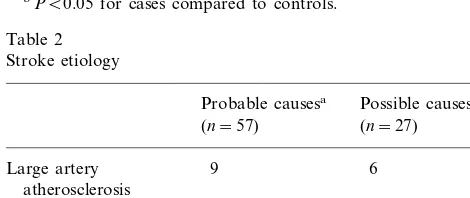
Etiology of stroke for probable and possible causes is shown in Table 2. Among 110 stroke patients, 57 had at least one probable cause, 27 had at least one possible cause, and the remaining 26 were indeterminate. ‘Other determined causes’ of stroke include hematologic disor-ders, non-atherosclerotic vasculopathy (e.g. vasculitis, dissection), migraine, drug abuse and stroke associated with pregnancy or the postpartum state.
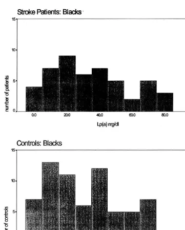
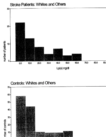
The distribution of lp (a) values differed markedly
between blacks and whites/other, both for cases and
controls (Figs. 1 and 2). Subsequent lp (a) and lipid analyses were done separately for these two groups. Mean and median lp (a) values were higher in blacks than whites/other, but within a racial group, lp (a) values were similar between cases and controls (Table 3). Lp (a) values for both racial groups were divided into quartiles, and the odds ratio for stroke was calcu-lated for each quartile compared to the lowest quartile (Table 4). No significant association of lp (a) with
stroke was seen for any quartile, and there did not appear to be a dose-response trend for higher lp (a) quartiles. We also analyzed our results using the me-dian lp (a) concentration for all cases (14 mg/dl) as a cut-point to stratify into high and low lp (a) groups. The crude odds ratio was similar between blacks (1.33, 95% CI 0.57 – 3.10) and whites (1.54, 95% CI 0.84 – 2.80). After adjustment for age, race, hypertension, diabetes, cigarette smoking, coronary artery disease, total cholesterol and HDL cholesterol, the odds ratio was 1.36 (95% CI 0.80 – 2.29). Power calculations were performed le6el to estimate a possible type II error due to insufficient sample size. The power of our study for detecting an lp (a)difference of 14 mg/dl between cases and controls was greater than 99%, and for detecting a difference of 6.1 mg/dl was 80%.
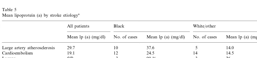
Mean lp (a) values for patients grouped by stroke etiologies are shown in Table 5. Among stroke patients, 15 were classified as having either a possible or proba-ble atherosclerotic cause for stroke. Atherosclerotic le-sions involving cerebral vessels were demonstrated by
cerebral angiography (n=10), carotid duplex
ultra-sound (n=4) and magnetic resonance angiography
(n=1). Location of the atherosclerotic lesion was in the extracranial carotid artery (n=9), intracranial carotid artery (n=2) and middle cerebral artery (n=4). The mean lp (a) for this subgroup of patients was 29.7 mg/dl (mean for blacks, 37.6 mg/dl; mean for whites/
other races, 14.0 mg/dl) and was not significantly differ-ent from controls (mean for blacks, 32.2 mg/dl; mean for whites/other races, 16.3 mg/dl).
Six patients had lacunar stroke in the absence of large artery atherosclerosis or cardiac source of em-bolism. Three black patients with lacunar stroke had markedly elevated lp (a) values (74, 76 and 120 mg/dl) resulting in a significantly elevated mean lp (a) value for that group. For other stroke etiologies, mean lp (a) values were not significantly different than controls.
4. Discussion
Most, [10 – 14,16 – 19] but not all, [15,20,26] previous cross-sectional studies have found elevated lp (a) con-centrations in patients with ischemic stroke or TIA as compared to controls. Several groups have found a particularly strong association of lp (a) with carotid artery atherosclerosis [19,23] or carotid intimal thicken-ing [22]. In contrast, Ridker reported in the only prospective study that elevated lp (a) concentrations did not predict future stroke in patients [25], and a number of cross-sectional studies have found an effect of lp (a) on stroke risk only in certain stroke subtypes [10,15,20]. Association with a particular stroke subtype, however, has not been consistent among studies.
Table 1 Hypertension (%) 29 (26.6)a
4 (1.9) 12 (10.9)a
Diabetes mellitus (%)
45 (41.3)
Current cigarette smoking (%) 67 (31.0) 15 (13.6)a 7 (3.2) Coronary artery disease (%)
Hypercholesterolemia (%) 32 (29.4)a 25 (11.9)
Post- or perimenopausal (%) 7 (6.4) 10 (4.6) 9 (4.2) Replacement estrogen use (%) 7 (6.4)
21 (9.7) 21 (18.9)b
Oral contraceptive use (%)
aP50.001 for cases compared to controls. bPB0.05 for cases compared to controls.
Table 2
Other determined cause 32 5
aThree patients had two probable, but only one cause is listed per patient using the following hierarchy: large artery atherosclerosis\ cardioembolism\lacune\other determined cause.
In our study of young women with stroke, we found no association of lp (a) concentrations with stroke risk, using either quartiles or median values for analysis. Calculated odds ratios were not significantly altered by adjustment for other risk factors. The distribution of lp (a) is different in blacks than whites, with blacks having higher mean and median values, and whites having a distribution skewed towards lower values. We found similar results in both cases and controls (Figs. 1 and 2), and within a racial group lp (a) concentrations were not significantly different between cases and controls. The risk of stroke is known to be higher in blacks than whites, even after adjusting for known risk factors [36]. A lipid component such as lp (a), which is genetically-influenced and is higher in blacks [18], would be an
ideal risk factor to explain this difference. At least in young women, our data do not support this hypothesis. Nagayama [17] reported significantly higher lp (a) concentrations in patients with atherothrombotic stroke as compared to controls and noted that this difference was more prominent in patients under the age of 45, suggesting lp (a) may be a more important risk factor in young stroke patients. The number of young stroke patients, however, was small (n=11). In our study, 15 patients were classified as having possible/probable stroke due to large artery atherosclerosis. Although the mean lp (a) concentration was higher in this subgroup as compared to controls, the difference was not statisti-cally significant. Mean lp (a) values were significantly elevated in the lacune group compared to controls, due
Fig. 2. The distribution of lp (a) values among white stroke patients and controls.
Table 3
Lipoprotein (a) and race
Blacks Whites/other
Controls (n=66)
Cases (n=49) Cases (n=61) Controls (n=150)
Mean lp (a) (SD) (mg/dl) 35.5 (25.8) 32.2 (28.3) 17.7 (18.7) 16.3 (19.7)
28.3 8.9
31.3 6.9
Median lp (a) (mg/dl)
to markedly elevated lp (a) values in three black pa-tients. While this finding is of interest, the very small number of patients involved suggests a possible spuri-ous result. No other stroke etiology was associated with lp (a) values that were significantly different from controls.
Table 4
Odds ratio for stroke by lipoprotein (a) quartile
Quartile Odds ratio for Odds ratio for whites/other blacks (95% CI) (95% CI)
1st quartile Reference group Reference group 2nd quartile 1.23 (0.24–6.34) 0.75 (0.34–1.66)
1.53 (0.70–3.36) 2.22 (0.48–10.2)
3rd quartile
4th quartile 1.13 (0.25–5.12) 1.16 (0.48–2.77)
who had blood draw before 2 months as compared to after 2 months, making timing of blood draw an un-likely source of bias.
The use of estrogen-containing compounds are known to lower lp (a) concentrations [34,35]. Although cases were more likely than controls to be on OCs at the time of stroke, the OCs were discontinued in all cases after the stroke, typically about a month prior to the time of blood draw for lp (a) and lipid measure-ments. The lp (a)-lowering effect of OCs would have been removed from these cases, therefore, resulting in relatively higher lp (a) concentrations for these patients. Despite this potential bias in favor of an association of high lp (a) with cases, our results still did not support such a finding. The proportion of women who were peri- or post menopausal or who were on replacement estrogen therapy was small and similar among cases and controls.
If the association of lp (a) with stroke were due to the promotion of premature atherosclerosis, we may have missed such an association because of a relatively small
number (n=15) of our cases having large-artery
atherosclerotic stroke. Nevertheless, to our knowledge, this is the largest study to date to investigate this question in young stroke patients. Our results with lacunar stroke patients are of interest, but the small number of cases make this finding a preliminary one, needing further confirmation. If lp (a) acts primarily as a hypercoagulable agent, however, one would expect an association with all mechanisms of stroke, which we did not find. Our study had 80% power to detect an lp (a) difference of 6.1 mg/dl between cases (using all stroke subtypes) and controls and greater than 99% power in detecting a difference of 14 mg/dl. These 6alues are comparable to the range of between 4 and 28 mg/dl differences in lp (a) reported in other studies [10 – 20]. Based upon our data, therefore, we cannot recommend routine measurement of lp (a) in young stroke patients at this time. Larger studies will be needed to determine subjects were African-American, whereas most previous
studies involved predominantly white or Asian popula-tions. In the Atherosclerosis Risk in Communities (ARIC) Study, lp (a) was found to be an independent risk factor for self-reported stroke or TIA in both blacks and whites, with similar odds ratios for both races (blacks 1.17, whites 1.19). Blacks in the United States as well as in Africa have a different distribution of lp (a) concentrations [37,38] and lp (a) polymor-phisms [38] compared to other races, and these differ-ences may influence the risk of atherosclerosis. We did not assess lp (a) polymorphisms in our subjects, but there is some evidence that certain polymorphisms are more atherogenic [19,39] or are more potent inhibitors of fibrinolysis [9].
The variable time interval between stroke and blood draw in cases is a potential limitation of our data. Lp (a) may act as an acute phase reactant and has been reported to rise within the first week of myocardial infarction and surgical procedures [40]. Other studies, however, have not confirmed significant changes in lp (a) after acute myocardial infarction [41]. Kargman et al. [42] recently reported serial lp(a) measurements o6er the first month after acute ischemic stroke and found no significant trend or fluctuation,suggesting the absence of an acute phase response. The majority of patients in our study had blood drawn for lp (a) more than 2 months after stroke. On retrospective analysis, we found no significance difference in mean lp (a) values in cases
Table 5
Mean lipoprotein (a) by stroke etiologya
All patients Black White/other
No. of cases Mean lp (a) (mg/dl) No. of cases Mean lp (a) (mg/dl) Mean lp (a) (mg/dl)
14.0 5
10
Large artery atherosclerosis 29.7 37.6
12 24.5 14
Other determined cause 23.3 10
14
Indeterminate 23.3 29.4 12 20.7
32.2
Controls 21.3 16.3
aPatients with multiple etiologies are listed only once, using the following hierarchy: large artery atherosclerosis\cardioembolism\lacune\ other determined cause\indeterminate.
if lp (a) is an important risk factor for certain mecha-nisms of stroke in young adults, such as those with atherosclerotic or lacunar stroke.
Acknowledgements
Supported in part by a Clinical Stroke Research Center Award from the National Institute of Neurolog-ical Disorders and Stroke (NS16332-11 to Drs Kittner, Hebel, Price and Sloan and to Feeser); by a Clinical Investigator Development Award from the National Institute of Neurological Disorders and Stroke (K08-NS01764-01A1 to Dr Wozniak); and by a National
Institutes of Health Training Grant (c T32AG00209
to Dr Jenner).
References
[1] Lip GY, Jones AF. Lipoprotein (a) and vascular disease: throm-bogenesis and atherogenesis. Q J Med 1995;88:529 – 39. [2] Mbewu AD, Durrington PN. Lipoprotein (a): structure,
proper-ties and possible involvement in thrombogenesis and atherogene-sis. Atherosclerosis 1990;85:1 – 14.
[3] Rath M, Niendorf A, Reblin T, Dietel M, Krebber H-J, Beisiegel U. Detection and quantification of lipoprotein (a) in the arterial wall of 107 coronary bypass patients. Arteriosclerosis 1989;9:579 – 92.
[4] Niendorf A, Rath M, Wolf K, Peters S, Hartmut A, Beisiegel U, Dietel M. Morphological detection and quantification of lipo-protein (a) deposition in atheromatous lesions of human aorta and coronary arteries. Vichows Archiv A Pathol Anat 1990;417:105 – 11.
[5] Jamieson DG, Usher DC, Rader DJ, Lavi E. Apolipoprotein (a) deposition in atherosclerotic plaques of cerebral vessels. A poten-tial role for endothelial cells in lesion formation. Am J Pathol 1995;147:1567 – 74.
[6] McLean JW, Tomlinson JE, Kuang W-J, Eaton DL, Chen EY, Fless GM, Scanu AM, Lawn RM. cDNA sequence of human apolipoprotein (a) is homologous to plasminogen. Nature 1987;330:132 – 7.
[7] Miles LA, Fless GM, Levin EG, Scanu AM, Plow EF. A potential basis for the thrombotic risks associated with lipo-protein (a). Nature 1989;339:301 – 3.
[8] Hajjar KA, Gavish D, Breslow JL, Nachman RL. Lipoprotein (a) modulation of endothelial cell surface fibrinolysis and its potential role in atherosclerosis. Nature 1989;339:303 – 5. [9] Hervio L, Chapman MJ, Thillet J, Loyau S, Angeles-Cano E.
Does apolipoprotein (a) heterogeneity influence lipoprotein (a) effects on fibrinolysis? Blood 1993;82:392 – 7.
[10] Murai A, Miyahara T, Fujimoto M, Matsuda M, Kameyama M. Lp (a) lipoprotein as a risk factor for coronary heart disease and cerebral infarction. Atherosclerosis 1986;59:199 – 204.
[11] Zenker G, Koltringer P, Bone G, Niederkorn K, Pfeiffer K, Jurgens G. Lipoprotein (a) as a strong indicator for cerebrovas-cular disease. Stroke 1986;17:942 – 5.
[12] Jurgens G, Koltringer P. Lipoprotein (a) in ischemic cerebrovas-cular disease: a new approach to the assessment of risk for stroke. Neurology 1987;37:513 – 5.
[13] Woo J, Lau E, Lam CW, Kay R, Teoh R, Wong HY, Prall WY, Kreel L, Nicholls MG. Hypertension, lipoprotein (a), and apolipoprotein A-I as risk factors for stroke in the Chinese. Stroke 1991;22:203 – 8.
[14] Pedro-Botet J, Senti M, Nogues X, Rubies-Prat J, Roquer J, L DO, Olive J. Lipoprotein and apolipoprotein profile in men with ischemic stroke. Role of lipoprotein (a), triglyceride-rich lipo-proteins, and apolipoprotein E polymorphism, Stroke 1992;23:1556 – 62.
[15] Lindgren A, Nilsson EP, Norrving B, Johansson BB. Plasma lipids and lipoproteins in subtypes of stroke. Acta Neurol Scand 1992;86:572 – 8.
[16] Shintani S, Kikuchi S, Hamaguchi H, Shiigai T. High serum lipoprotein (a) levels are an independent risk factor for cerebral infarction. Stroke 1993;24:965 – 9.
[17] Nagayama M, Shinohara Y, Nagayama T. Lipoprotein (a) and ischemic cerebrovascular disease in young adults. Stroke 1994;25:74 – 8.
[18] Schreiner PJ, Chambless LE, Brown SA, Watson RL, Toole J, Heiss G. Lipoprotein (a) as a correlate of stroke and transient ischemic attack prevalence in a biracial cohort: the ARIC study. Ann Epidemiol 1994;4:351 – 9.
[19] Jurgens G, Taddei-Peters WC, Koltringer P, Petek W, Chen Q, Greilberger J, Macomber PF, Butman BT, Stead AG, Ransom JH. Lipoprotein (a) serum concentration and apolipoprotein (a) phenotype correlate with severity and presence of ischemic cere-brovascular disease. Stroke 1995;26:1841 – 8.
[20] van Kooten F, van Krimpen J, Dippel DW, Hoogerbrugge N, Koudstaal PJ. Lipoprotein (a) in patients with acute cerebral ischemia. Stroke 1996;27:1231 – 5.
[21] Koltringer P, Jurgens G. A dominant role of lipoprotein (a) in the investigation and evaluation of parameters indicating the development of cervical atherosclerosis. Atherosclerosis 1985;58:187 – 98.
[22] Schreiner PJ, Morrisett JD, Sharrett AR, Patsch W, Tyroler HA, Wu K, Heiss G. Lipoprotein (a) as a risk factor for preclinical atherosclerosis. Arterioscler Thromb 1993;13:826 – 33.
[23] Watts GF, Mazurkiewicz JC, Tonge K, Nelson V, Warburton FG, Slavin BM. Lipoprotein (a) as a determinant of the severity of angiographically defined carotid atherosclerosis. Q J Med 1995;88:321 – 6.
[24] Willeit J, Kiechl S, Santer P, Oberhollenzer F, Egger G, Jarosch E, Mair A. Lipoprotein (a) and asymptomatic carotid artery disease. Evidence of a prominent role in the evolution of ad-vanced carotid plaques: the Bruneck study. Stroke 1995;26:1582 – 7.
[25] Ridker PM, Stampfer MJ, Hennekens CH. Plasma concentration of lipoprotein (a) and the risk of future stroke. JAMA 1995;273:1269 – 73.
[26] Hachinski V, Graffagnino C, Beaudry M, Bernier G, Buck C, Donner A, Spence JD, Doig G, Wolfe BM. Lipids and stroke: a paradox resolved. Arch Neurol 1996;53:303 – 8.
[27] Rosengren A, Wilhelmsen L, Eriksson E, Risberg B, Wedel H. Lipoprotein (a) and coronary heart disease: a prospective case-control study in a general population sample of middle aged men. Br Med J 1990;301:1248 – 51.
[28] Jauhiainen M, Koskinen P, Ehnholm C, Frick MH, Manttari M, Manninen V, Huttunen JK. Lipoprotein (a) and coronary heart disease risk: a nested case-control study of Helsinki Heart Study participants. Atherosclerosis 1991;89:59 – 67.
[29] Ridker PM, Hennekens CH, Stampfer MJ. A prospective study of lipoprotein (a) and the risk of myocardial infarction. JAMA 1993;270:2195 – 9.
[30] Schaefer EJ, Lamon-Fava S, Jenner JL, McNamara JR, Ordovas JM, Davis CE, Abolafia JM, Lippel K, Levy RI. Lipoprotein (a) levels and risk of coronary heart disease in men. The Lipid Research Clinics Coronary Primary Prevention Trial. JAMA 1994;271:999 – 1003.
[32] Kittner SJ, Stern BJ, Feeser BR, Hebel JR, Nageu DA, Buchholz DW, Earley CJ, Johnson CJ, Macko RF, Sloan MA, Wityk RJ, Wozniak MA. Pregnancy and the risk of stroke. New Engl J Med 1996;335:768 – 74.
[33] Warnick G, Russell J, Albers JJ. Dextran sulfate-Mg2+
precipi-tation procedures for quantification of high density-lipoprotein cholesterol. In: Cooper GR, editor. Selected Methods of Clinical Chemistry, Washington, DC, 1983:91 – 9.
[34] Soma MR, Osnago GI, Paoletti R, Fumagalli R, Morrisett JD, Meschia M, Crosignani P. The lowering of lipoprotein (a) induced by estrogen plus progesterone replacement therapy in postmenopausal women. Arch Intern Med 1993;153:1462 – 8.
[35] Jenner JL, Ordovas JM, Lamon FS, Schaefer MM, Wilson PW, Castelli WP, Schaefer EJ. Effects of age, sex, and menopausal status on plasma lipoprotein (a) levels. The Framingham offspring study. Circulation 1993;87:1135 – 41.
[36] Kittner SJ, White LR, Losonczy KG, Wolf PA, Hebel JR. Black – white differences in stroke incidence in a national sample. The contribution of hypertension and diabetes mellitus. JAMA 1990;264:1267 – 70.
[37] Parra H-J, Luyeye I, Bouramoue C, Demarquilly C, Fruchart J-C.
Black – white differences in serum lp (a) lipoprotein levels. Clin Chim Acta 1987;167:27 – 31.
[38] Marcovina SM, Albers JJ, Jacobs DJ, Perkins LL, Lewis CE, Howard BV, Savage P. Lipoprotein (a) concentrations and apolipoprotein (a) phenotypes in Caucasians and African Amer-icans. The CARDIA study. Arterioscler Thromb 1993;13:1037 – 45. [39] Seed M, Hoppichler F, Reaveley D, McCarthy S, Thompson GR, Boerwinkle E, Utermann G. Relation of serum lipoprotein (a) concentration and apolipoprotein (a) phenotype to coronary heart disease in patients with familial hypercholesterolemia. New Eng J Med 1990;322:1494 – 8.
[40] Maeda S, Abe A, Seishima M, Makino K, Noma A, Kawade M. Transient changes of serum lipoprotein (a) as an acute phase protein. Atherosclerosis 1989;78:145 – 50.
[41] Andreassen A, Berg K, Torsvik H. Changes in Lp(a) lipoprotein and other plasma proteins during acute myocardial infarction. Clin Gen 1994;46:410 – 6.
[42] Kargman DE, Tuck C, Berglund L, Lin IF, Mukherjee RS, Thompson EV, Jones J, Boden-Albala B, Paik MC, Sacco RL. Lipid and lipoprotein levels remain stable in acute ischemic stroke: the Northern Manhattan Stroke Study. Atherosclerosis 1998;139:391 – 9.
.