Administration of gonadotropin-releasing
hormone during metoestrus in cattle:
influence on luteal function and cycle length
J. Taponen
a,∗, H. Rodr´ıguez-Mart´ınez
b, T. Katila
a aDepartment of Clinical Veterinary Sciences, University of Helsinki,Pohjoinen Pikatie 800, FI-04920 Saarentaus, Finland
bDepartment of Obstetrics and Gynaecology, Swedish University of Agricultural Sciences, S-750 07 Uppsala, Sweden
Received 19 January 2000; received in revised form 6 June 2000; accepted 4 September 2000
Abstract
Gonadotropreleasing hormone (GnRH) has been used to warrant the success of artificial in-semination by accurately timing occurrence of ovulation. In practical conditions, GnRH may be administered too late, after ovulation, with an eventual reduction in pregnancy rate. The aim of this study was to investigate whether GnRH administration after ovulation would have a negative effect on luteal function.
Three cows and six heifers of the Finnish Ayrshire breed were used. Oestruses were synchronised. After detection of ovulation, one of the following treatments was implemented: gonadorelin (250mg, i.m.) at either 0–24 h (T1) or 24–48 h (T2) post-ovulation or control (no gonadorelin, C). Every animal was assigned once to each of these three manipulations. Ultrasonography was performed on days 1, 4 or 5, 7 or 8, 11 or 12, 14 or 15 post-ovulation and daily from the beginning of the next oestrous signs until ovulation (day 0 =day of ovulation). Blood samples for progesterone (P4)
determinations were collected daily from day 1 after the occurrence of ovulation until recording of the next oestrus.
Administration of GnRH during metoestrus did not induce ovulation of either large or small follicles and, thus, no accessory corpora lutea (CL) were formed. In T1, on day 14 or 15, the diameter of CL was 1.3±0.3 mm smaller than in C (P <0.01), but no differences were found either on days 11 or 12 or on the same days of the T2 and C treatments. No significant differences in levels or profiles of P4curves were found between GnRH treatments and control. Neither had
the treatments any effects on the length of the oestrous cycle.
∗Corresponding author. Tel.:+358-19-5295-308; fax:+358-19-6851-181. E-mail address:[email protected] (J. Taponen).
In conclusion, GnRH treatment during metoestrus does not seem to alter subsequent luteal function and, thus, this does not explain previous reports of reduced fertility post-treatment. © 2000 Elsevier Science B.V. All rights reserved.
Keywords:Cattle-endocrinology; Gonadotropin-releasing hormone; Corpus luteum; Progesterone; Cycle length
1. Introduction
Gonadotropin-releasing hormone (GnRH) has been indicated as a tool for control of ovarian function in cattle when applied close to the time of insemination to induce ovulation and enhance conception rate (review by Thatcher et al. (1993), Morgan and Lean (1993) and Shaw (1999)). During recent years, emphasis has been put on the use of GnRH during the luteal phase to support CL-function and thus, possibly, to avoid early embryonic mortality around the time of maternal recognition of pregnancy (review by Thatcher et al. (1993) and Shaw (1999)). In most of these studies, GnRH has been given during early or mid-dioestrus as single or multiple injections.
As Taponen et al. (1999) have described earlier, in tie-stall conditions, especially during the long wintertime in the north, occurrence of silent heat is one of the most important causes for sub-fertility. Therefore, GnRH treatment is often used by practitioners to induce ovula-tion as close as possible to artificial inseminaovula-tion (AI) hoping to enhance concepovula-tion rates. Under these circumstances, GnRH treatment may be clearly given before the physiological luteinizing hormone (LH) surge, but also after ovulation, e.g. during metoestrus.
The effects of GnRH given during the luteal phase on luteal function and oestrous cycle length have been studied intensively (review by Thatcher et al. (1993)). In some studies, increased luteal lifespan and cycle length and/or progesterone (P4) production have been
found (Milvae et al., 1984; Macmillan et al., 1985; Stevenson et al., 1993), but the results have been inconsistent. This is probably due to different experimental designs (natural or synthetic GnRH, dose, cycle day of treatment, single or repeated injections). Despite this intensive research, very few studies have been published on GnRH treatments given during metoestrus in cattle. Macmillan et al. (1986) reported a reduction in pregnancy rate by 10.9% units when buserelin (5 microg) was administered 1–3 days after insemination. Ford and Stormshak (1978) found a significantly reduced P4 concentration when GnRH (100
microg) was given 55 h after the detected oestrus. Later, Rodger and Stormshak (1986) and Martin et al. (1990) studied the influence of GnRH (100mg) given on day 2 post-oestrus (day of oestrus =day 0) on subsequent luteal function. In both studies, a tendency for a reduction in P4concentration was found. It has been shown that early embryonic loss is
probably associated with a complex endocrine syndrome, which could be manifested by a lower capacity to synthesise and/or release CL-progesterone (Lamming et al., 1989). It is, however, questionable that the reduced fertility reported by Macmillan et al. (1986) can be explained by an impaired luteal function, because the reasons behind the decline of P4
given slightly post-ovulation on the development and function of the corpus luteum and the length of the oestrous cycle in cattle.
2. Materials and methods
2.1. Animals
Three cows (1–2 calvings) and six heifers of the Finnish Ayrshire breed were used. The cows had calved 9–13 months earlier. The heifers were between 12 and 15 months of age at the beginning of the experiment. All animals had shown normal oestrous cycles and were clinically healthy. They were stanchioned and fed grass silage, dry hay, and corn according to Finnish standards. The experiment lasted 5 months, from March to July.
2.2. Experimental procedures
The experiment included two different GnRH treatments (T1, T2) and one control ma-nipulation (C). Every animal was assigned once to each of these three mama-nipulations. These were performed in random order for each animal. In all cases a spontaneous ovulation preceded the manipulation period.
Oestrus was induced with 0.5 mg of cloprostenol (PG) (Estrumat® 0.25 mg/ml, Mallinck-rodt Veterinary Ltd., Harefield, Uxbridge, England) administered intramuscularly (i.m.) in 18 cases. In nine cases, oestrus was induced with an intravaginal device containing 1.9 mg of progesterone and 10 mg of encapsulated oestradiol benzoate, attached to the device (EAZI-BREEDTM CIDR® device, InterAg, Hamilton, New Zealand; Cidirol® capsules, InterAg, Hamilton, New Zealand). The device was inserted for 12 days. From the second day after PG or device removal, the animals were examined daily by transrectal ultrasound to determine the occurrence of ovulation.
After detection of ovulation, the animals were given 250mg of gonadorelin i.m. (Fertagyl® 0.1 mg/ml, Intervet International B.V., Boxmeer, Holland) as follows: 0–24 h after ovulation (T1), 24–48 h after ovulation (T2), or no gonadorelin (control manipulation, C).
Ultrasonographic examinations were performed daily from the second day after PG treat-ment or device removal until ovulation. After this, examinations were carried out on days 1, 4 or 5, 7 or 8, 11 or 12, 14 or 15 and daily from the beginning of the next oestrous signs until ovulation (day 0=day of ovulation).
Blood samples for hormone determinations were collected by vacuum puncture of a tail blood vessel into silicone plain tubes (Venoject) by 20 G needles (Vacutainer). Samples were taken daily from day 1 after ovulation until the next oestrus. Serum was harvested, frozen and stored in plastic tubes at−20◦C until analysed. All procedures were performed in the morning, from 08.00 to 10.00 h.
2.3. Ovarian examinations
the corpus luteum (CL) and of possible secondary corpora lutea, and to determine the day of ovulation and diameter of the ovulatory follicle as described by Assey et al. (1993). All examinations were carried out by the same operator.
The ovaries were scanned several times in lateromedial and dorsoventral planes to de-termine the largest cross-section of follicles and/or a CL. By freezing the image, the largest and the smallest diameters were measured and recorded, with the average diam-eter calculated later. The central cavities of CLs were measured and recorded in the same way.
All follicles equal to or larger than 8 mm were measured. Follicles smaller than 8 mm were only counted and divided according to their size into groups of small (less than 5 mm) and medium (5–8 mm) follicles. Day of ovulation (day 0) was determined to be the last day when the follicle was intact according to ultrasound scanning in the morn-ing before the subsequent examination the next mornmorn-ing when the follicle had disap-peared.
2.4. Hormone determinations
The concentration of progesterone in the serum was measured in all samples by radioim-munoassay by use of a commercial kit (Coat-A-Count® Progesterone, Diagnostic Products Corporation, Los Angeles, USA). The intra-assay coefficient of variation for progesterone was 6.31% when calculated from duplicates of measurements between 1.0 and 20.0 nmol/l (n =380). The inter-assay coefficient of variation was 6.74% (81.9 nmol/l,n =8). The detection limit of the assay was 0.3 nmol/l.
2.5. Statistical analysis
Data were analysed using the statistical analysis system software (SAS, 1987). Pro-gesterone concentrations were analysed by repeated measures analysis of variance, with manipulation and day as the within-subject factors. Significance of day effects and day by manipulation interaction effects was evaluated by use of Greenhouse–Geisser-adjusted
P-values. Effects of manipulations on interoestrous intervals and on size of CLs were anal-ysed by repeated measures analysis of variance, with manipulation as the within-subject factor. Differences between animals in interoestrous intervals were tested by one-way anal-ysis of variance. Differences were considered significant atP <0.05.
3. Results
3.1. Follicles
On day 1 (0–24 h after ovulation), follicles larger than 8 mm were found in four cases, two times in T1, once in T2 and once in C. GnRH treatment did not induce ovulation of larger or smaller follicles. Thus, no accessory corpora lutea were found later.
3.2. Corpus luteum
The size of CL was analysed on days 11 or 12 and 14 or 15 after ovulation. Only the diameter of CL was included in the analysis. In T1, T2 and C, on day 11 or 12, the diameters (mean±S.E.M.) were 22.2±0.7, 22.7±1.1 and 22.7±1.1 mm, respectively; and on day 14 or 15, 21.2±0.7, 22.1±1.0 and 22.6±0.7 mm, respectively. In T1, on day 14 or 15, CL was 1.3±0.3 mm smaller than in C (P < 0.01), but no significant differences were found on day 11 or 12. Neither were there any differences found between T2 and C.
3.3. Progesterone
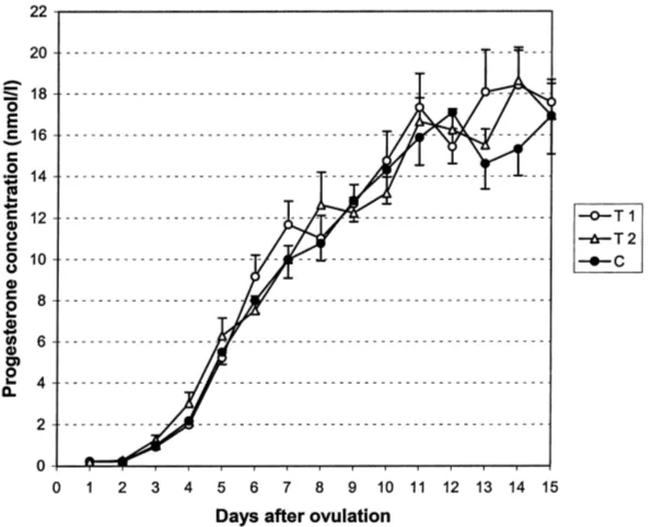
The P4curves of T1, T2 and C from days 1–15 are shown in Fig. 1. A significant effect of
day was observed on P4concentration in all manipulations (P <0.001). P4had begun to rise
on day 3. The rise continued steadily until day 11. The level remained thereafter unchanged. The mean estimated daily rise in P4concentration from days 2 to 11 was 1.8±0.1 nmol/l
(mean±S.E.M.). In T1, T2, and C, the rises were 1.9±0.2, 1.8±0.1 and 1.7±0.1 nmol/l,
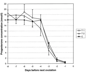
Fig. 2. Mean (±S.E.M.) P4concentration during 8 days before next ovulation, when GnRH was given 0–24 h (T1) or 24–48 h (T2) after preceding ovulation, or with no GnRH (C) administered.
respectively. There were no significant differences in levels or profiles of P4curves between
GnRH treatments and control manipulation.
In C, P4concentration rose continuously from days 2 to 11, but a slight decline in P4
concentration was noticed in GnRH treatments between days 7 and 8 after ovulation in T1 and between days 8 and 9 in T2. In both treatment groups (T1 and T2), the decline occurred between days 6 and 7 after the GnRH injection (Fig. 1). In individual animals, this decline occurred in 8/9 animals in T1 and T2, and in 2/9 in C between days 5 and 9 after ovulation. The decline occurred in six cases between days 7 and 8 in T1; in T2 there was more inconsistency.
P4curves of T1, T2 and C during the last 8 days before the next ovulation are shown
in Fig. 2. A significant decline in P4concentration was detected between days−4 and−3
(P <0.01) in T1, T2 and C. There were no significant differences in the levels or profiles of P4curves during these days between GnRH treatments and control manipulation.
3.4. Length of the oestrous cycle
4. Discussion
The mean maximum diameter of the ovulatory follicle during the last 24 h before ovu-lation has been reported to be between 15.3 and 20.3 mm in two-wave cycles and between 12.8 and 18.6 mm in three-wave cycles after natural luteolysis (Savio et al., 1988; Sirois and Fortune, 1988; Knopf et al., 1989; Ginther et al., 1989a,b) and between 13.7 and 19.5 mm after prostaglandin F2a-induced luteolysis (Kastelic et al., 1990a; Kastelic and Ginther, 1991). Our findings of 15.4 mm were in good accordance with these. However, this was 1.3 mm smaller than our earlier results with the same herd (Taponen et al., 1999).
In 4/27 cases, a follicle larger than 8 mm was detected during the first 24 h after the ovu-lation. These follicles were most probably regressing dominant follicles from the preceding anovulatory follicle waves, as can be seen from the studies of Ginther et al. (1989a) and Knopf et al. (1989). None of these follicles ovulated after GnRH treatment, which confirms their suggested atretic nature.
Kastelic et al. (1990b) have shown that the correlation between size of CL and plasma P4concentration is between 0.7 and 0.8. Thus, the size of CL can be used as a method for
assessment of luteal function. In our study, the difference of 1.3 mm in the diameter of the CL on day 14 or 15 between T1 and C was statistically significant, implying a difference of about 20% in spherical volume. The difference is, however, so small and close to the precision of measurement that its practical significance may be questionable.
Although there were no significant differences in levels or profiles of P4curves between
GnRH and control treatments in our study, a slight decrease in P4concentration was noticed
in T1 beginning from day 8 and in T2 from day 9 (7 days after the GnRH administration), but not in C. This finding is in agreement with the results reported by Rodger and Stormshak (1986) and Martin et al. (1990). Although the differences in P4 concentrations were so
small that statistical significance could not be reached, the similarity of the findings shows that GnRH given during metoestrus can cause a reduction in P4production after about 1
week. Martin et al. (1990) have suggested that luteinizing hormone secreted in response to GnRH acts on the CL to promote the conversion of small luteal cells to large cells. Occurrence of such a phenomenon could explain alterations in P4production. Although
follicular growth was not followed in the present study, it is obvious that the normal pattern of follicular growth was likely perturbed by GnRH administration, and thus, the rise in oestradiol concentration was delayed. Also this may have some effects on the development and function of CL.
can be assessed from the experimental protocol that the interval was, in most cases, most probably less than 24 h. In our experiment, the time interval from spontaneous LH surge to GnRH administration was 30–54 h in T1 and 54–78 h in T2. Due to these reasons, it is not probable that the weak LH response to GnRH administration after the spontaneous LH surge in our study could explain the non-altered P4production.
In practice, based on the findings of Macmillan et al. (1986), GnRH may cause reduced fertility and should not be administered during at least the first 3 days after ovulation. In the present study, exogenous GnRH given 0–48 h after ovulation had no influence on P4
concentrations during the subsequent oestrous cycle, nor on the length of the cycle. The only significant effect was the slight decrease in the size of CL. These findings are not relevant enough to explain the reduced pregnancy rate after the GnRH treatment given 1–3 days after insemination, as reported by Macmillan et al. (1986). In conclusion, administration of GnRH after ovulation, considering previous and the present results, should be avoided in practice.
Acknowledgements
We wish to thank Dr. Satu Sankari for supervising the hormonal analyses.
References
Assey, R.J., Purwantara, B., Greve, T., Hyttel, P., Schmidt, M.H., 1993. Corpus luteum size and plasma progesterone levels in cattle after cloprostenol-induced luteolysis. Theriogenology 39, 1321–1330.
Chenault, J.R., Kratzer, D.D., Rzepkowski, R.A., Goodwin, M.C., 1990. LH and FSH response of Holstein heifers to fertirelin acetate, gonadorelin and buserelin. Theriogenology 34, 81–98.
Ford, S.P., Stormshak, F., 1978. Bovine ovarian and pituitary responses to PMS and GnRH administered during metoestrus. J. Anim. Sci. 46, 1701–1706.
Ginther, O.J., Kastelic, J.P., Knopf, L., 1989a. Composition and characteristics of follicular waves during the bovine estrous cycle. Anim. Reprod. Sci. 20, 187–200.
Ginther, O.J., Knopf, L., Kastelic, J.P., 1989b. Temporal associations among ovarian events in cattle during oestrous cycles with two and three follicular waves. J. Reprod. Fertil. 87, 223–230.
Kastelic, J.P., Ginther, O.J., 1991. Factors affecting the origin of the ovulatory follicle in heifers with induced luteolysis. Anim. Reprod. Sci. 26, 13–24.
Kastelic, J.P., Knopf, L., Ginther, O.J., 1990a. Effect of day of prostaglandin F2 alpha treatment on selection and development of the ovulatory follicle in heifers. Anim. Reprod. Sci. 23, 169–180.
Kastelic, J.P., Bergfelt, D.R., Ginther, O.J., 1990b. Relationship between ultrasonic assessment of the corpus luteum and plasma progesterone concentration in heifers. Theriogenology 33, 1269–1278.
Kesner, J.S., Convey, E.M., Anderson, C.R., 1981. Evidence that estradiol induces the preovulatory LH surge in cattle by increasing pituitary sensitivity to LHRH and then increasing LHRH release. Endocrinology 108, 1386–1391.
Knopf, L., Kastelic, J.P., Schallenberger, E., Ginther, O.J., 1989. Ovarian follicular dynamics in heifers: test of two-wave hypothesis by ultrasonically monitoring individual follicles. Dom. Anim. Endocrinol. 6, 111–119. Lamming, G.E., Darwash, A.O., Back, H.L., 1989. Corpus luteum function in dairy cows and embryo mortality.
J. Reprod. Fertil. Suppl. 37, 245–252.
Macmillan, K.L., Day, A.M., Taufa, V.K., Gibb, M., Pearce, M.G., 1985. Effects of an agonist of gonadotrophin releasing hormone in cattle. I. Hormone concentrations and oestrous cycle length. Anim. Reprod. Sci. 8, 203– 212.
Macmillan, K.L., Taufa, V.K., Day, A.M., 1986. Effects of an agonist of gonadotrophin releasing hormone (Buserelin) in cattle. III. Pregnancy rates after a post-insemination injection during metoestrus or dioestrus. Anim. Reprod. Sci. 11, 1–10.
Martin, T.L., Swanson, L.V., Appell, L.H., Rowe, K.E., Stormshak, F., 1990. Response of the bovine corpus luteum to increased secretion of luteinizing hormone induced by exogenous gonadotropin releasing hormone. Dom. Anim. Endocrinol. 7, 27–34.
Milvae, R.A., Murphy, B.D., Hansel, W., 1984. Prolongation of the bovine estrous cycle with a gonadotropin-releasing hormone analog. Biol. Reprod. 31, 664–670.
Morgan, W.F., Lean, I.J., 1993. Gonadotrophin-releasing hormone treatment in cattle: a meta-analysis of the effects on conception at the time of insemination. Aust. Vet. J. 70, 205–209.
Rodger, L.D., Stormshak, F., 1986. Gonadotropin-releasing hormone-induced alteration of bovine corpus luteum function. Biol. Reprod. 35, 149–156.
Rosenberg, M., Chun, S.Y., Kaim, M., Herz, Z., Folman, Y., 1991. The effect of GnRH administered to dairy cows during oestrus on plasma LH and conception in relation to the time of treatment and insemination. Anim. Reprod. Sci. 24, 13–24.
SAS Institute, 1987. SAS/STATTMGuide for Personal Computers, 6th Edition. SAS Institute, Cary, NC, 1028 pp. Savio, J.D., Keenan, L., Boland, M.P., Roche, J.F., 1988. Pattern of growth of dominant follicles during the oestrous
cycle of heifers. J. Reprod. Fertil. 83, 663–671.
Shaw, D.W., 1999. Use of GnRH to enhance pregnancy rates and shorten the postpartum interestrus interval in dairy cattle. Newsletter of the Society for Theriogenology, February 1999, pp. 9–10.
Sirois, J., Fortune, J.E., 1988. Ovarian follicular dynamics during the estrous cycle in heifers monitored by real-time ultrasonography. Biol. Reprod. 39, 308–317.
Stevenson, J.S., Phatak, A.P., Rettmer, I., Stewart, R.E., 1993. Postinsemination administration of receptal: follicular dynamics, duration of cycle, hormonal responses, and pregnancy rates. J. Dairy Sci. 76, 2536–2547. Taponen, J., Katila, T., Rodr´ıguez-Mart´ınez, H., 1999. Induction of ovulation with gonadotropin-releasing hormone during proestrus in cattle: influence on subsequent follicular growth and luteal function. Anim. Reprod. Sci. 55, 91–105.
Thatcher, W.W., Drost, M., Savio, J.D., Macmillan, K.L., Entwistle, K.W., Schmitt, E.J., de la Sota, R.L., Morris, G.R., 1993. New clinical uses of GnRH and its analogues in cattle. Anim. Reprod. Sci. 33, 27–49.