Insect Biochemistry and Molecular Biology 31 (2001) 7–17
www.elsevier.com/locate/ibmb
Review
Lipid storage and mobilization in insects: current status and future
directions
Estela L. Arrese
1, Lilian E. Canavoso, Zeina E. Jouni, James E. Pennington,
Kozo Tsuchida
2, Michael A. Wells
*Department of Biochemistry and Center for Insect Science, Biological Sciences West, The University of Arizona, Tucson, AZ 85721-0088, USA
Received 25 January 2000; received in revised form 26 April 2000; accepted 26 April 2000
Abstract
In this paper we review the current status of research on fatty acid absorption and conversion to diacylglycerol in the midgut. We further discuss how diacylglycerol may leave the midgut and associate with lipophorin in hemolymph. We review the present understanding of the role of the lipid transfer particle and lipophorin receptors in lipid delivery between lipophorin and tissues. Finally, we discuss recent studies on the mobilization of diacylglycerol from the fat body in response to adipokinetic hormone. Several suggestions for exciting areas of future research are described.2001 Elsevier Science Ltd. All rights reserved.
Keywords:Insect; Lipophorin; Lipid mobilization; Lipid storage; Receptor; Lipid transport particle; Adipokinetic hormone
1. Introduction
The pace of gene and genome sequencing seems to increase almost daily. As we try to cope with exponen-tially expanding databases, there is, perhaps, a tendency to lose sight of the fact that biochemistry and physiology provide the context within which we can interpret the functions of all these genes and their protein products. We have written this review in the hope that it will stimulate insect scientists to explore new ways to com-bine the power of molecular biology with biochemical and physiological studies that will ultimately lead to a more complete understanding of the regulation of lipid storage and mobilization.
For reasons of space and interest, we have chosen to focus this review on the metabolic pathways by which dietary fatty acids are converted to stored triacylglycerol (TAG) in the fat body and then mobilized to support metabolic needs. If the readers think that this material is already well understood and holds no exciting
ques-* Corresponding author. Tel.:+1-520-621-3847; fax:+ 1-520-621-9288.
E-mail address:[email protected] (M.A. Wells).
1 Department of Biochemistry and Molecular Biology, Oklahoma
State University, Stillwater, OK, USA.
2 National Institute for Infectious Diseases, Tokyo, Japan.
0965-1748/01/$ - see front matter2001 Elsevier Science Ltd. All rights reserved. PII: S 0 9 6 5 - 1 7 4 8 ( 0 0 ) 0 0 1 0 2 - 8
tions for future research, consider the life history of the tobacco hornworm, Manduca sexta. Larvae feed con-stantly (Manduca means glutton in Latin) and this behavior is only interrupted during the molt (Ziegler, 1985). As a result, the content of fat body TAG increases from a few milligrams at hatching up to 300 mg at the end of the larval stage (Fernando-Warnakulasuriya et al., 1988). During the pupal period, these lipid stores are used to support metamorphosis and the adult moth util-izes the stores to support the energy demands imposed by reproduction and flight (Ziegler, 1991). In short, the larva accumulates reserves while the adult consumes them. Thus, during development the fat body changes from a lipid-storing tissue to a lipid-mobilizing tissue (Tsuchida and Wells, 1988); however, at present, there is no information about how this profound transition in the metabolic conditions of the fat body is regulated, but clearly it must be regulated.
examination, as we shall see, reveals many critical gaps. Thus, the purpose of this review is to describe this broad picture and point out where, in our opinion, the gaps lie and to suggest how those gaps might be filled.
2. Unique properties of lipid transport in insects
Before proceeding to the heart of this review, it is worth reminding the reader about several unique proper-ties of lipid transport in insects that will be a common thread for much of what follows. Lipophorin is the major lipoprotein found in insect hemolymph and serves as a reusable, non-internalized shuttle (Chino and Kitazawa, 1981; Chino, 1985; Tsuchida and Wells, 1988). Lipo-phorins are named according to their density: low-den-sity lipophorin (LDLp), high-denlow-den-sity lipophorin (HDLp), and very high-density lipophorin (VHDLp) (Beenakkers et al., 1985). Lipophorin is a nearly spherical particle with a surface composed of phospholipids and proteins. In contrast to vertebrate lipoproteins, diacylglycerol (DAG) is the predominant core neutral lipid of lipopho-rin with lesser amounts of sterols, hydrocarbons, carot-enoids and other acylglycerols—a comprehensive tabu-lation of lipophorin properties has been published (Soulages and Wells, 1994). Most of the fatty acids, whether in transit to storage or utilization sites, are trans-ported as DAG in lipophorin. One hallmark of lipid transport in insects is the tissue specificity of lipid deliv-ery. Thus, the same lipophorin molecule can selectively deliver DAG to the fat body, and hydrocarbon and carot-enoids to the cuticle in larvalM. sexta, while inBombyx mori lipophorin also delivers carotenoids to the middle silk gland.
3. Digestion, absorption and transport of lipids from the midgut
One of the major functions of the midgut is to digest dietary lipids, and absorb and process the digestion pro-ducts for export into the hemolymph. TAG is a major lipid component of the diet and the major form for fatty acid storage (Beenakkers et al., 1985; Downer, 1985; Turunen and Crailsheim, 1996). Lipid export from enterocytes in insects differs from that in vertebrates in two important ways: (1) DAG is the main lipid exported and (2) DAG release does not involve the biosynthesis of a lipoprotein particle in the midgut, but rather the DAG is released directly into existing lipophorin in the hemolymph (Chino, 1985; Prasad et al., 1986; Shapiro et al., 1988; Ryan, 1990; Soulages and Wells, 1994). Understanding the regulation of the export of DAG from the midgut involves integrating several processes: (1) the mechanisms of lipid digestion and absorption within the midgut lumen; (2) the pathway(s) for DAG synthesis in
the enterocyte; and (3) the mechanism of transfer of DAG from the enterocyte to lipophorin in the hemo-lymph.
The digestive process has been sufficiently charac-terized to suggest two models of lipolysis in the midgut lumen, the complete hydrolysis of TAG to fatty acids and glycerol (Weintrab and Tietz, 1973, Weintrab and Tietz, 1978; Tsuchida and Wells, 1988) and the forma-tion of fatty acids and monoacylglycerol (MAG) (Hoffman and Downer, 1979; Male and Storey, 1981). Triacylglycerol lipase has been studied only in crude preparations from a few species. The enzyme released fatty acids from the 1- and 3-positions and showed a preference for unsaturated fatty acid (Bollade et al., 1970; Weintrab and Tietz, 1973; Hoffman and Downer, 1979). When 2-MAG is the product of hydrolysis, it may be absorbed as inPeriplaneta americana(Bollade et al., 1970), or further hydrolyzed as seen in Locusta
migratoria (Weintrab and Tietz, 1973). The complete
hydrolysis of TAG to fatty acids and glycerol, involving migration of the fatty acid from 2-MAG to 1-MAG, was suggested to occur in midgut ofM. sexta(Tsuchida and Wells, 1988). It is known that absorption of fatty acids in insect midgut is efficient (Tsuchida and Wells, 1988; Law et al., 1992; Soulages and Wells, 1994), but nothing is known about the possible role of fatty acid trans-porters in the lumenal membrane of the enterocyte (Hui and Bernlohr, 1997; Stahl et al., 1999).
Lipid transport within the midgut epithelial cell may involve specific intracellular proteins. Two fatty acid-binding proteins were isolated from the midgut of M. sexta (Smith et al., 1992) and a lutein-binding protein was isolated from midgut of silkworm B. mori (Jouni and Wells, 1996). However, the role(s) of these proteins remains unclear in both insect and vertebrate systems. It is possible that these proteins prevent accumulation of toxic levels of free fatty acids in the cell or that the fatty acid bound to the protein is directed into specific meta-bolic pathways.
In vivo and in vitro studies using several insect orders demonstrated that DAG is the main lipid appearing in the hemolymph after glyceride digestion (Weintrab and Tietz 1973, 1978; Turunen, 1975; Reisser-Bollade, 1976; Turunen and Chipendale, 1977; Chino and Downer, 1979; Tsuchida and Wells, 1988). In the midgut of M.
sexta, Tsuchida and Wells (1988) showed that nearly
90% of the fed labeled triolein was absorbed after 4 h and, of that absorbed, more than 70% was found in fat body as TAG. In the hemolymph, more than 90% of the label was in DAG and all the hemolymph-DAG was present in lipophorin.
E.L. Arrese et al. / Insect Biochemistry and Molecular Biology 31 (2001) 7–17
of mammals the monoacylglycerol and phosphatidic acid pathways contribute about 80% and 20%, respectively, to chylomicron-TAG synthesis (Yang and Kuksis, 1991). In Pieris brassicae larvae, dietary glycerol was used in the midgut cells in the synthesis of sn-1,2-DAG, indicating the presence of the phosphatidic acid pathway (Turunen, 1993). In M. sextalarvae, we have observed that DAG is synthesized via the phosphatidic acid path-way and that there is no contribution from the MAG pathway (L.E. Canavoso and M.A. Wells, 2000). Although it is possible that the newly synthesized DAG could be released immediately to hemolymph lipophorin, we observed significant labeling of the midgut TAG pool. These results are consistent with the suggestion that absorbed fatty acids are first converted to TAG, which serves as a reservoir from which DAG is released at a rate consistent with its movement from the cell into lipophorin. This mechanism assures maximal absorption of fatty acid from the lumen, while maintaining a low intracellular concentration of DAG, which can be toxic at high concentrations. TAG accumulation was also reported in the midgut of Aeshna cyanea (Komnick et al., 1984). In M. sexta, the conversion of TAG to sn-1,2-DAG is accomplished by a cytosolic lipase, which shows preferential hydrolysis of the sn-3 fatty acid from TAG to form sn-1,2 DAG (E.R. Rubiolo and M.A. Wells, unpublished).
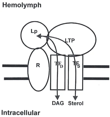
The roles of lipophorin receptors and lipid transfer particles (LTPs) appear to be critical in understanding the lipid transfer process in midgut and its regulation. Both aspects will be discussed in detail in other sections of this review.
3.1. Future directions
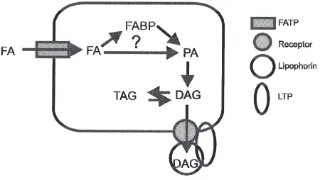
There are still many unanswered questions regarding lipid digestion and absorption. Fig. 1 shows a possible
Fig. 1. A scheme showing the possible steps involved in fatty acid (FA) absorption into the midgut enterocyte, its transformation into diacylglycerol (DAG) and export of DAG from the cell into the hemo-lymph. The scheme emphasizes the need for more studies on fatty acid uptake into the enterocyte and the potential role of the fatty acid trans-port protein (FATP), the role of intracellular binding proteins such as the fatty acid binding protein (FABP), the pathway for DAG biosynth-esis, and the mechanism of DAG export from the enterocyte.
scheme that illustrates several areas for future studies. Among these are:
O understanding the mechanism of fatty acid uptake into the midgut cell;
O delineating the role of intracellular lipid-binding pro-teins, such as fatty acid-binding protein and lutein-binding protein;
O elucidating the mechanism by which DAG is exported into hemolymph lipophorin.
4. Lipophorin receptors
Despite the fact that lipophorin receptor activity has been characterized in several organisms, little is known about the function of the receptors in the transfer of lipid between lipophorin and cells. A membrane protein with lipophorin binding activity has been identified in the lar-val fat body of M. sexta (Tsuchida and Wells, 1990). The purification of this receptor activity from solubilized membranes yielded a 120-kDa protein with a Kd for HDLp of 4.1±0.19×1028 M. Binding of HDLp to this
receptor showed a dependence on Ca2+and inhibition by
suramin. Lipophorin binding to the midgut of M. sexta
larvae was characterized in a midgut membrane prep-aration (Gondim and Wells, 2000). Specific binding of lipophorin to the midgut membrane was a saturable pro-cess with Kd=1.5±0.2×1027 M and, in contrast to the
receptor from the fat body, binding did not depend on calcium. The lipid content of the lipophorin did not sig-nificantly affect the affinity of the membrane preparation for lipoprotein. Immunocytochemical studies in A. cyanea revealed that lipophorin binds to the surface of midgut and fat body cells (Bauerfeind and Komnick, 1992a,b). However, it is not yet known whether a mem-brane-associated protein mediates this binding.
delipid-ated lipophorin has not been reported in hemolymph, although it might occur at low levels if reloading is rapid.
Endocytosis of lipophorin by fat body cells has been shown inL. migratoria(Dantuma et al. 1997, 1998), and
A. cyanea (Bauerfeind and Komnick, 1992b). In A.
cyanea, lipophorin was shown to be bound to the larval fat body cell surface as well as being located intracellu-larly. The intracellular localization revealed the presence of lipophorin in the secretory pathway (endoplasmic reticulum, Golgi apparatus andtransGolgi network), as would be expected for a secreted protein. Interestingly, lipophorin was also found in endosomes, which would be consistent with an endocytotic uptake of lipophorin from the hemolymph. In a similar study, the authors were unable to find lipophorin associated with endo-somes in the midgut (Bauerfeind and Komnick, 1992b). Bauerfeind and Komnick postulated that endocytosis of lipophorin in the fat body may be involved in the removal of old or damaged lipophorin. The authors also postulated that a mechanism of HDLp endocytosis into the fat body, lipid delivery, and recycling of the delipid-ated particle into the hemolymph might exist. Other than the existence of endosomal associated lipophorin, no evidence for such a unique and complex mechanism exists in A. cyanea.
In L. migratoria, a single fat body binding site for lipophorin was characterized (Dantuma et al., 1996). This binding activity differs from that ofM. sextain that it does not require divalent cations and seems to have a broader specificity as it can bind human low-density lipoprotein (LDL). In a later study (Dantuma et al., 1997), the lipophorin binding activity in L. migratoria
fat body was found to be involved in the endocytosis of lipophorin. The authors found that lipophorin, even though it is endocytosed, does not appear to accumulate in the cell and an insignificant amount of lipophorin is degraded. On the other hand, the lipid moiety of HDLp is able to accumulate intracellularly. Furthermore, by using ammonium chloride, an endocytosis inhibitor, the authors demonstrated that the inhibition of endocytosis did not significantly affect the rate of exchange of DAG and cholesterol between the fat body and HDLp in vitro. Based on these observations, the authors concluded that the endocytosis of lipophorin is not necessary for the transfer of DAG and cholesterol. The authors also postu-lated that the majority of the endocytosed lipophorin is not lysosomally degraded and may be resecreted after endocytosis. The physiological relevance of the endo-cytosis and subsequent secretion of lipophorin is still unclear, but it is clear that it does not play a significant role in the transfer of the major neutral lipids.
A putative CDNA for the receptor involved in the endocytosis of lipophorin has been cloned from the fat body of L. migratoria (Dantuma et al., 1999). By per-forming polymerase chain reaction (PCR) using
degenerate primers designed against the conserved regions of the LDL receptor family, the authors were able to obtain a product that was used to probe a cDNA library. The cDNA coded for an 883-residue integral membrane protein belonging to the LDL receptor family. This receptor is expressed in several tissues, including oocytes, brain, and midgut, in addition to fat body. When expressed in vertebrate cells, the receptor mediated the endocytosis of L. migratoriaHDLp from the media.
A homologue of the vertebrate macrophage-specific scavenger receptor has been cloned and characterized from Drosophila melanogaster L2 cells (Pearson et al., 1995). Macrophage scavenger receptors have broad ligand specificity and are able to bind and assist in the endocytosis of molecules as part of a clearance mech-anism to remove damaged or foreign molecules (Abrams et al., 1992). Using a technique known as expression cloning, in which an expression library transfected in COS-M6 cells was probed with fluorescent labeled ace-tylated human LDL, the authors isolated a cDNA for this receptor, named dSR-C1. The cDNA codes for a 609-residue membrane protein, but little is known what role, if any, this receptor might play in lipid metabolism in insects. In addition, little is known about the binding characteristics of this receptor for endogenous insect lipoproteins.
4.1. Future directions
More research is needed to determine the mechanism of lipid transfer between lipophorin and tissues, and the function of lipophorin receptors in this transfer. What is clearly needed is more information about lipophorin receptors, including:
O the types of receptors present—docking and/or endo-cytotic;
O sequence and structure of the various receptors. This information can possibly be obtained using an expression cloning technique such as the one used by Pearson et al. (1995). There are several significant benefits of this approach:
O it would retrieve only clones that are functional; O it would retrieve every type of lipophorin binding
protein that can bind labeled lipophorin;
O it does not depend on any assumptions about the nature of the receptors, as is required when cloning using PCR and sequences of receptors charac-terized from vertebrates.
E.L. Arrese et al. / Insect Biochemistry and Molecular Biology 31 (2001) 7–17
receptors function (docking or endocytosis) and if other factors, such as lipid specific active transporters or LTPs, are necessary to facilitate the transfer of lipid.
5. Lipid transfer particle
LTP was first purified from the hemolymph of larval
M. sexta(Ryan et al., 1986a,b). LTP is a very high-den-sity lipoprotein with a molecular mass of more than 670 kDa. It contains 14% lipids and three glycosylated apoli-poproteins, apoLTP-I, -II and -III of 350, 85, and 60 kDa respectively. LTP has also been identified and purified from the hemolymph of other species, including L.
migratoria (Hirayama and Chino, 1990), P. americana
(Takeuchi and Chino, 1993), Musca domestica(Capurro and De Bianchi, 1990) and B. mori (Tsuchida et al., 1997). In M. sexta, LTP is synthesized in the fat body and secreted into the hemolymph (Van Heusden et al., 1996). LTP is also present in oocytes where it plays a role in the conversion of adult HDLp to egg VHDLp (Liu and Ryan, 1991). Whether LTP is synthesized in oocytes or is transported into oocytes has not been determined.
Electron microscopy showed that LTP consists of a spherical head and an elongated, hinged, cylindrical tail (Ryan et al., 1990a). The location of the three apolipop-roteins within the particle structure, their stoichiometry and their specific functions are not yet known, although existing data demonstrate that the three apoproteins are required for LTP activity (Van Heusden et al., 1996).
The physiological function of LTP is not completely elucidated but in vitro studies have shown that LTP cata-lyzes the exchange and/or transfer of DAG: (1) between lipophorins and human lipoproteins (Blacklock and Ryan, 1994); (2) from fat body to HDLp resulting in the formation of LDLp (Van Heusden and Law, 1989); (3) from the insect midgut to HDLp (L.E. Canavoso and M.A. Wells, unpublished); and (4) from HDLp or LDLp to vitellogenin, a major female specific lipoprotein in insect hemolymph (Tsuchida et al., 1997). LTP also facilitates the transfer of other lipids from HDLp to LDLp, including phospholipids (Tsuchida et al., 1997), carotenoids (Tsuchida et al., 1998) and hydrocarbons (Takeuchi and Chino, 1993).
It is interesting to focus on the role of LTP in DAG transfer between cells and lipophorin. In M. sexta, in vitro assays using either TAG-labeled adult fat bodies (Van Heusden and Law, 1989) or DAG-labeled larval midgut sacs (L.E. Canavoso and M.A. Wells, unpublished) showed similar features: (a) the release of labeled DAG from the tissues was inhibited by treating fat bodies or midgut sacs with anti-LTP, and (b) in both systems, the DAG transfer activity could be restored by adding LTP to the media. While these results indicate that LTP plays an essential role in promoting DAG
transfer from the cells to lipophorin and supports the model depicted in Fig. 2, the mechanism is unknown. One possibility is that LTP plays a role in the formation of the lipophorin–receptor complex. Alternatively, LTP might act to enhance the function of the tissue-specific lipid transfer factors, which in turn could determine the direction of the lipid transfer. In vitro experiments using anti-receptor antibodies could provide a useful approach to address this point.
In vitro, it has been demonstrated that LTP can pro-mote the net transfer of lipids between lipophorins obtained from different life stages ofM. sexta, e.g. larval HDLp and adult LDLp (Ryan et al., 1987), and of M. domestica(Capurro and De Bianchi, 1990). The ability
of M. sexta LTP to convert adult LDLp particles into
two new lipoproteins species with different sizes, den-sities, and apolipoprotein contents has been demon-strated in vitro (Ryan et al., 1990b) and LTP converts a partially delipidated lipophorin into HDLp and VHDLp (Z.E. Jouni and M.A. Wells, unpublished). However, the ability of LTP to promote net lipid transfer was absent when lipophorins were obtained from the same life stages, e.g. adult L. migratoria (Hirayama and Chino, 1990), P. americana (Takeuchi and Chino, 1993) and
M. sexta(Tsuchida et al., 1997). This later result is con-sistent with the fact that HDLp and LDLp coexist in adult hemolymph, which would not be possible if LTP transferred lipids between the two lipophorins in vivo.
Thus, the physiological significance of net lipid transfer between lipophorin species remains unclear.
5.1. Future directions
There are many unanswered questions about the physiological role of LTP, including the following.
O The mechanism by which LTP mediates the transfer of lipids is clearly an important area for future study. O LTP biosynthesis and assembly in the fat body have not been studied. Nor has the role of the various apoli-poproteins in LTP function been studied in detail. Being a very large protein, LTP imposes difficulties in exploring and answering many questions, because cloning and sequencing the cDNA may be a formi-dable task if apoLTP-I, -II, and -III are made from a single gene, as in the case of apolipophorin-I and -II. O The role of LTP in specific lipid transfer is also unknown. A possible lipid transfer complex is depicted in Fig. 2, in which LTP is proposed to mediate the transfer of any lipid with specificity being determined by the properties of putative lipid transfer factors in the cell membrane. Although no such fac-tors have yet been characterized, their existence is supported by genetic evidence from the white cocoon mutants in B. mori, in which the normal carotenoid pigment of the cocoon, lutein, is absent (Tazima, 1978). There are two important points: (1) lutein is a lipid and (2) in one mutant, the I mutant, lutein trans-fer from the midgut to lipophorin is inhibited—lutein accumulates within midgut epithelia cells. Importantly, in the I mutant the transfer of other lip-ids, DAG and sterol, from the midgut to lipophorin is normal. Thus, only the transfer of lutein is affected. One possible explanation for these observations is that the I mutation is in the lutein-transfer factor. The presence, or absence, of such transfer factors is then proposed to account for the specificity of cellular lipid uptake. Much work will be required to prove such a mechanism.
6. Lipid mobilization
Chino and Gilbert (1964) first demonstrated that the fat body mobilizes lipids as DAG, and this has been con-firmed in many insect species (Beenakkers et al., 1985). Adipokinetic hormones (AKHs) are a large family of 8– 10-amino-acid peptides secreted by the neurosecretory cells of the corpora cardiaca that control lipid mobiliz-ation from the fat body (Beenakkers et al., 1985; Gold-sworthy and Mordue, 1989; GoldGold-sworthy et al., 1997). Thus, the injection of AKH into adult insects such as
Acheta domesticus, L. migratoria and M. sexta
stimu-lates the formation of DAG which accumustimu-lates in the hemolymph associated with lipophorin (Ga¨de and Been-akkers, 1977; Shapiro and Law, 1983; Woodring et al., 1989; Strobel et al., 1990). For example, after hormonal stimulation of adultM. sexta, the content of hemolymph DAG doubles and DAG comprises about 95% of the total hemolymph lipids, whereas FFA constitutes less than 1% (Arrese and Wells, 1997).
In L. migratoria (Tietz et al., 1975) and M. sexta
(Arrese and Wells, 1997) the fat body synthesizes prim-arily sn-1,2-DAG and only trace amounts of the sn-1,3-and sn-2,3-isomers have been detected. DAG is secreted into the hemolymph and loaded into pre-existing HDLp. As a result, lipophorin becomes considerably lower in density and an LDLp is formed. This particle, in addition to apoLp-I and apoLp-II, also contains apoLp-III, which binds to surface patches enriched in DAG and stabilizes the particle (Soulages and Wells, 1994). LDLp transports DAG to the sites of utilization, the flight muscle and ovaries, where it is hydrolyzed to FFA by a lipophorin-lipase (Van Antwerpen and Law, 1992; Van Heusden and Law, 1989; Van Antwerpen et al., 1998).
Most of the current information about lipid mobiliz-ation in insects comes from studies carried out usingL.
migratoria and M. sexta. Since Tietz et al. (1975)
described the presence of a microsomal monoacylgly-cerol acyltransferase (MGAT) activity in the fat body of L. migratoria, it had been widely speculated that the synthesis of DAG involves the hydrolysis of TAG into 2-monoacylglycerol (2-MAG) followed by its reacyl-ation to DAG catalyzed by MGAT. However, more recent studies inM. sextasupport the conclusion that the stored TAG is converted directly to DAG (Arrese and Wells, 1997). In other words, the direct stereospecific hydrolysis of TAG into sn-1,2-DAG is the pathway for the synthesis of DAG that is released into the hemo-lymph as result of AKH action. This conclusion is based on the following results.
O Although microsomal MGAT activity was found in
M. sexta fat body, AKH did not stimulate its activity (Arrese et al., 1996a). Given the rapid response in DAG production as a result of AKH injection, it seems highly unlikely that AKH activates MGAT gene expression.
O The content of MAG in the microsomes or in the whole fat body remains unchanged after activation of DAG synthesis by AKH. Thus, AKH does not modify the synthesis of sn-1,2-DAG through the activation of MGAT.
E.L. Arrese et al. / Insect Biochemistry and Molecular Biology 31 (2001) 7–17
and phosphatidic acid pool sizes remained unchanged (Arrese and Wells, 1997).
O It is important to note that the metabolic fate of the fatty acid removed from the sn-3 position of TAG is unknown.
Altogether, these observations support the conclusion that AKH-stimulated synthesis of sn-1,2-DAG in the fat body involves the stereospecific hydrolysis of the TAG stores into DAG, a step that must be catalyzed by a TAG-lipase. At present, the only fat body TAG-lipase that has been purified is that from the fat body of adult
M. sexta(Arrese and Wells, 1994). The enzyme is a sin-gle polypeptide of 76 kDa that has several properties in common with the vertebrate hormone-sensitive lipase (HSL), which catalyzes the rate-limiting step in mobiliz-ation of adipose tissue fatty acids. Like HSL, the M. sextafat body TAG-lipase is a phosphorylatable enzyme. In adult M. sexta, the activation of the fat body TAG-lipase precedes the appearance of DAG in the hemo-lymph, suggesting that AKH stimulates DAG secretion by activating the fat body TAG-lipase (Arrese et al., 1996b).
The sequence of events leading to the stimulation of lipolysis induced by AKH is still controversial. The present data support a model in which the initial event involves the binding of AKH to its receptor (Ziegler et al., 1995), which induces a rapid and sustained increase in Ca2+ influx and an activation of adenylate cyclase.
These processes give rise to two intracellular messen-gers, Ca2+and cAMP (Ga¨de and Holwerda, 1976;
Spen-cer and Candy, 1976; Lum and Chino, 1990; Wang et al., 1990; Arrese et al., 1999). In addition to the evidence in favor of Ca2+and cAMP as second messengers in the
action of AKH-mobilizing lipids, recent reports showed that AKH increases levels of inositol (1,4,5)-triphosph-ate (InP3) in the fat body of two locusts, Schistocerca
gregaria (Stagg and Candy, 1996) and L. migratoria
(Van Marrewijk et al., 1996; Vroemen et al., 1998). Interestingly, the report onS. gregariashowed a differ-ence in sensitivity between cAMP and the InP3
responses. The latter exhibited an EC50 value for AKH
that was 100-fold higher that the corresponding value for cAMP. In any case, these findings raise the question of whether the increase in Ca2+ influx mentioned above
is due to a direct stimulation by AKH on calcium entry or a secondary consequence resulting from the depletion of internal stores (e.g. InP3-sensitive stores) initiated
by AKH.
How is the presence of these second messengers related? Two possibilities can be envisaged: (1) the accumulation of one of the messengers induces the appearance of the other; or (2) the AKH receptor acti-vates multiple signal transduction pathways leading to the accumulation of both second messengers. In ver-tebrate tissues, different patterns of cross-talk between
the Ca2+ and cAMP signal transduction systems have
been established. For example, intracellular free calcium can affect cAMP levels by modulation of adenylate cyclase activity or phosphodiesterase activities (Beavo and Reifsnyder, 1990; Choi et al., 1993). On the other hand, A-kinase or cAMP can affect intracellular Ca2+
levels by regulating Ca2+ channel activity (Hell et al.,
1994). Thus, unlike the vertebrate system in which lip-olysis is regulated solely by the cAMP system, in the insect fat body both cAMP and calcium are second messengers in the hormone-stimulated production of sn-1,2-DAG, suggesting that a novel signal transduction mechanism for activating lipolysis could be present in insects.
Recently, it has been shown that AKH rapidly induces an increase in the A-kinase activity of fat body cells from the adultM. sextaand that this increase in A-kinase activity precedes activation of the fat body TAG-lipase and the mobilization of DAG into the hemolymph (Arrese et al., 1999). The substrates for the M. sextafat body A-kinase are under investigation, but we know that A-kinase purified from theM. sextafat body phosphoryl-ates the fat body TAG-lipase as well as other proteins associated with the fat droplets (E.L. Arrese and M.A. Wells, unpublished). A possible scenario is that acti-vation of A-kinase by cAMP results in the phosphoryl-ation of the TAG-lipase, which, similar to the vertebrate system (Egan et al., 1992), leads to its activation and/or translocation to the substrate stores in the fat droplets.
levels of free DAG from accumulating in the cell (Weisiger, 1996). In contrast to the transport of DAG in the hemolymph by lipophorin, which has been the focus of extensive studies, nothing is known about the intra-cellular transport of DAG. This is also an unsolved ques-tion for mammalian cells in which DAG, acting as second messengers, undergoes intracellular translo-cation.
Starvation is another condition that leads to increased hemolymph lipid levels in both the adult L. migratoria
and larval M. sexta(Jutsum et al., 1975; Ziegler, 1991). Because starved cardiocectomized insects mobilize lip-ids as well as the controls, it has been concluded that the mechanism by which starvation induces lipid mobil-ization in these systems cannot be attributed to AKH but to some other factor (Cheesman and Goldsworthy, 1979; Ziegler, 1991). The brain could secrete this factor and one possible candidate is octopamine, the monohydroxy-phenolic analog of noradrenaline, which has been shown to stimulate lipid mobilization inL. migratoria(Orchard, 1987) and A. domesticus (Fields and Woodring, 1991). Octopamine also shows a moderate response, compared to AKH, in stimulating lipid mobilizing in adultM. sexta
(E.L. Arrese and M.A. Wells, unpublished results). Dav-enport and Evans (1984) showed that the level of octopa-mine in the hemolymph of L. migratoria that has been deprived of food increased and that the increased levels persisted for as long as the insects were unable to feed. Therefore, it is possible that octopamine could be involved in the regulation of lipid mobilization induced by starvation, as has been suggested by Downer (1985).
6.1. Future directions
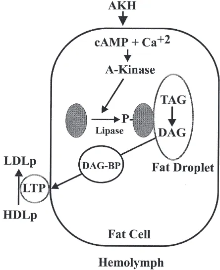
Fig. 3 shows a proposed model for mobilization of DAG from the fat body in response to AKH. There are several points in this model that need to be clarified by future experiments.
O How do cAMP and Ca2+ interact to transduce the AKH effect? Not shown in the model but also requir-ing clarification is the possible role of IP3.
O How does phosphorylation of the TAG-lipase lead to activation? How does the lipase express its stereos-pecificity?
O How is DAG transported from the fat droplet to the plasma membrane? Is a DAG-binding protein involved?
O How does DAG leave the fat body cell and enter HDLp to produce LDLp?
7. Summary
In this brief review we have presented a few of what we consider to be interesting areas related to lipid
stor-Fig. 3. A model for adipokinetic hormone (AKH)-stimulated diacyl-glycerol (DAG) production and secretion in the fat body. AKH increases the intracellular concentrations of cAMP and Ca2+. cAMP,
with or without Ca2+, then activates the cAMP-dependent protein
kin-ase (A-Kinkin-ase), which in turn phosphorylates the lipkin-ase, causing it to translocate to the surface of the fat droplet. On the surface of the fat droplet the lipase produces DAG, which is then transported to the plasma membrane via a DAG-binding protein (DAG-BP). Once in the membrane the DAG leaves the cell and is added to high-density lipo-phorin (HDLp) to produce low-density lipolipo-phorin (LDLp).
age and mobilization in insects. It is our hope that others will be stimulated to use biochemical, molecular biologi-cal and physiologibiologi-cal approaches to prove, or disprove, the models we have presented. Regardless of the out-come of those experiments, if some of you are encour-aged to enter this fascinating field, then our purpose will have been well served.
Acknowledgements
The work in the laboratory of Dr M.A. Wells was supported by NIH grants GM50008 and GM51296.
References
Abrams, J.M., Lux, A., Steller, H., Krieger, M., 1992. Macrophages in Drosophilaembryos and L2 cells exhibit scavenger receptor-mediated endocytosis. Proc. Natl. Acad. Sci. USA 89, 10375– 10379.
E.L. Arrese et al. / Insect Biochemistry and Molecular Biology 31 (2001) 7–17
Arrese, E.L., Wells, M.A., 1997. Adipokinetic hormone-induced lip-olysis in the fat body of an insectManduca sexta: synthesis of sn-1,2-diacylglycerols. J. Lipid Res. 38, 68–76.
Arrese, E.L., Rojas-Rivas, B.I., Wells, M.A., 1996a. Synthesis of sn-1,2-diacylglycerol by monoacylglycerol-acyltransferase from Man-duca sextafat body. Arch. Insect Biochem. Physiol. 31, 325–335. Arrese, E.L., Rojas-Rivas, B.I., Wells, M.A., 1996b. The use of decapi-tated insects to study lipid mobilization in adult Manduca sexta: effects of adipokinetic hormone and trehalose on fat body lipase activity. Insect Biochem. Mol. Biol. 26, 775–782.
Arrese, E.L., Flowers, M.T., Gazard, J.L., Wells, M.A., 1999. Calcium and cAMP are second messenger in the adipokinetic hormone-induced lipolysis of triacylglycerols inManduca sextafat body. J. Lipid Res. 40, 556–564.
Bauerfeind, R., Komnick, H., 1992a. Lipid-loading and unloading of lipophorin in the midgut epithelium of dragonfly larvae,Aeshna cyanea. J. Insect Physiol. 38, 147–160.
Bauerfeind, R., Komnick, H., 1992b. Immunocytochemical localiz-ation of lipophorin in the fat body of dragonfly larvae, Aeshna cyanea. J. Insect Physiol. 38, 185–198.
Beavo, J.A., Reifsnyder, D.H., 1990. Primary sequence of cyclic nucle-otide phosphodiesterase isozymes and the design of selective inhibitors. Trends Pharmacol. Sci. 11, 150–155.
Beenakkers, A.M.TH., Van der Host, D., Van Marrewijk, W.J.A., 1985. Insect lipids and lipoproteins, and their role in physiological processes. Prog. Lipid Res. 24, 19–67.
Blacklock, B.J., Ryan, R.O., 1994. Hemolymph lipid transport. Insect Biochem. Mol. Biol. 24, 855–873.
Bollade, D., Paris, R., Moulins, M., 1970. Origine et mode d’action de la lipase intestinale chez lez blattles. J. Insect Physiol. 16, 45–53. Capurro, M.DE L., De Bianchi, A.G., 1990. A lipid transfer particle inMusca domesticahaemolymph. Comp. Biochem. Physiol. 97B, 649–653.
Cheesman, P., Goldsworthy, G.J., 1979. The release of adipokinetic hormone during flight and starvation in Locusta. Gen. Comp. Endocrinol. 37, 35–43.
Chino, H., 1985. Lipid transport, biochemistry of hemolymph lipopho-rin. In: Kerkut, G.A., Gilbert, L.I. (Eds.), Comprehensive Insect Physiology, Biochemistry and Pharmacology, vol. 10. Pergamon Press, Oxford, pp. 115–135.
Chino, H., Downer, R.G.H., 1979. The role of diacylglycerol in absorp-tion of dietary glyceride in the American cockroach,Periplaneta americana. Insect Biochem. 9, 379–382.
Chino, H., Gilbert, L.I., 1964. Diglyceride release from insect fat body. Science 143, 359–361.
Chino, H., Kitazawa, K., 1981. Diacylglycerol-carrying lipoprotein of hemolymph of the locust and some insects. J. Lipid Res. 22, 1042–1052.
Choi, E.J., Xia, Z., Villacres, E., Storm, D., 1993. The regulatory diver-sity of the mammalian adenylyl cyclases. Curr. Opin. Cell Biol. 5, 269–273.
Dantuma, N.P., Van Marrewijk, W.J.A., Wynne, H.J., Van der Horst, D.J., 1996. Interaction of an insect lipoprotein with its binding site at the fat body. J. Lipid Res. 37, 1345–1355.
Dantuma, N.P., Pijnenburg, M.A.P., Diederen, J.H.B., Van der Horst, D.J., 1997. Developmental down-regulation of receptor-mediated endocytosis of an insect lipoprotein. J. Lipid Res. 38, 254–265. Dantuma, N.P., Pijnenburg, M.A.P., Diederen, J.H.B., Van der Horst,
D.J., 1998. Electron microscopic visualization of receptor-mediated endocytosis of Dil-labeled lipoproteins by diaminobenzidine photo-conversion. J. Histochem. Cytochem. 46, 1085–1089.
Dantuma, N.P., Potters, M., De Winther, M.P.J., Tensen, C.P., Kooi-man, F.P., Bogerd, J. et al., 1999. An insect homolog of the ver-tebrate very low density lipoprotein receptor mediates endocytosis of lipophorins. J. Lipid Res. 40, 973–978.
Davenport, A.P., Evans, P.D., 1984. Changes in haemolymph
octopa-mine levels associated with food deprivation in the locust, Schisto-cerca gregaria. Physiol. Entomol. 9, 269–274.
Downer, R.G.H., 1985. Lipid metabolism. In: Kerkut, G.A., Gilbert, L.I. (Eds.), Comprehensive Insect Physiology, Biochemistry and Pharmacology, vol. 10. Pergamon Press, Oxford, pp. 77–113. Egan, J.J., Greenberg, A.S., Chang, M., Wek, S., Moos, M.C. Jr.,
Londos, C., 1992. Mechanism of hormone-stimulated lipolysis in adipocytes, translocation of hormone-sensitive lipase to the lipid storage droplet. Proc. Natl. Acad. Sci. USA 89, 8538–8545. Fernando-Warnakulasuriya, G., Tsuchida, K., Wells, M.A., 1988.
Effect of dietary lipid content on lipid transport and storage during larval development ofManduca sexta. Insect Biochem. 18, 211– 214.
Fields, P.E., Woodring, J.P., 1991. Octopamine mobilization of lipids and carbohydrates in the house cricket,Acheta domesticus. J. Insect Physiol. 37, 193–199.
Ga¨de, G., Beenakkers, A.M.Th., 1977. Adipokinetic hormone-induced lipid mobilization and cyclic AMP accumulation in the fat body of Locusta migratoriaduring development. Gen. Comp. Endocrinol. 32, 481–487.
Ga¨de, G., Holwerda, D.A., 1976. Involvement of adenosine 39,59 -cyc-lic monophosphate in lipid mobilization in Locusta migratoria. Insect Biochem. 6, 535–540.
Goldsworthy, G.J., Mordue, W., 1989. Adipokinetic hormones, func-tions and structures. Biol. Bull. 177, 218–224.
Goldsworthy, G.J., Lee, M.J., Luswata, R., Drake, A.F., Hyde, D., 1997. Structures, assays and receptors for locust adipokinetic hor-mones. Comp. Biochem. Physiol. 117B, 483–496.
Gondim, K.C., Wells, M.A., 2000. Characterization of lipophorin bind-ing to the midgut of larvalManduca Sexta. Insect Biochem. Mol. Biol. 30, 405–413.
Hell, J.W., Westenbrok, R.E., Elliot, E.M., Catterall, W.A., 1994. Dif-ferential phosphorylation, localization, and function of distinct alpha 1 subunits of neuronal calcium channels. Two size forms for class B, C and D alpha 1 subunits with different COOH-termini. Ann. N. Y. Acad. Sci. 747, 282–293.
Hirayama, Y., Chino, H., 1990. Lipid transfer particle in locust hemo-lymph, purification and characterization. J. Lipid Res. 31, 793–799. Hoffman, A.G.D., Downer, R.G.H., 1979. End product specificity of triacylglycerol lipases from intestine, fat body, muscle and hemo-lymph of the American cockroach,Periplaneta americanaL. Lip-ids 14, 893–899.
Hui, T.Y., Bernlohr, D.A., 1997. Fatty acid transporters in animal cells. Frontiers in Bioscience 2, 222–231.
Jouni, Z.E., Wells, M.A., 1996. Purification and partial characterization of a lutein-binding protein from the midgut of the silkworm, Bom-byx mori. J. Biol. Chem. 271, 14722–14726.
Jutsum, A.R., Agarwal, H.C., Goldsworthy, G.J., 1975. Starvation and haemolymph lipids in Locusta migratoria migratorioides, R&F. Acrida 4, 47–56.
Komnick, H., Kukulies, J., Bongers, J., Fischer, W., 1984. Absorption of dietary triacylglycerol by lipolysis and lipid resynthesis in the mesenteron of larvalAeshna cyanea, Insecta, Odonata. Protoplasma 123, 57–69.
Law, J.H., Ribeiro, J.M., Wells, M.A., 1992. Biochemical insights derived from insect diversity. Annu. Rev. Biochem. 61, 87–111. Liu, H., Ryan, R.O., 1991. Role of lipid transfer particle in
transform-ation of lipophorin in insect oocytes. Biochim. Biophys. Acta 1085, 112–118.
Lum, P.Y., Chino, H., 1990. Primary role of adipokinetic hormone in the formation of low density lipophorin in locusts. J. Lipid Res. 31, 2039–2044.
Orchard, I., 1987. Adipokinetic hormones—an update. J. Insect Phy-siol. 33, 451–463.
Pearson, A., Lux, A., Krieger, M., 1995. Expression cloning of dSR-CI, a class C macrophage-specific scavenger receptor from Droso-phila melanogaster. Proc. Natl. Acad. Sci. USA 92, 4056–4060. Prasad, S.V., Fernando-Warnakulasuriya, G.P.P., Sumida, M., Law,
J.H., Wells, M.A., 1986. Lipoprotein biosynthesis in the larvae of the tobacco hornworm, Manduca sexta. J. Biol. Chem. 261, 17174–17176.
Reisser-Bollade, D., 1976. Exogenous lipid transport by the hemo-lymph lipoproteins inPeriplaneta americana. Insect Biochem. 6, 241–246.
Ryan, R.O., 1990. Dynamics of insect lipophorin metabolism. J. Lipid Res. 31, 1725–1739.
Ryan, R.O., Prasad, S.V., Henriksen, E.J., Wells, M.A., Law, J.H., 1986a. Lipoprotein interconversions in an insect,Manduca sexta. Evidence for a lipid transfer factor in the hemolymph. J. Biol. Chem. 261, 563–568.
Ryan, R.O., Wells, M.A., Law, J.H., 1986b. Lipid transfer protein from Manduca sexta hemolymph. Biochem. Biophys. Res. Commun. 136, 260–265.
Ryan, R.O., Wells, M.A., Law, J.H., 1987. The dynamics of lipophorin interconversions inManduca sexta. In: Law, J.H. (Ed.), Molecular Entomology. UCLA Symposia on Molecular and Cellular Biology, New Series, vol. 49. Alan R. Liss, New York, pp. 257–266. Ryan, R.O., Howe, A., Scraba, D.G., 1990a. Studies on the
mor-phology and structure of the plasma lipid transfer particle from the tobacco hornworm,Manduca sexta. J. Lipid Res. 31, 871–879. Ryan, R.O., Van Antwerpen, R., Van der Horst, D.J., Beenakkers,
A.M.Th., Law, J., 1990b.Manduca sextalipid transfer particle acts upon a lipoprotein to catalyze lipid and apoprotein dispro-portionation. J. Biol. Chem. 265, 546–552.
Shapiro, J.P., Law, J.H., 1983. Locust adipokinetic hormone stimulates lipid mobilization inManduca sexta. Biochem. Biophys. Res. Com-mun. 115, 924–931.
Shapiro, J.P., Law, J.H., Wells, M.A., 1988. Lipid transport in insects. Annu. Rev. Entomol. 33, 297–318.
Smith, A.F., Tsuchida, K., Hanneman, E., Suzuki, T.C., Wells, M.A., 1992. Isolation, characterization, and cDNA sequence of two fatty acid-binding proteins from the midgut ofManduca sexta. J. Biol. Chem. 267, 380–384.
Soulages, J.L., Wells, M.A., 1994. Lipophorin, the structure of an insect lipoprotein and its role in lipid transport in insects. Adv. Prot. Chem. 45, 371–415.
Spencer, I.M., Candy, D.J., 1976. Hormonal control of diacylglycerol mobilization from fat body of the desert locust,Schistocerca grega-ria. Insect Biochem. 6, 289–296.
Stagg, L.E., Candy, D.J., 1996. The effect of adipokinetic hormones on the levels of inositol phosphates and cyclic AMP in the fat body of the desert locustSchistocerca gregaria. Insect Biochem. Mol. Biol. 26, 537–544.
Stahl, A., Hirsch, D.J., Gimeno, R.E., Punreddy, S., Ge, P., Watson, N. et al., 1999. Identification of the major intestinal fatty acid transport protein. Mol. Cell 4, 299–308.
Strobel, L.M., Kanost, M.R., Ziegler, R., Wells, M.A., 1990. Adipoki-netic hormone causes formation of a low density lipophorin in the house cricket,Acheta Domesticus. Insect Biochem. 20, 859–863. Takeuchi, N., Chino, H., 1993. Lipid transfer particle in the
hemo-lymph of the American cockroach, evidence for its capacity to transfer hydrocarbons between lipophorin particles. J. Lipid Res. 34, 543–551.
Tazima, Y., 1978. The Silkworm: An Important Laboratory Tool. National Institute of Genetics, Mishima, Japan.
Tietz, A., Weintraub, H., Peled, Y., 1975. Utilization of 2-acyl-sn-glycerol locust fat body microsomes. Specificity of the acyl-trans-ferase system. Biochem. Biophys. Acta 618, 80–87.
Tsuchida, K., Wells, M.A., 1988. Digestion, absorption, transport and
storage of fat during the last larval stadium of Manduca sexta. Changes in the role of lipophorin in the delivery of dietary lipid to the fat body. Insect Biochem. 18, 263–268.
Tsuchida, K., Wells, M.A., 1990. Isolation and characterization of a lipoprotein receptor from the fat body of an insect,Manduca sexta. J. Biol. Chem. 265, 5761–5767.
Tsuchida, K., Soulages, J.L., Moribayashi, A., Suzuki, K., Maekawa, H., Wells, M.A., 1997. Purification and properties of a lipid transfer particle fromBombyx mori. Comparison to the lipid transfer par-ticle fromManduca sexta. Biochim. Biophys. Acta. 1337, 57–65. Tsuchida, K., Arai, M., Tanaka, Y., Ishihara, R., Ryan, R.O., Maek-awa, H., 1998. Lipid transfer particle catalyzes transfer of caroteno-ids between lipophorins of Bombyx mori. Insect Biochem. Mol. Biol. 28, 927–934.
Turunen, S., 1975. Absorption and transport of dietary lipids inPieris brassicae. J. Insect Physiol. 21, 1521–1529.
Turunen, S., 1993. Metabolic pathways in the midgut epithelium of Pieris brassicaeduring carbohydrate and lipid assimilation. Insect Biochem. Mol. Biol. 23, 681–689.
Turunen, S., Chipendale, G.M., 1977. Lipid absorption and transport, sectorial analysis of the larval midgut of southwestern corn borer, Diatraea grandiossella. Insect Biochem. 7, 203–208.
Turunen, S., Crailsheim, K., 1996. Lipid and sugar absorption. In: Lehane, M.J., Billingsley, P.F. (Eds.), Biology of Insect Midgut. Chapman and Hall, London, pp. 293–320.
Van Antwerpen, R., Law, J.H., 1992. Lipophorin lipase from the yolk ofManduca sextaeggs, identification and partial characterization. Arch. Insect Biochem. Physiol. 20, 1–12.
Van Antwerpen, R., Salvador, K., Tolman, K., Gentry, C., 1998. Uptake of lipids by developing oocytes of the hawkmothManduca sexta. The possible role of lipoprotein lipase. Insect Biochem. Mol. Biol. 28, 399–408.
Van Heusden, M.C., Law, J.H., 1989. Characterization and identifi-cation of a lipoprotein lipase from Manduca sextaflight muscle. Insect Biochem. Mol. Biol. 23, 785–792.
Van Heusden, M.C., Yepiz-Plascencia, G.M., Walker, A.M., Law, J.H., 1996.Manduca sextalipid transfer particle, synthesis by fat body and occurrence in hemolymph. Arch. Insect Biochem. Phy-siol. 31, 39–51.
Van Marrewijk, W.J.A., Van den Broek, A.T.M., Gielbert, M.L., Van der Host, D.J., 1996. Insect adipokinetic hormone stimulates inosi-tol phosphate metabolism, roles for both Ins, 1,4,5.P3 and Ins,
1,3,4,5.P4 in signal transduction? Mol. Cell. Endocrinol. 122,
141–150.
Vroemen, S.F., Van der Horst, D.J., Van Marewijk, W.J.A., 1998. New insights into adipokinetic hormone signaling. Mol. Cell. Endocri-nol. 141, 7–12.
Wang, Z., Hayakawa, Y., Downer, R.G.H., 1990. Factors influencing cyclic AMP and diacylglycerol levels in fat body of Locusta migratoria. Insect Biochem. 20, 325–330.
Weintrab, H., Tietz, A., 1973. Triglyceride digestion and absorption in the locust,Locusta migratoria. Biochem. Biophys. Acta 306, 31–41.
Weintrab, H., Tietz, A., 1978. Lipid absorption by isolated intestinal preparations. Insect Biochem. 8, 267–274.
Weisiger, R.A., 1996. Cytoplasmic transport of lipids: role of binding proteins. Comp. Biochem. Physiol. 115B, 319–331.
Woodring, J.P., Fescemeyer, H.W., Lockwood, J.A., Hammond, A.M., Ga¨de, G., 1989. Adipokinetic hormone mobilization of lipids and carbohydrates in the house cricket, Acheta domesticus. Comp. Biochem. Physiol. 92A, 65–70.
Yang, L., Kuksis, A., 1991. Apparent convergence at 2-monoacylgly-cerol level of phosphatidic acid and 2-monoacylgly2-monoacylgly-cerol pathways of synthesis of chylomicron triacylglycerols. J. Lipid Res. 32, 1173–1186.
E.L. Arrese et al. / Insect Biochemistry and Molecular Biology 31 (2001) 7–17
Hoffmann, K.H. (Ed.), Environmental Physiology and Biochemis-try of Insects. Springer, Berlin, pp. 95–118.
Ziegler, R., 1991. Changes in lipid and carbohydrate metabolism dur-ing starvation in adultManduca sexta. J. Comp. Physiol. B 161, 125–131.