90
Preliminary Investigation on The Patient and Occupational Doses
in Interventional Procedures in Indonesia
Eri Hiswara, Nunung Nuraeni, Kri Yudi Pati Sandy, Helfi Yuliati and Dyah D. Kusumawati
Center for Technology of Radiation Safety and Metrology, National Nuclear Energy Agency of Indonesia, Jalan Lebak Bulus Raya No. 49, Kotak Pos 7043 JKSKL, Jakarta 12070, E-mail: [email protected]
Abstract.The medical use of ionizing radiation is known to offer great benefit to patients, yet contribute significantly to radiation
exposure of individuals and populations. According to UNSCEAR, the increase of medical examination procedures involving fluoroscopic and interventional radiology resulted in both modalities become the biggest contribution to the radiation doses received by population. This study attempts to investigate the patient and occupational doses in interventional procedures in Indonesia. Patient doses were estimated by using individually packed three TLD-100 chips attached in the x-ray tube, while occupational doses were measured also by using individually packed three chips of TLD-100 placed in over- and under-thyroid shield used by medical staff, over- and under-apron in waist position, inside a special ‘eye-D’ holder, and inside a ring holder. All TLDs were calibrated in the Secondary Standard Dosimetry Laboratory (SSDL) Jakarta. The results show that coiling procedure in interventional radiology produced the highest dose to patient compared with other procedures, while in interventional cardiology, the patient received the highest dose when undergoing catheterization followed by stenting procedure. In the case of occupational exposure, the.medical staff received the highest dose when conducting the coiling procedure and catheter followed by stenting procedure in interventional radiology and interventional cardiology, respectively. The results of measurement were also in good agreement with some other published data.
Keywords: Patient doses, occupational doses, fluoroscopy, interventional radiology, interventional cardiology
Introduction
The medical use of ionizing radiation is known to offer great benefit to patients, yet contribute significantly to radiation exposure of individuals and populations. According to UNSCEAR (2010), more than 80% of radiation dose received by world population from man-made radiation originated from medical application, particularly from fluoroscopic and interventional radiology. The main reason for this contribution was the increase of examination procedures involving both modalities. UNSCEAR estimated that the total radio-logical examination during 1991-1996 was 1,900 million, or around 330 examinations for every 1.000 population around the world, while during 1985-1990, the total number was 1,600 million or around 300 examinations per 1.000 population.
As for interventional radiology, this procedure provides many unwanted radiation doses to both medical staffs and patients. Many specialists who carry out catheterization are not aware that this procedure delivers radiation doses to patients with a higher level than is commonly received during chest X-rays. Mettler, et.al (2008) reported that coronary percutaneous transluminal angioplasty, stent placement or radiofrequency ablation examination would deliver effective dose to patient as much as 15 mSv, compared to only 0.02 mSv for PA chest.
The higher and noticeable radiation doses received by patient and medical staffs have also been confirmed by several studies conducted in different countries, e.g. by AlSuwaidi et.al (2015) in UAE, Cui et.al (2013) in China, Molyvda-Athanasopoulou et.al (2011) in Greece, Szumska et.al (2016) in Poland, and Ubeda et.al (2013)
in Chile. Some suggestions and recommendations on how to reduce both patient and medical occupational doses have been expressed by Leyton et.al (2014) and Vaz (2014).
The results of measurement of patient doses are eventually used for establishing diagnostic reference levels (DRLS) for interventional procedures. Some authors were already attempts to do so. Korir et.al (2014) has made an attempt to establish DRLs in Kenya, while local DRLs for paediatric interventional cardiology have been calculated by McFadden et.al (2013).
Moreover, the dose-response relationship for cataract, i.e. opacity of the lens of the eye, has also been studied extensively in recent years. These studies include those from Antic et.al (2013), Carinou et.al (2011), Geber et.al (2011), McVey et.al (2013) and Ubeda et.al (2013). The results, however, seem vary one to the other. While Geber et.al (2011) stated that the best position for eye lens dosimeter to be at the side of the head, for example, McVey et.al (2013) concluded that the forehead of the wearer provides the most robust position to site a dosimeter to estimate eye doses.
Materials and Methods
The study was carried out in Medistra Hospital, National Center for Brain Hospital, and HasanSadikin Hospital in the cities of Jakarta and Bandung that provide fluoroscopy, interventional radiology and inter-ventional cardiology services. A total of 32 patients– consisting of 19 fluoroscopic and interventional radio-logy patients and 13 interventional cardioradio-logy patients, and 60 medical staffs–consisting of 31 fluoroscopic and interventional radiology workers and 29 interventional cardiology workers, participated in this study.
Patient doses were estimated by using individually packed three TLD-100 chips manufactured by Thermo Scientific Harshaw. As the position of patient against the X-ray beam was moved intermittently, the TLDs then were attached in the X-rays tube. Patient doses were calculated by considering the distance between the focus of X-rays and the surface of patient’s couch.
Measurements of occupational doses were made also using individually packed three chips of TLD-100 from Thermo Scientific Harshaw. The chips were placed in over-and under-thyroid shield used by medical staff, over- and under-apron in waist position, inside a special ‘eye-D’ holder, and inside a ring holder. Figure 1 shows the arrangement of placing TLD in the body of medical staff.
The readout of TLD chip under each position has the following function:
a. those placed under-thyroid shield was used to estimate thyroid dose;
b. those placed under-apron in waist was used to estimate gonad/ovary dose;
c. those placed in temple inside a special ‘eye-D’ holder was used to estimate lens eye dose, Hp(3);
Figure 1. The ‘eye-D’ holder used to measure eye lens dose (left) and the ring holder to measure extremity dose (right).
d. those placed in finger inside a ring holder was used to estimate extremity dose, Hp(0.07); and
e. those placed over-thyroid shield and under-apron waist was used to estimate effective dose, E, which was calculated by using the following equation suggested by Niklason et.al. (1994):
WhereHosis the over-thyroid shield dose andHuis the under-apron waist dose.
All TLDs were calibrated in the Secondary Standard Dosimetry Laboratory (SSDL) Jakarta. The standard deviation of the TLD batch was of the order of 5%, with the overall uncertainty was 20% at the 95% confidence level.
Results and Discussion
Table 1 and Table 2 show the patient doses received during fluoroscopy and interventional radio-logy, and interventional cardiology procedures, respect-tively. The values of DAP (dose-area product) shown were those that are provided by the x-ray system.
As can be seen in Table 1, the patient received a quite higher dose when undergoing coiling procedure, with a mean dose of 235.15 mGy. In interventional radiography, coiling is a rather complex procedure in which a catheter is passed through the groin up into the artery containing the aneurysm. The goal of the treatment is to safely seal off the aneurysm and stop further blood from entering into the aneurysm and increasing the risk of rupture or possibly rebleeding. Complexity of the procedure reflected in the duration of fluoro time of 17-18 min each, which is longer than that of other treatment of mostly less than 3 min.
DSA cerebral was the most practiced procedure observed during the course of this study. With fluoro time ranging from 7-17 min, this particular procedure give radiation dose to patient around 97.48-286.53 mGy. This result was relatively higher than that reported by Manninen et.al (2012) of 2.71 mSv. However, the latter was obtained from irradiation of an anthropomorphic phantom, which can be different in terms of some parameters (e.g. tube voltage, mAs, fluoro time and projection) as used in this study.
In interventional cardiology, as can be seen in Table 2, catheterization followed by stenting procedure produ-ced the highest dose to patient of 3488 mGy with a long fluoro time of achieving 45 min. The catheter procedure itself, however, can be carried out in short time and gave smaller dose to patient as also shown in the Table 2. This brings the notion that it is stenting procedure that produce high doses to patient. As can be seen in the table, the dose of 1125.50 mGy received by patient undergoing stenting procedure can be regarded as a prove that this procedure delivers a high dose to patient.
Table 1. Patient doses received during fluoroscopy and interventional radiology.
Procedure No. of patient DAP
(mGy cm2)
Range of fluoro time
(min.) Mean dose (mGy)
Fistolography 2 - 1.30-7.26 101.58 (30.94-172.22)
Urethrocystography 2 - 2.22-2.26 47.21 (42.83-51.59)
Cystography 1 - 1.65 5.88
Upper gastrointestinal 1 - 1.8 5.07
Cholangography 1 - 1.36 23.78
OMD Oesephagus 1 - 3.98 62.65
Prostal colostomy 1 - 2.00 35.42
Colostomy proximal 1 - 8.30 114.47
DSA cerebral 7 - 7.10-17.49 97.48-286.53
Coiling 2 50723 17.56-18.14 235.15 (189.22-281.09)
Table 2. Patient doses received during interventional cardiology procedures.
Procedure No. of patient DAP
(mGy m2)
Range of fluoro time
(min.) Mean dose (mGy)
Stenting 3 52384 9.41-12.15 1125.50 (439.42 – 1871.58)
LAA/heart appendix 1 1645 5.55 81.81
Catheter+stenting 2 53588 2.14-45.41 3488.86
Catheter+stenting 3 positions
1 56345 49.19
-Angiofibroma embolization
1 25646 14.35 1381.01
Coronary angiography 2 42071-53625 3.55-4.30 168.56 (153.29-183.83)
Catheterization 2 15725-20416 4.03-5.04 77.76 (69.82-85.70)
TAE embolization 1 11.58 1502.21
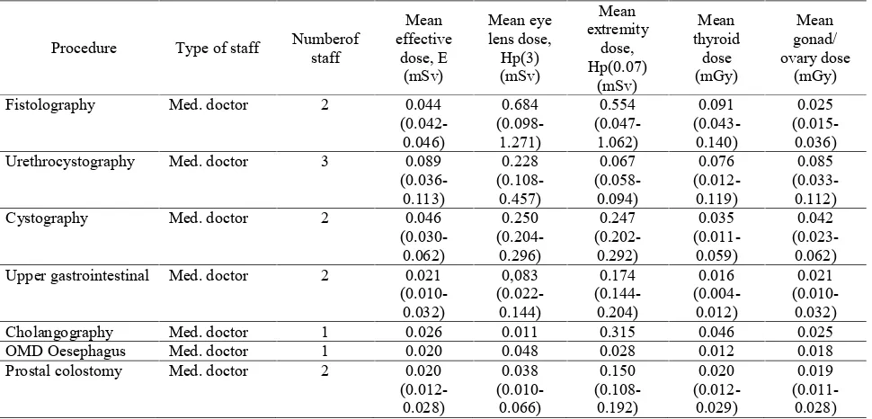
Table 3. Occupational doses during fluoroscopy and interventional radiology procedures.
Procedure Type of staff Numberof staff
Fistolography Med. doctor 2 0.044 (0.042-Urethrocystography Med. doctor 3 0.089
(0.036-Cystography Med. doctor 2 0.046
(0.030-Upper gastrointestinal Med. doctor 2 0.021
(0.010-Cholangography Med. doctor 1 0.026 0.011 0.315 0.046 0.025 OMD Oesephagus Med. doctor 1 0.020 0.048 0.028 0.012 0.018 Prostal colostomy Med. doctor 2 0.020
(0.012-Colostomy proximal Med. doctor 3 0.029 DSA cerebral Med. doctor 2 0.096
(0.033-Radiographer 1 0.056 - 1.369 0.051 0.045
Coiling Med. doctor 4 0.065
(0.016-Radiographer 1 0.014 - 0.106 0.019 0.014
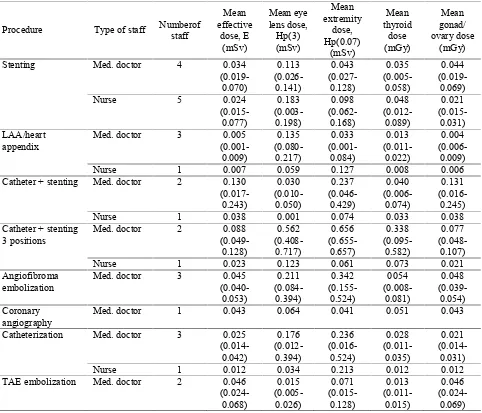
Table 4. Occupational doses during interventional cardiology procedures.
Procedure Type of staff Numberof
staff
Stenting Med. doctor 4 0.034
(0.019-Med. doctor 3 0.005
(0.001-Nurse 1 0.007 0.059 0.127 0.008 0.006
Catheter + stenting Med. doctor 2 0.130
(0.017-Nurse 1 0.038 0.001 0.074 0.033 0.038
Catheter + stenting 3 positions
Med. doctor 2 0.088
(0.049-Nurse 1 0.023 0.123 0.061 0.073 0.021
Angiofibroma embolization
Med. doctor 3 0.045
(0.040-Med. doctor 1 0.043 0.064 0.041 0.051 0.043
Catheterization Med. doctor 3 0.025
(0.014-Nurse 1 0.012 0.034 0.213 0.012 0.012
TAE embolization Med. doctor 2 0.046
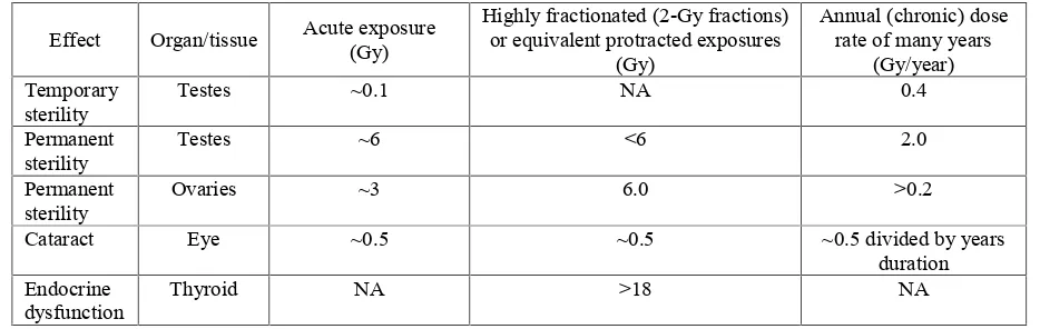
(0.024-94 Table 5. Estimates of the threshold doses in tissues and organs in adults exposed to acute, fractionated or protracted,
and chronic irradiation (ICRP, 2012).
Effect Organ/tissue Acute exposure (Gy)
Highly fractionated (2-Gy fractions) or equivalent protracted exposures
(Gy)
Annual (chronic) dose rate of many years
(Gy/year) Temporary
sterility
Testes ~0.1 NA 0.4
Permanent sterility
Testes ~6 <6 2.0
Permanent sterility
Ovaries ~3 6.0 >0.2
Cataract Eye ~0.5 ~0.5 ~0.5 divided by years
duration Endocrine
dysfunction
Thyroid NA >18 NA
The effective dose, eye lens dose, extremity dose, thyroid dose and gonad/ovary dose received by medical staff in fluoroscopy and interventional radiology in Indonesia was in general highest when performing coiling and DSA cerebral procedures. This is no wonder since both are quite complex procedures that require relatively longer fluoro time than that required by other procedures.
In the case of interventional cardiology, it was detected that the procedure of catheterization followed by stenting in three positions gave the highest dose to medical staff for every type of dose measured (i.e. effective dose, eye lens dose, extremity dose, thyroid and gonad/ovary dose) in this study. From Table 2 it can be seen that this catheterization and stenting in three positions required the longest fluoro time compared with that required by any other procedures.
For coronary angiography, Table 4 shows that the highest eye lens dose was received by doctor with a value of 0.064 mSv, or 64 µSv. This result was in a fair good agreement with the result reported by Szumska et.al (2016) of 73 µSv. As mentioned by Szumska et.al (2016), consi-derable variation exists among studies on eye lens dose due to some factors as type and comple-xity of the procedure undertaken, the skill and experience of the operators, the shielding equipment used, the angiographic equipment and the exposure settings.
Savitri and Susanto (2014) previously reported a study on occupational exposure in interventional radiology facilities in 7 hospitals in Indonesia. The mean thyroid shield, under-apron waist, and over-apron waist doses were found to be 0.06 mSv, 0.024 mSv and 0.103 mSv, respectively. In the case of over-thyroid shield dose, by comparing with the results of this study of mean thyroid dose as shown in Table 4, it can be seen that most of the results of this study are lower than that of Savitri and Susanto (2012). Both
results can be regarded as the same as this study performed the measurement in under-thyroid shield.
The mean under-apron waist dose obtained by Savitri and Susanto (2012 also in the same range as the mean gonad/ovary dose measured in this study.
The dose limit applied in Indonesia for occupational dose to limit the probability of the occurrence of stochastic effect is effective dose of 20 mSv per year. As can be seen in Table 3 and Table 4, each procedure produces a certain value of effective dose. Based on the data presented we cannot, however, make a conclusion that the dose received by a certain medical staff has exceeded the dose limit, nor we can restrict the medical staff to conducting one particular procedure in a year with a fear that the dose he/she received will exceed the dose limit. These data of radiation dose received by each medical staff presented in the tables can only be used as a guidance on how many time each of them can perform a certain procedure. He/she should be able to consider by him/herself which procedure that can be performed in a year and how many times in maximum he/she can be involved in performing that procedure.
As tissue, eye lens, thyroid and gonad/ovary can suffer a damage if they receive radiation doses that exceeding their dose limits. Table 5 shows estimation of some threshold doses in tissues and organs in adults exposed to acute, fractionated or protracted, and chronic irradiation (ICRP, 2012).Testes are tissues contained in gonad, while ovaries are those contained in ovary.
95 thoroughly with a view to find the way to reduce the
radiation dose received by this particular medical staff.
The data presented in this paper are preliminary, as the study will be continued until 2019. However, the big picture is not expected to change much. The medical procedure in hospitals in Indonesia usually are conducted in the same manner, nurses and radiographers mostly underwent the same education and training, and most of the x-ray machine used are manufactured by only a few companies.
Conclusion
The results show that coiling procedure in interventional radiology produced the highest dose to patient compared with other procedures. In interventional cardiology, the patient received the highest dose when undergoing catheterization followed by stenting procedure. The occupational exposure also follow this pattern, i.e. medical staff received the highest dose when conducting the coiling procedure and catheter followed by stenting procedure in interventional radiology and interventional cardiology, respectively. The results of measurement were also in good agreement with some other published data.
Acknowledgements
The authors wish to thank to management and staff of Medistra Hospital, National Center for Brain Hospital, and Hasan Sadikin Hospital for granting permission to carry out the study in their respective hospital. This study was funded by the research grant of PTKMR BATAN.
References
Al Suwaidi, J.S., Almazrouei, N.K., Pottybindu, S. et.al. (2015). Patient dose monitoring in Dubai in radiography and interventional procedures.Proceedings of the Second International Symposium on the System of Radiological Protection.Ann. ICRP 44(1S), 249-258.
Antic, V, Ciraj-Bjelac, O, Rehani, M. et.al. (2013). Eye lens dosimetry in interventional cardiology: results of staff dose measurements and link to patient dose levels.Radiat. Prot. Dosim. 154(3),276-284.
Carinou, E, Brodecki, M, Domienik, J. et.al (2011). Recommendations to reduce extremity and eye lens doses in interventional radiology and cardiology. Radiat. Meas. 46,1324-1329.
Cui, Y, Zhang, H, Zheng, J, et.al (2013). An investigation of patient doses during coronary interventional procedures in China.Radiat.Prot.Dosim. 156(3),296-302.
Geber, T, Gunnarsson, M, Mattsson, S (2011). Eye lens dosimetry for interventional procedures-Relation between the absorbed dose to the lens and dose at measurement positions.Radiat. Meas. 46,1248-1251.
ICRP (2012).ICRP Statement on Tissue Reactions / Early and Late Effects of Radiation in Normal Tissues and Organs–Threshold Doses for Tissue Reactions in a Radiation Protection Context.ICRP Publication 118. Ann. ICRP 41(1/2).
Korir, G.K, Wambani, J.S, Yuko-Jowi, C.A. et.al (2014). Establishing diagnostic reference levels for interventional procedures in Kenya.Radiography, 20, 148-152.
Leyton, F., Canevaro, L., Dourado, A. et.al. (2014). Radiation risks and the importance of radiological protection in interventional cardiology: A systematic review.Rev.Bras. Cardiol. Invasiva, 22(1), 87-98. Manninen, A.-L, Isokangas, J.-M, Karttunen, A, et.al
(2012).A comparison of radiation exposure between diagnostic CTA and DSA examinations of cerebral and cervicocerebral vessels.Am. J.Neuroradiol. 33,2038-2042 McFadden, S, Hughes, C, D’Helft, C.I. et.al (2013). The
establishment of local diagnostic reference levels for paediatric interventional cardiology.Radiography, 19, 295-301.
McVey, S, Sandison, A and Sutton, D.G (2013).An assessment of lead eyewear in interventional radiology.J. Radiol. Prot. 33,647-659.
MettlerJr, F.A, Huda, W, Yoshizumi, T.T, et.al (2008). Effective dose in radiology and diagnostic nuclear medicine: A catalog.Radiology, 248(1),254-263.
Miller, D.L., Balter, S., Dixon, R.G. et.al.(2012). Quality improvement guidelines for recording patient radiation dose in the medical record for fluoroscopically guided procedures.J.Vasc.Interv.Radiol. 23, 11-18.
Molyvda-Athanasopoulou, E., Karlatira, M., Gotzamani-Psarrakou, A. et.al.(2011). Radiation exposure to patients and radiologists during interventional procedures. Radiat. Prot.Dosim. 147(1-2), 86-89.
Niklason, L.T., Marx, M. Victoria & Chan, H-P. (1994). The estimation of occupational effective dose in diagnostic radiology with two dosimeters. Health Phys. 67(6), 611-615.
Savitri, L. and Suwanto, W. (2014). Controlling occupational exposure in interventional radiology facilities. Proc. Seminar on Nuclear Safety.BAPETEN, Jakarta, 34-38 (in Indonesian).
Szumska, A., Kopeć, R & Budzanowski, M. (2016). Occupational doses of medical staff and their relation to patient exposure incurred in coronary angiography and intervention.Radiat.Meas.84, 34-40.
Ubeda, C, Vano, E, Gonzalez, L. et.al. (2013). Evaluation of patient doses and lens radiation doses to interventional cardiologists in a nationwide survey in Chile.Radiat. Prot.Dosim. 157(1),36-43.
96
Effects of Atomic Radiation. UNSCEAR 2008 Report to the General Assembly. Volume I. New York: United Nations.
Vaz, P (2014). Radiation protection and dosimetry issues in the medical applications of ionizing radiation. Radiat. Phys. Chem. 104, 23-30.
Discussion
Q : Dwi Ramadhani
Based on your opinion, it is possible that the medical staff in interventional radiology and cardiology receive the stochastic effect because of occupational radiation exposure.
A : Eri Hiswara
Talking about stochastic effect is actually talking about probability; and from this point of view, it is possible that they may receive the effect. The issue is that how much the nominal risk (in %) for the effect particularly for medical staff.
Q : Dwi Ramadhani
It is possible to assess the biological effects in medical staff, to ensure that there was no a significant negative effect induced by occupational radiation exposure?
A : Eri Hiswara
The biological effect can be assesses through cytogenetic study for these staffs.
Q : Setiawan Soetopo
According to the research, what is your suggestion for doctor who are doing interventional radiology or cardio-logy and suggestion for stakeholders in the concept of radiation protection.
A : Eri Hiswara