Molecular and cellular mechanism
in peritoneal adhesion formation
Tri Hanggono Achmad
Department of Biochemistry and Health Research Unit
School of Medicine Padjadjaran University / Hasan Sadikin General Hospital
Introduction
Intra-abdominal adhesions, as a result of damage to the peritoneum, continue to be a central and current problem in abdominal surgery. By far the majority of adhesion remains symptom-free or even promotes intra-abdominal healing processes. However, 12% of adhesions cause recurring, chronic abdominal complaints and over 3% lead to serious symptoms requiring repeat laparatomies. The most severe complication, adhesion-related ileus, accounts for a considerable proportion of emergency operations (1).
Different to the process-like wound healing of external body surfaces, the visceral and parietal peritoneum healing process follows its own rules. Large serous defects are not covered in a stepwise manner starting at the wound edge, but quickly and synchronously over the entire surface by mesothelial cells. These cells come from the underlying connective tissue. The peritoneum is thus capable of reserosing even larger defects in a short period of time. The underlying tissue, however, lags behind this surface restitution, and scars remain after healing (2,3).
Progress and development in bio-molecular sciences enabled us to explore and explain the genesis and process of diseases, like wound healing, in more detailed, to cellular as well as to molecular level. Discoveries of molecules involve in the pathophysiology of some diseases lead researchers to focus on the interaction between cells and in the cells mediated by these signal-molecules. Wound healing represents a complex cascade of biochemical and cellular events designed to achieve regeneration or restoration of tissue integrity following injury. This paper will discuss the molecular and cellular mechanism in the generation of peritoneal adhesion.
General consideration in peritoneal adhesive formation
Adhesions were thought to be due to peritoneal injury healing by scar formation, which developed adhesions between adjacent viscera. Specific stimuli were identified which were known to cause adhesion production. These included trauma, infection and ischaemia. These stimuli created an acute inflammatory intraperitoneal response, which resulted in a fibrin-rich inflammatory exudate. Fibrin was laid down and produced fibrinous adhesions (4).
themselves. This step is prerequisite for rapid reserosing and thus for adhesion-free healing. In the absence of a healthy peritoneum, the fibrinous adhesions persisted and became organized to develop fibrous adhesions. Thus, central importance in the pathophysiology of adhesions is the shift dynamic balance between fibrinolysis and fibrin formation (1).
This theory has been labeled the classical pathway for adhesion formation (Fig. 1).
trauma
Insult infection FIBRIN RICH EXUDATE
Ischaemia
FIBRINOUS ADHESIONS
INTACT PERITONEUM LOWERED trauma
PLASMINOGEN ischaemia
ACTIVATOR
FIBRINOLYSIS ACTIVITY
ORGANISATION ADHESION FREE
HEALING
ADHESIONS
Fig.1. Pathway of adhesion formation
Cellular involvement pathway in peritoneal adhesive formation
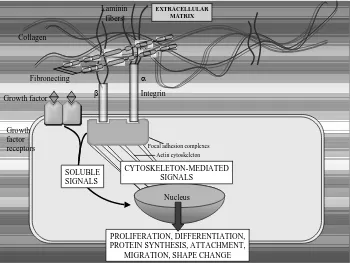
Though the serial cellular events that transpire following a peritoneal injury have been defined, the exact mechanism in peritoneal repair and adhesion formation has only been partially elucidated.The cellular events in tissue repair are regulated by growth signals, including migration, proliferation, and phenotype modulation of connective tissue cells, epithelial, and endothelial cells. Circulating platelets and inflammatory cells are the source of such endogenous signals controlling the normal process of wound healing. In later stages of tissue repair, signals are released by tissue repair cells themselves and act in an autocrine or paracrine fashion. Distinct surface receptors for several ligands have been identified. Binding of growth factors to specific cell membrane receptors activates tyrosine kinase activity and initiates intracellular signal transduction.
Fig. 2. Intercellular signaling of cytokines
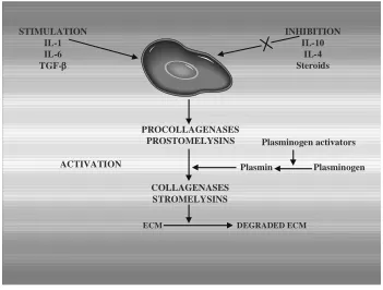
Preliminary finding demonstrating that numerous cytokines such as interleukin (IL)-1 (6), IL-2, transforming growth factor (TGF)-, and platelet-derived growth factor (PDGF)- (7) are signaling molecules, which acts in the formation of postoperative adhesion, while IL-10 (cytokine synthesis-inhibiting factor, CSIF) significantly inhibits such adhesion formation (8). The cellular events appear to be orchestrated at least in part by signals that function as chemoattractants and immunostimulants. Interleukin (IL)-6 (9), transforming
IL-1 IL-6 TGF-
tPA uPA
reduce postoperative adhesion formation. IL-10, also known as cytokine synthesis inhibiting factor, may be one of these centrally active mediators that inhibits the expression of cytokines such as LI-1, TNF-, or IL-6. Another cytokine that may have anti-adhesive effects is IL-4, with its inhibiting effects on monocytes and macrophages (11). In vitro findings demonstrate that IL-10 and IL-4 inhibit production of cytokines (e.g. adhesiogenic IL-6) in monocytes and macrophages by different mechanism (12). A thorough elucidation of the role and characteristics of centrally active cytokines might make possible the development of adhesion-preventing agent by targeted immunomodulation.
Fig. 3. Cytokines actions in adhesions material formation
There are multiple mechanisms by which IL-10 can reduce adhesion formation. Interleukin-10, produced by type-2 helper T cells, inhibits profibrotic cytokine production by type-1 helper T cells, peripheral blood mononuclear cells, and macrophages. One of these adhesiogenic cytokines suppressed by IL-10 is IL-2, which is known to stimulate the synthesis of two highly fibrinogenic cytokines, TGF- and platelet-derived growth factor B. Similarly, IL-10 acts on monocytes and macrophages to suppress the synthesis of the potent adhesion-forming mediators IL-1 and IL-6. Further reduction in IL-1 activity may be effected by the IL-10 induced up regulation of the anti inflammatory IL-1 receptor antagonist (13,14).
STIMULATION IL-1 IL-6 TGF-
INHIBITION IL-10
IL-4 Steroids
PROCOLLAGENASES PROSTOMELYSINS
ACTIVATION
Plasminogen activators
Plasmin Plasminogen
COLLAGENASES STROMELYSINS
Numerous studies in both humans and animals have demonstrated that IL-4 inhibits the productions of adhesiogenic cytokines such as TNF-, or IL-1 by monocytes and macrophages. Interleukin-4 further up-regulates the expression of the anti-inflammatory IL-1 receptor antagonist. Differential and synergistic effects between IL-10 and IL-4 have been described in particular for the reduction of IL-6 expression (15,16).
Molecular activity in peritoneal adhesion formation
Peritoneal healing exhibits characteristics of “too much repair”, although the healing mechanism following peritoneal injury differs substantially from that in the skin, as the entire peritoneal surface becomes reepithelialized, not only from the borders but also at the center of the defect. Regardless of the differences in reepithealization, the formation of peritoneal adhesion displays many phenomena, which can also be observed in other tissues (17).
It is now concerned as verified that, in addition to mechanical damage of the serosa, antibodies, necroses, residual blood, bacteria, and toxins as well as physical and chemical noxae lead to tissue damage. Following damage to very sensitive mesothelial cells, there is a flow of mediators, such as serotonin, bradykinin, histamine and prostaglandin, which leads to an increase in vessel permeability and results in the extravasation of serosanguine liquid in the abdominal cavity. Through the concomitantly extravasated fibrinogen, fibrin develops via the influence of released tissue thrombokinase, which goes on to form a loose three-dimensional network. This finally causes the adhesion of the adjacent serosal surfaces (5,18).
The studies by Buckman in 1976 identified the inhibition of fibrinolysis on a fibrin plate by the presence of ischaemic tissue (18). This suggested that, rather than there being a simple absence of PAA in the presence of ischaemia, there was active inhibition of activity . This was not followed up until 1990, when Vipond et al. identified the presence of plasminogen activator inhibitor-1 (PAI-1) in inflamed and ischaemic peritoneum (19). In 1993, Whawell et al. found that PAI-2 was present in similar tissue samples, but that tissue plasminogen activator (t-PA) was also present in normal quantities (20). Evaluation of the presence of PAI-1 or PAI-2 after simple trauma has not been carried out, but measurements by Buckman failed to indicate PAA inhibition after simple trauma. It would appear that after simple trauma PAA is reduced secondary to the reduction in the presence of t-PA. In the presence of inflammation or ischaemia there are normal levels of t-PA, but there is inhibition of PAA by PAI-1 and PAI-2. This suggests that there are two variations on the pathogenesis of adhesions; indeed, there may be two types of adhesions.
surgery may be beneficial, as they can support a precarious anastomosis, introduce a new blood supply to ischaemic tissue and wall off areas of sepsis to prevent spread. These adhesions are probably facilities by suppression of PAA by PAI-1 and PAI-2. It may be possible to selectively inhibit the formation of adhesions that may produce intestinal obstruction and to permit the formation of adhesions that are beneficial (21).
Fig. 4. Intracellular signalling pathway in adhesions formation
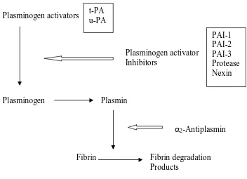
For many years it has been known that peritoneal tissue has fibrinolytic activity (22). Experimental studies have shown that injury to the peritoneum reduces its fibrinolytic capacity, and that the same injuries also produce intra-abdominal adhesions (18). The fibrinolytic system is an enzymatic cascade with a number of activators and inhibitors (fig. 2). In the presence of bacterial peritonitis or tissue ischaemia, there was a marked reduction in human peritoneal plasminogen-activating activity (23).
The plasminogen activator in human tissue has been identified as tissue plasminogen activator (tPA) using antibody inhibition studies and antigenic immunoassay techniques (19). Inflamed human peritoneal tissue contained levels of tPA comparable with normal peritoneum. The loss of fibrinolytic activity in inflamed peritoneal tissue was found to be the result of plasminogen-activator inhibitor (PAI) production. High levels of both PAI-1 and PAI-2 were found in inflamed human peritoneal tissue (20).
Plasminogen activators
Plasminogen activator Inhibitors
Plasminogen Plasmin
2-Antiplasmin
Fibrin Fibrin degradation Products
Fig. 5. The fibrinolytic system
Peritoneal injury either by operation or bacterial peritonitis results in a high concentration peritoneal pro inflammatory cytokine response particularly TNF, IL-1 and IL-6 (24). The concentration of these cytokins in the peritoneum is several hundred fold that seen in the plasma of these patients. Studies of human mesothelial cells, either derived from the omentum or from a cell line have shown that these pro-inflammatory cytokines individually and synergistically stimulate the production of PAI-1 in vitro. The time course of this cytokine response seen in the peritoneal exudate is also compatible with a cytokine-induced stimulation of PAI production.
In summary fibrous adhesions are formed within the peritoneal cavity when the normal fibrinolytic activity of the peritoneum is lost. This fibrinolytic activity is tPA dependent and its loss is the result of the production of PAI-1 and 2 by the mesothelium and sub-medothelial tissues. This production of PAI-s appears to be the result of pro-inflammatory cytokine stimulation of mesothelial and other cells.
References
1. Lorenz EPM, Zuehlke HV, Kange R, and Savvas V, Pathophysiology and classification of adhesions, In: Treitner KH and Schumpelick V Ed. Peritoneal adhesions, Springer-Verlag, Berlin, 1997, pp29-34
2. Ellis H, Wound repair: reaction of the peritoneum to injury. Ann R Coll surg Engl., t-PA
u-PA
3. Renvall SY, Peritoneal metabolism an dintra-abdomunal adhesion formation during experimental peritonitis. Acta Chir Scand (Supl), 1980;508: 4-48.
4. Menzies D, Aetio-pathogenesis of peritoneal adhesions with respect to post-traumatic fibrinolitic activity, In: Treitner KH and Schumpelick V Ed. Peritoneal adhesions, Springer-Verlag, Berlin, 1997, pp 105-110
5. Ellis H, Harrison W, and Hugh TB, The healing of peritoneum under normal and pathological conditions. Br J Surg. 1965;52:471-476.
6. McBride WH, Mason K, Withers HR, and Davis C., Effect of ionterleukin 1, inflammation, and surgery on the incidence of adhesion formation and death after abdominal irradiation in mice. Cancer Res., 1989;49: 169-173.
7. Kovacs EJ, Brockk B, Silber IE, Neuman JE. Production of fibrogenic cytokines by IL-2 treated peripheral blood leukocytes:expression of TGF- and PDGF-B chain genes. Obstet Gynecol 1993;82:29-36.
8. Montz FJ, Holschneider CH, Bozuk M, Gotlieb WH, and Martinez-Maza O, Interleukin-10: ability to minimize postoperative intraperitoneal adhesion formation in amurine model. Fertil. Steril, 1994;61:1136-1140
9. Saba AA, Kaidi AA, Godziachvili V, Dombi GW, dawe EJ, Libcke JH, et al. Effects of interleukin-6 and its neutralizing antibodies on peritoneal adhesion formation and wound healing. Am Surg 1996;62:569-72.
10. Chegini N, Simms J, Williams RS, and Masterson BJ. Identification of epidermal growth factor, transforming growth factor-, and epidermal growth factor receptor in surgically induced pelvic adhesions in the rat and intraperitoneal adhesions in the human. Am J Obstet Gynecol 1994;171:321-7.
11. Te Velde AA, Huijbens RJF, Heije K, de Vries JE, and Figdor CG. Interleukin-4 (IL-4) inhibits secretion of IL-1, tumor necrosis factor , and IL-6 by human monocytes. Blood 1990;76:1392-7.
12. Takeshita S, Gage JR, Kishimoto T, Vredevoe DL, and Martinez-Maza O. Differential regulation of IL-6 gene transcription and expressionn by IL-4 and IL-10 in human monocytic cell lines. J Immunol 1996;156:2591-8.
13. Fiorentino DF, Zlotnik A, Mosmann TR, Howard M, and O’Garra A. IL-10 inhibits cytokine production by activated macrophages. J Immunol 1991;147:3815-22.
15. Hart PH, Ahern MJ, Smith MD, and Finlay-Jones JJ. Comparison of the supressive effects of interleukin-10 an dinterleukin-4 on synovial fluid.
16. Zissel G, Schlaak J, Schlaak M, and Muller-Quernheim J. Regulation of cytokine release by alveolar macrophages treated with interleukin-4, interleukin-10, or transforming growth factor beta. Eur. Cytokine Netw., 1996;7:59-66.
17. Fukasawa M, Campeau JD, Yanagihara DL, Rodges KE, and Di Zerega GS Mitogenic and protein synthetic activity of tissue repair cells: control by the postsurgical macrophage. J Invest Surg., 1980;2: 169-180.
18. Buckman RF, Woods M, Sargent L, and Gervin AS, A unifying pathogenetic mechanism in the etiology of intraperitoneal adhesions. J Surg Res., 1976;20:1-5.
19. Vipond MN, Whawell SA, Thompson JN, and Dudley HAF Peritoneal fibrinolytic activity and intra-abdominal adhesions. Lancet, 1990;335:1120-1122.
20. Whawell SA, Vipond MN, Scott-Coombes DM, and Thompson JN, Plasminogen activator inhibitor 2 reduces peritoneal fibrinolytic activity in inflammation. Br J Surg., 1993;80;107-109.
21. Menzies D, and Ellis H, Intestinal obstruction from adhesions – how big is the problem? Ann R Coll Surg Engl., 1990;72:60-63.
22. Myrhe-Jensen O, Larsen SB, and Astrup T; Fibrinolytic activity in serosal and synovial membranes. Arch Pathol 1969;88:623-630.
23. Thompson JN, Patterson-Brown S, Harbourne T, Whawell SA, Kalodki E, and Dudley HAF; Reduced human peritoneal plasminogen activating activity: As possible mechanism of adhesion formation. Br J Surg 1989; 76: 32-384.