S Hasan Arshad
North Staffordshire University Hospital
Stoke-on-Trent, Staffordshire, UK
Stephen T Holgate
Southampton General Hospital
Southampton, UK
N Franklin Adkinson, Jr
John Hopkins Asthma and Allergy Centre
Baltimore, MD, USA
K Suresh Babu
Southampton General Hospital
Southampton, UK
CLINICAL PUBLISHING
OXFORD
Distributed worldwide by
Taylor & Francis Ltd
Mill Street, Oxford OX2 0JX, UK
Tel: +44 1865 811116 Fax: +44 1865 251550 Web: http://www.clinicalpublishing.co.uk/ Distributed by:
Taylor & Francis Ltd 6000 Broken Sound Parkway NW, Suite 300 Boca Raton, FL 33487, USA E-mail: orders@crcpress.com
Taylor & Francis Ltd 23–25 Blades Court Deodar Road London SW15 2NU, UK E-mail: uk.tandf@thomsonpublishingservices.co.uk
© Atlas Medical Publishing Ltd 2005 First published 2005
This edition published in the Taylor & Francis e-Library, 2006.
“ To purchase your own copy of this or any of Taylor & Francis or Routledge’s collection of thousands of eBooks please go to http://www.ebookstore.tandf.co.uk/.”
All rights reserved. No part of this publication may be reproduced, stored in a retrieval system, or transmitted, in any form or by any means, without the prior permission in writing of Clinical
Publishing or Atlas Medical Publishing Ltd.
Although every effort has been made to ensure that all owners of copyright material have been acknowledged in this publication, we would be glad to acknowledge in subsequent reprints or
editions any omissions brought to our attention.
A catalogue record for this book is available from the British Library
ISBN 0-203-49280-3 Master e-book ISBN
ISBN 0-203-62586-2 (Adobe e-Reader Format) ISBN 1 904392 24 5 (Print Edition)
The publisher makes no representation, express or implied, that the dosages in this book are correct. Readers must therefore always check the product information and clinical procedures with the most up-to-date published product information and data sheets provided by the manufacturers and the most recent codes of conduct and safety regulations. The authors and the publisher do not accept any liability for any errors in the text or for the
Preface vi
Acknowledgements vii
Abbreviations viiI
1
Introduction 1
2
Assessment 30
3
Asthma 50
4
Allergic rhinitis 79
5
Atopic dermatitis 95
6
Contact dermatitis 107
7
Urticaria and angioedema 115
8
Food allergy 125
9
Insect allergy 138
10
Drug allergy 152
11
Anaphylaxis 162
The last 30 years have witnessed an unprecedented increase in the prevalence of all allergic disease that includes asthma, allergic rhinoconjunctivitis, drug and food allergy, anaphylaxis, occupational allergy, and allergic skin disease manifesting as atopic eczema and urticaria. While there is much debate about why this ‘epidemic’ has occurred, the highest prevalence and greatest change in demography appears to be linked to aspects of the Western lifestyle that not only includes altered exposure to micro-organisms, which is frequently cited as the cause for the changing trends, but also changes in housing, drug use, and diet, as well as indoor and outdoor air pollutants. It is most likely that more than one factor is involved in driving the trends.
Irrespective of the underlying cause(s), allergic disease has now assumed public health proportions affecting all age groups and often manifesting as multiple organ disease. Indeed, complex allergy often presents a diagnostic and therapeutic challenge. Since allergy has such protean manifestations it often falls not only within the domain of organ-based specialists, but also general practitioners, immunologists, and those specializing in allergy. All such professionals will find this Atlas an aid to the diagnosis and management of allergy.
With the aid of clear diagrams and ample clinical and pathological illustrations, this Atlas describes the mechanisms, epidemiology, diagnosis, and treatment of all of the common allergies. Great care has been taken to draw out the most salient features of each of the disorders and to present them in a way that will enhance learning and recall memory.
While there are many books on allergy, both small and large, this Atlas fills a unique niche in providing an accessible source of up-to-date information. This book is of special value to medical students and those in post-graduate training, as well as to practitioners who wish for an easy-to-use, illustrated reference source.
We are extremely grateful to our patients who co-operated in this effort and it is their interest that has made this unique book possible. We hope you find its content valuable. We are always pleased to receive comments, especially if you have any useful suggestions for future revisions.
ACD allergic contact dematitis AD atopic dermatitis ADR adverse drug reaction AHR airway hyper-responsiveness AIDS aquired immunodeficiency syndrome
ALL acute lymphoblastic leukaemia APC antigen-presenting cell ARDS adult respiratory distress syndrome
ARIA Allergic Rhinitis and its Impact on Asthma (Study) BALF bronchoalveolar lavage fluid
BHR bronchial hyper-responsiveness BPT bronchial provocation test
CD contact dermatitis
CLA cutaneous lymphocyte-associated antigen CLL chronic lymphocytic leukaemia
CS corticosteroids
DBPCFC double blind, placebo controlled food challenge DH dermatitis herpetiformis
DNA deoxyribonucleic acid DPI dry powder inhaler ECP eosinophil cationic protein
ECRHS European Community Respiratory Health Survey EDN eosinophil-derived neurotoxin
ELISA enzyme linked immunosorbent assay EPO eosinophil peroxidase FEV1 forced expiratory volume in 1 second
FGF fibroblast growth factor FVC forced vital capacity GALT gut associated lymphoid tissue
GI gastrointestinal
GM-CSF granulocytic monocyte colony stimulating factor
H/O history of HPA hypothalamopituitary axis ICAM intercellular adhesion molecule
ID intradermal (test)
Ig immunoglobulin
IL interleukin
i.m. intramuscular
IMN infectious mononucleosis
INF interferon
ISAAC International Study of Asthma and Allergies in Childhood
i.v. intravenous
LABA long-acting beta agonist
LT leukotriene
MBP major basic protein MCP monocyte chemotactic protein MCT mast cell tryptase MCTC mast cell tryptase, chymase
MDI metered-dose inhaler MHC major histocompatibility complex MIP 1α macrophage inflammatory protein 1 alpha NARES non-allergic rhinitis with eosinophilic syndrome
NK natural killer (-cell)
NO nitric oxide
(i)NOS (inducible) nitric oxide synthase NSAID non-steroidal anti-inflammatory drug
OCP oral contraceptive pill PAF platelet activating factor PAR perennial allergic rhinitis
PC20 provocation concentration (20% decrease in FEV1)
PD20 provocation dose (20% decrease in FEV1)
PDE phosphodiesterase PEF peak expiratory flow PEFR peak expiratory flow rate
RAST radio allergen sorbent test SALT skin associated lymphoid tissue
SAR seasonal allergic rhinitis
s.c. subcutaneous
SCF stem cell factor SIT specific injection immunotherapy SJS Steven-Johnson syndrome
SPT skin prick test
sRaw specific airway resistance
TCR T-cell receptor
TEN toxic epidermal necrolysis TGF-β tissue growth factor-beta
THO T-helper (cells) TNF-α tumour necrosis factor-alpha VCAM vascular cell adhesion molecule
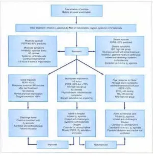
Introduction
An allergy is an immunologically-mediated adverse reaction to a foreign substance, usually a protein. Allergic reactions can affect almost any tissue or organ in the body, with clinical manifestations depending on the target organ. For example, asthma is the result of an allergic reaction affecting lower airways. When the nose and eyes are targeted, it manifests clinically as allergic rhinoconjunctivitis. Skin manifestations include: allergic maculopapular rash, urticaria, and angioedema. Systemic reactions may result in anaphylaxis.
Clinical manifestations of allergic diseases
Common clinical manifestations of allergy include asthma, atopic dermatitis, allergic rhinitis, and urticaria/angioedema.
Asthma
Experts from the National Institutes of Health defined asthma as:
‘…a chronic inflammatory disorder of the airways in which many cells and cellular elements play a role, in particular, mast cells, eosinophils, T lymphocytes, macrophages, neutrophils, and epithelial cells. In susceptible individuals, this inflammation causes recurrent episodes of wheezing, breathlessness, chest tightness, and coughing, particularly at night or in the early morning. These episodes are usually associated with widespread but variable airway obstruction that is often reversible either spontaneously or with treatment. The inflammation also causes an associated increase in the existing bronchial hyper-responsiveness to a variety of stimuli.’ (National Institutes of Health [1997] Guidelines for the Diagnosis and Management of Asthma. NIH Publication 97–4051 A, Bethesda, Maryland.)
In severe asthma, airway obstruction can be extreme with occasionally fatal outcome
1.1
Airway in a normal subject and in
those with severe asthma.
Definition of atopy and sensitization
Atopy is the genetic predisposition to produce Immunoglobulin E (IgE) antibodies on exposure to allergens.
Sensitization is the state where, following exposure to the allergens, the immune system is primed to produce IgE antibodies specific to that allergen.
Atopic dermatitis
Atopic dermatitis (AD) or atopic eczema is a chronic relapsing inflammatory skin condition with pruritis, and with typical morphology (erythematous vesicular lesions) and distribution (flexural areas, trunk) (1.2). However, both morphology and distribution can vary depending on the severity and duration of the disease, and age of the patient. It is often, but not always, associated with atopy.
Allergic rhinitis
Rhinitis is defined as inflammation of the nasal mucosa. It is characterized by nasal congestion, rhinorrhoea, sneezing, itching of the nose, and/or post-nasal drainage. It may be associated with ocular symptoms such as watering, congestion, and redness (rhinoconjunctivitis). Rhinitis may be allergic or non-allergic. Allergic rhinitis is diagnosed when symptoms are triggered by a recognized allergen. Nasal polyposis is a common complication of allergic rhinitis (1.3).
Urticaria and angioedema
Urticaria is an erythematous, palpable, itchy rash with distinct margins that may be localized to one area of the skin or generalized (1.4). The rash varies in shape and size, and may involve any area of the skin. Angioedema is subcutaneous or submucous oedema without distinct margins. It may occur with urticaria or as an isolated phenomenon. Typical episodes are of sudden onset, often itchy, and resolve to leave normal looking skin. Repeated episodes may occur (intermittent urticaria/angioedema) and if these continue beyond 6 weeks, it is termed chronic recurrent urticaria/angioedema.
1.3
Right ethmoidal polyp due to
allergic rhinitis. (Courtesy of Dr SH
Abid.)
Prevalence of allergy
Food allergy and atopic eczema is common in early childhood. These atopic children are at a higher risk of developing asthma and rhinitis in later childhood and early adult life. Allergic manifestations can be life-threatening such as severe asthma and anaphylactic reactions.
Asthma affects 10–15% of children and 5–10% of adults
1.4
A patient with generalized
urticaria.
(1.3 million children and 1.8 million adults in the UK are affected). Allergic rhinitis is observed in 10–20% of unselected population and atopic eczema in 5–10%, depending on the age and population studied. Perceived food allergy is extremely common and reported by 20–25% of the population. However, this is confirmed by double blind food challenges in only 2–7%. The true prevalence depends on the age and it is more common in young children.
Figures 1.5 and 1.6 show data from longitudinal follow-up studies of a whole population birth cohort. Children were
1.6
Sensitization to common allergens
at ages 4 and 10 years in an unselected
population. (HDM, house dust mite.)
(Data from the Isle of Wight birth
cohort.)
asthma, allergic rhinoconjunctivitis,
and atopic eczema: ISAAC.
The
Lancet,
351(9111):
1225–33.)
seen at regular intervals from birth to 10 years of age, thus reducing selection and re-call bias. They were also skin prick tested for 14 common food and inhalant allergens at 4 and 10 years of age. This prospective study confirmed a higher lifetime prevalence of allergic diseases during the first decade of life. Sensitization to at least one allergen was 20% at 4 years and increased to nearly 30% at 10 years of age.
1.8
Twelve-month prevalences of
allergic rhinoconjuctivitis symptoms.
(Data derived from Beasley
et al.
[1998]. Worldwide variation in
prevalence of symptoms of asthma,
allergic rhinoconjunctivitis, and atopic
eczema: ISAAC.
The Lancet,
World wide prevalence
As noted above, the prevalence of asthma and allergy showed wide variations in different populations, but these studies were not directly comparable, as study tools were often neither standardized nor validated. The inception of ISAAC (International Study of Asthma and Allergies in Childhood) using standardized questionnaire material has helped to reduce this problem. During the 1990s, the prevalence of asthma, atopic dermatitis, and rhinitis was
1.9
Twelve-month prevalences of
atopic eczema symptoms. (Data
derived from Beasley
et al.
[1998].
Worldwide variation in prevalence of
symptoms of asthma, allergic
rhinoconjunctivitis, and atopic eczema:
ISAAC.
The Lancet,
351(9111):
1225–
33.)
assessed in several countries.
data from different countries and populations could be compared with a degree of confidence. This study
1.10
Prevalence of asthma in adults. In
adults, as in children, asthma
prevalence varied widely with
Australia, Denmark, and UK showing
highest prevalences, whereas
developing countries such as India and
Algeria showed lowest prevalence of
asthma. Unfortunately, only a few
countries outside Europe and
Australia/New Zealand were included
in the original study. However, several
reports from countries around the
world have since been published, using
the European Community Respiratory
Health Survey questionnaire,
confirming differing prevalence across
the globe. (Data derived from
European Community Respiratory
Health Survey [1996],
Eur Respir J,
9:
687–95.)
confirmed a wide variation in prevalence of all allergic manifestations. For example, asthma prevalence varied from <5% (Indonesia and Albania) to >35% (UK).
Rise in prevalence
Studies have consistently shown an increasing prevalence of allergic disease in recent decades. This is independent of the increasing awareness of allergy (and thus increase in reported symptoms) and changes in diagnostic criteria. Although there is considerable variation in the prevalence between countries (presumably due to different diagnostic criteria), the two observations in the same population used similar methodologies and yet showed a consistent pattern of rise in prevalence (1.11).
Recent reports, however, indicate that the prevalence may have reached a peak; the last few years have shown a slight decline in prevalence in some developed countries. Figure
1.12 shows data from a recent Canadian study. Asthma prevalence was highest in the pre-school age children, and continued to show a slight increase in prevalence over the study period. However, in other age groups there seems to be a trend of increased prevalence from 1991–1995 and then stabilization between 1995–1998. It may be too early to say if the rise in asthma prevalence, observed consistently during the 1960s through the early 1990s, has reached a plateau.
1.12
Stabilization of an increasing
trend in physician-diagnosed asthma
prevalence in Saskatchewan, Canada,
1991–98. (Data derived from
Senthilselvan A,
et al.
[2003].
Stabilization of an increasing trend in
physician-diagnosed asthma
prevalence in Saskatchewan, 1991 to
1998.
Chest,
124(2):
438–48.)
Expression of allergic diseases
Development of an allergy depends on genetic and environmental factors. Genetic susceptibility is a prerequisite for the development of asthma and other allergic disorders, but the precise nature and location of these genes is still not fully understood. Allergic disorders are polygenetic diseases, and a large number of genes on several different chromosomes have been implicated. They may share genes which regulate atopic immune responses, such as those for interleukin (IL)-4, IL-13 and interferon gamma (INF-γ). Other relevant genes may be related to the target organ’s structure or function. For example, for asthma these may determine airway responsiveness or regulate airway epithelial and mesenchymal function. A certain combination or series of combinations of genes or genetic polymorphisms may be required for the phenotypic expression.
and number of allergic manifestations, and may also influence the degree of severity and prognosis (1.13).
Initial allergen exposure causes sensitization in individuals who are genetically predisposed. Atopy or sensitization to allergens is common and may occur in 40–50% of the general population. Those with genetic predisposition to a particular target organ disease, such as asthma or rhinitis, develop a specific clinical disease. Exposure to certain environmental factors may promote or protect against this development. Further exposure to allergens or pollutant may result in symptoms or exacerbation. (1.14).
It is important to note that the degree of allergen exposure required to cause sensitization is not known, and may vary from allergen to allergen. In some cases, a higher exposure may indeed be protective by stimulating immune tolerance mechanisms. Recent studies suggest that a higher exposure to the cat allergen may protect against the development of sensitization. However, the same cannot be said about other common allergens, such as pollens, house dust mites, or cockroaches.
Atopy, as defined by positive skin prick test or presence of specific IgE to common allergens, is an important risk factor for the development of asthma and allergic disease
(1.15,1.16). However, atopy can be present without clinical manifestation (asymptomatic sensitization). These asymptomatically sensitized children, when followed over a period of several years, show a high rate of development of clinical disease. It is not known why some of these children develop clinical manifestations, whereas others remain asymptomatic.
1.13
Genetic and environmental risk
factors for the development of asthma.
(BHR, bronchial
hyper-responsiveness.)
1.15
The risk of asthma (odds ratios
and their 95% confidence intervals) in
10-year-old children sensitized (on
skin prick test) to three common
allergens: house dust mites, cats, and
dogs.
1.16
The risk of atopic dermatitis
(odds ratios and their 95% confidence
intervals) in 4-year-old children
sensitized (on skin prick test) to four
common allergens: house dust mites,
grasses, cats, and peanuts.
Allergic immune responses
Regulation of stem cell differentiation occurs through interaction with various cell surface receptors and cytokines. Cytokines have pleiotropic effects on the haematopoetic and lymphoid cell development, affecting both the growth and maintenance of the pluripotent stem cells, as well as the differentiation of specific lineages.
1.17
The differentiation of the
haematopoetic cells. (NK, natural
killer; CFU, colony forming unit;
GEMM, granulocyte, erythroid,
monocytic-dendritic, and
megakaryocytic lineage; BFU-E, blast
forming unit erythroid; Eo,
eosinophil.)
Antigen-presenting cells
The three types of APCs present different sets of antigens and also serve to activate T-cells at different points during the immune response. Most (including B-T-cells, macrophages, and dendritic cells), express major histocompatibility complex (MHC) class II antigens on their surface. These are important for communication with T-cells and subsequent T-cell activation. Although many cells do not normally express MHC class II antigens, certain cytokines can induce their expression and thereby potentiate antigen presentation. In addition to antigen presentation, the APCs provide co-stimulatory signals via B7.1 and B7.2.
Cells of the monocyte-macrophage system exist in the blood. Macrophages are more differentiated monocytes that are resident in the tissues. These cells express MHC class II molecules on their surface. They have phagocytic and cytotoxic functions. The microscopic shape of the macrophages depends upon their function. They have a flexible cytoskeleton that makes them ovoid during transit, and stellate, with engulfing surface projections, during phagocytosis.
B-cells are produced in the bone marrow and are distributed through the body in the lymph nodes. B-cells respond to the ‘foreign’ antigens of a pathogen by producing specific antibodies. B-cell receptors bind soluble antigens. The bound antigen molecules are engulfed into the B-cell by receptor-mediated endocytosis. The antigen is then digested into fragments which are displayed at the cell surface, nestled inside a class II histocompatibility molecule. Helper T-cells, specific to this structure, bind the B-cell and secrete lymphokines.
Eosinophils and mast cells
1.18
Antigen-presenting cells involved
in allergic immune response.
The pro-inflammatory effector function of eosinophils has been supported by an extensive number of studies. One of the important observations is that the treatment of monkeys with an anti-ICAM-1 monoclonal antibody clearly inhibited allergen-induced lung eosinophil influx and also prevented airway hyper-responsiveness. Similar findings were obtained following treatment with an anti-IL-5 antibody (TRFK-5), reinforcing the interpretation that there is indeed a causal link between eosinophilia and airway hyper-reactivity. However, there is evidence opposing this concept. It has been determined that blocking the effects of IL-5 by an anti-IL-5 antibody, mepolizumab, or a soluble IL-5 receptor, suppresses allergen-induced bronchoalveolar lavage fluid (BALF) eosinophilia with little effect on bronchial hyper-reactivity. This puts into question the functional role of eosinophils in allergic diseases.
Mast cells (1.19) are found only in tissues and they contain abundant granules. The stem cell factor is essential for mast cell development and survival, and influences mast cell function. Mast cells are 6–12 µm in diameter and the cytoplasm contains granules and crystals alone, or in combination. The surface of mast cells have receptors for IgE, cytokines, growth factors, and cell adhesion structures. There are two different types of mast cells which are designated mucosal or connective tissue, based on their location. Mast cells contain granules which store mediators of inflammation. Degranulation of mast cells can be induced by physical trauma, temperature, toxins, proteases, and immune-mediated mechanisms involving the aggregation of IgE bound to high-affinity receptors (FcεRI) on the surface of these cells. The effects of degranulation by cross-linking of cell surface IgE by antigens releases heparin, histamine, and other mediators to initiate an immediate allergic response. Upon stimulation, mast cells release cytokines, including tumour necrosis factor-alpha (TNF-α) and IL-4, that can modulate adhesion molecules on endothelial cells.
Mast cells originate from CD34+ progenitor cells in the bone marrow. Mast cells are widely distributed throughout the body in both connective tissue and at mucosal surfaces. The predominant mast cell subtype in the lung contains tryptase (MCT), although tryptase and chymase mast cells (MCTC) are also present. Mast cell activation occurs when IgE, bound to the high-affinity IgE Fc receptor (FcεRI) on mast cells, is cross-linked by allergens triggering mast cell degranulation. This results in the release of preformed granule-derived mediators and neutral proteases, the synthesis and release of newly formed lipid products, and the transcription of numerous cytokines (Table 1.1,
indicate that allergens, such as Der p 1 (a serine protease), as well as neuropeptides, eosinophil products, defensins, and changes in osmolality can induce mast cell histamine and cytokine secretion directly through IgE-independent mechanisms. Antigen-specific mast cell activation results in an immediate response while the late-phase response is a consequence of cytokines and other mediators derived from these cells or other cells recruited to the site of inflammation (1.20).
1.19
Effector cells of the allergic
immune responses: eosinophils and
mast cells.
1.20
The effects of mast cell
Lymphocytes
Lymphocytes are divided into T- and B-cells. In the early stages of development, T-cell precursors migrate to the thymus. T-cells are divided into subsets based on their surface expression of CD4 and CD8. CD4 T-cells recognize antigens in the context of MHC class II molecules and CD8 cells recognize antigens presented by class I molecules. T-cells express a clonal antigen-specific receptor. The T-cell receptor (TCR) is in fact very similar to immunoglobulin. It has two paired polypeptide chains both of which have constant and variable portions and both of which are composed of immunoglobulin-like domain. B-cells represent 5–10% of lymphocytes and their cytoplasm is characterized by scattered ribosomes and isolated rough endoplasmic reticula. Plasma cells are the mature form of B-lymphocytes that secrete antibodies.
A small fraction (~2%) of the lymphocytes circulating in the blood are neither T- nor B-cells. Most of these are called NK-cells because they are already specialized to kill certain types of target cells, especially host cells that have become infected with a virus or have become cancerous (1.21). They express CD 16 and/or CD56 antigens. They are found in the liver, spleen, lungs, blood, and gastrointestinal (GI) tract. NK-cells are activated by IL-2, IL-12, IL-15, or IL-18.
Table 1.1
Mast cell mediators in allergic disease
Mediators Biological effects
Histamine Bronchoconstriction, tissue oedema, mucus secretion, fibroblast and endothelial proliferation
Heparin Anticoagulant, storage matrix for mast cell mediators, fibroblast activation Tryptase Generates c3a and bradykinin, increases BHR, activates collagenase, fibroblast
proliferation
Chymase Mucus secretion, extracellular matrix degradation PGD2 Bronchoconstriction, tissue oedema, mucus secretion
LTC4 Bronchoconstriction, tissue oedema, mucus secretion
TNF-α Neutrophil chemotaxis, MHC class II expression, increased expression of adhesion molecules, mucus secretion, increases IL-8 and IL-6 synthesis in fibroblasts
IL-4, IL-13 B-cell proliferation, increase IgE synthesis, activation of eosinophils IL-5, GM-CSF Eosinophil recruitment, activation, and survival
IL-6 IgE synthesis, differentiation of T-cells, mucus secretion IL-8 Neutrophil chemotaxis
IL-16 T-cell chemotaxis
SCF Growth, differentiation, and survival of mast cells Basic FGF Angiogenesis, fibroblast proliferation
MIP-1α Macrophage differentiation, neutrophil chemotaxis
BHR, bronchial hyper-responsiveness; FGF, fibroblast growth factor; GM-CSF, granulocyte monocyte colony stimulating factor; Ig, immunoglobulin; IL, interleukin; LT, leukotriene; MCP, monocyte chemotactic protein; MHC, major histocompatibility complex; MIP, macrophage inflammatory protein; PG, prostaglandin; SCF, stem cell factor; TNF-α, tumour necrosis factor-alpha
1.21
Lymphocytes orchestrate the
immune responses in both allergic and
non-allergic inflammation.
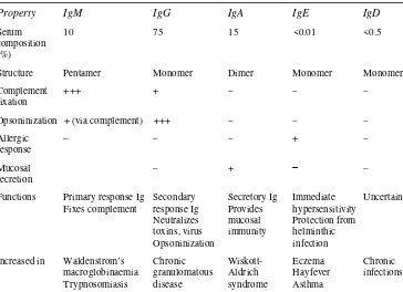
Table 1.2 Characteristics of different classes of
immunoglobulins
Property IgM IgG IgA IgE IgD
Serum composition (%)
10 75 15 <0.01 <0.5
Structure Pentamer Monomer Dimer Monomer Monomer Complement
Functions Primary response Ig Fixes complement
Actinomycosis
ALL, acute lymphoblastic leukaemia; CLL, chronic lymphocytic leukaemia; Ig, immunoglobulin; IMN, infectious mononucleosis; SLE, systemic lupus erythematosus
Immunoglobulins
Immunoglobulins are glycoprotein molecules which are produced by plasma cells and which function as antibodies. The immunoglobulins derive their name from the finding that when antibody-containing serum is placed in an electrical field, the antibodies responsible for immunity migrated with the globular proteins. All immunoglobulins have a four chain structure as their basic unit. They are composed of two identical light chains (23 kD) and two identical heavy chains (50–70 kD). The heavy and light chains can be divided into two regions based on variability in the amino acid sequences: the variable and the constant regions. Subtle structural differences in their antigen combining sites, or variable region, account for their unique antigen-binding specificities. There are five different classes of immunoglobulins (Table 1.2).
1.23
Class switching to IgE and B-cell
proliferation.
Antigen uptake
When a T- or B-cell engages an antigen through its antigen receptor an intra-cellular signal is generated (1.22). The first signal, which gives specificity to the immune response., is provided by the interaction of antigenic peptide-MHC complex with the TCR. The second antigen-independent co-stimulatory signal is delivered to T-cells by APCs to promote T-cell clonal expansion, cytokine secretion, and effector function. In the absence of the second signal, antigen-specific lymphocytes fail to respond effectively, and are functionally inactivated and resistant to subsequent activation to the antigen.
Production of immunoglobulin E
Immunoglobulin E
IgE is the immunoglobulin involved in allergic reactions. IgE is commonly known as the ‘reaginic’ antibody. IgE is the least common immunoglobulin, since it binds very tightly to Fc receptors on basophils and mast cells, even before interacting with antigens and, secondly, due to the very small amount synthesized. Like other antibodies, IgE is comprised of two identical light chains and two identical heavy chains, each chain made up of 110 amino acids called immunoglobulin domains, covalently linked by disulphide
1.24
Structure of IgE.
bonds (1.24). The L-chain has one N-terminal variable (VL) domain and one constant (CL) domain. Likewise, the H-chain consists of one N-terminal V (VH) domain and four C (CH) domains. The antibody class is determined by the CH sequence designated as Cε for IgE. A given B-cell produces an antibody with one specificity, as defined by the VL and VH combination, but during an antibody response, it can ‘switch’ classes. IgE has a profound effector function due to the fact that basophils and mast cells have high affinity receptors for the Fc portion of the molecule. IgE also binds to Fc receptors on the surface of eosinophils. This allows eosinophils to participate in antibody-dependent, cell-mediated cytotoxicity reactions against parasitic helminths.
IgE receptors and their interaction
1.25
IgE binding to high- and
low-affinity receptors.
either wraps around a single Cε3 domain to make contact with both sides, or it interacts with opposite faces of the Cε3 domains on one side of IgE. The β-chain spans the plasma membrane four times and the two γ-chains extend a considerable distance into the cytoplasm. Allergen-mediated cross-linkage of the bound IgE results in aggregation of the FcεRI receptors and rapid tyrosine phosphorylation, which initiates the process of mast cell degranulation.
FcεRII is the low-affinity IgE receptor with a kD of 1× 10⁻⁶ M and is specific for the CH3/CH3 domain of the IgE. It belongs to the family of C-type lectins. CD23 is a 45-kD polypeptide chain with extracellular structural motifs, a transmembrane sequence, and a cytoplasmic tail. The cytoplasmic tail can be either of two types: CD23a and CD23b. Allergen cross-linkage of IgE bound to the FcεRII receptors results in activation of B-cells, eosinophils, and alveolar macrophages. Blockage of this receptor with a monoclonal antibody leads to diminished IgE secretion by the B-cells. Interestingly, CD23 appears to act both in the up-regulation and down-regulation of IgE synthesis, and atopic individuals have higher levels of CD23 on their lymphocytes and macrophages. CD23-IgE interaction provides an important mechanism whereby allergen-specific IgE can augment cellular and humoral immune responses in settings of recurrent allergen exposure.
Summary
in turn, leads to anaphylaxis (1.26). Subcutaneous administration causes a local inflammatory reaction. Inhaled allergens activate mucosal mast cells leading to broncho-constriction and increased mucous secretion. Ingested allergens lead to food allergy.
1.26
Type of allergic reaction/disease
may depend on the characteristics of
allergen and the route of
administration.
Allergens
A number of allergens are present in the air (aeroallergens) and, partly depending on the size of the particle, may cause upper or lower airway allergies. Systemic reactions to aeroallergens are rare. Allergens from food sources tend to cause GI or skin manifestations but, depending on the nature of the food allergen and sensitivity of the subject, may result in a systemic reaction. Allergens injected into the body, such as insect venom or drugs, often result in a systemic reaction. Allergens coming in contact with the skin may cause urticaria or contact dermatitis. However, there is considerable overlap and aeroallergens, such as house dust mites, may exacerbate atopic dermatitis in a sensitized individual, and food allergens may rarely cause asthma.
Aeroallergens
The most frequently encountered aeroallergens are: house dust mites, pollens (trees, grasses, and weeds), mould spores, and allergens of animal origin.
House dust mites
mattresses, blankets, and pillows, as well as furniture and soft toys. House dust provides them with a habitat and contains their food source in the form of sloughed human skin. They are approximately 0.3 mm in length and are visible only by microscopic examination of the dust (1.27). Humidity and temperature are the most critical factors in the survival of the house dust mite population. There are two groups of important allergens derived from the common species of house dust mites: Group 1 (Der p 1 and Der f 1) and Group 2 (Der p 2 and Der f 2), isolated from Dermatophagoides pteronyssinus and Der. farinae, respectively. It is estimated that 20–30% of the European population is sensitized to Der. pteronyssinus. The presence of house dust mite sensitization is strongly associated with asthma and perennial allergic rhinitis. The role of house dust mites in atopic dermatitis remains unclear.
1.27
An electron microscopic
photograph of a common house dust
mite,
Dermatophagoides
pteronyssinus
.
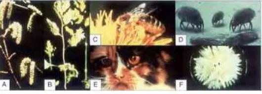
Pollen
1.28
Common species of
pollen-bearing plants:
A
timothy grass
(Phleum pratense);
B
rye grass
(Lolium perenne);
C
meadow fescue
(Festuca pratensis);
D
olive
(Olea
europaea);
E
grey alder
(Alnus
incana);
F
silver birch
(Betula
pendula);
G
hazel
(Corylus avellana);
1.29
American cockroach,
Periplaneta
americana
. (Courtesy of H Robertson,
Iziko Museum of Cape Town, South
Africa.)
Cockroach
Cockroaches are widely present in urban or inner-city areas and cockroach proteins are an important indoor allergen in these places. Two species of cockroaches, the American
(Periplaneta americana)(1.29) and German (Blattella germanica) commonly inhabit the home and have a potential to cause sensitization. Important cockroach allergens are Bla g 1, with a molecular weight of 30 kD, and Bla g 2, with a molecular weight of 36 kD. Cockroach allergens can be detected anywhere in the house, but highest levels are usually found in kitchens and bathrooms. Sensitization to cockroach allergen has been shown to be an important risk factor for the development of asthma among inner-city residents.
Animal
1.30
Cats and dogs are the most
common furry pets responsible for
allergy to animals.
Foodallergens
Food allergens are almost always proteins. Potentially, any food can provoke a reaction. However, some foods are more allergenic than others, and these are responsible for most of the food allergic reactions. Common allergenic foods include: milk, egg, wheat, fish, and shellfish (1.31). Food allergic reactions may be IgE-mediated or non-IgE-mediated.
Cow’smilk and eggs
1.31
Common allergenic foods.
Fish and shellfish
Seafoods are composed of diverse sea organisms and many of them cause allergic reactions (1.32). Tropomyosin is a major allergen in many shellfish, especially Crustacea and molluscs.
Peanuts
Peanuts are the main cause of food-induced anaphylactic reactions. Ara h 1 has been identified as the major peanut allergenic protein. Peanuts (Arachis hypogaea) belong to the legume family. However, clinical features of a peanut allergy are more closely related to those of a tree nut allergy, rather than other legumes, such as peas. Peanut allergy occurs in about 0.5–1% of the general population. It is characterized by immediate onset, often with severe symptoms on minimal contact. There is considerable cross-reactivity with one or more tree nuts. Nearly 30% of patients that are allergic to peanuts are also allergic to tree nuts.
Treenuts
Commonly encountered tree nuts causing allergy include hazelnuts, walnuts, Brazil nuts, almonds, cashews, pecans, chestnuts, pine nuts, pistachios, and coconuts. Allergic reactions to tree nuts can be serious and life-threatening. Attempts have been made to identify and characterize allergenic proteins. Major seed storage proteins include legumins, vicilins, and 2S albumins.
Wheat
Wheat is one of the common allergens, especially in children. Both IgE-mediated and non-IgE-mediated reactions are common. Gliadins are said to be major wheat allergens. IgE antibodies to purified gliadin in children correlate with clinical symptoms on wheat challenge.
Insect allergens
Common insects causing allergic reactions are honeybees, wasps, hornets, yellow jackets, and fire ants. Allergy to bees and wasps occurs in most parts of the world. Reactions may vary from mild localized swelling to systemic reactions and anaphylaxis. Insect stings account for 25% of the cases of severe systemic anaphylaxis referred to an allergy clinic.
Latex
Exposure to latex increased worldwide during the 1990s, with a parallel increase in the incidence of latex allergy among the general population, as well as high-risk groups. Latex (natural rubber latex) is encountered widely in rubber-containing products, such as gloves, balloons, catheters, condoms, and surgical instruments (1.33). Thus, health care workers and those undergoing frequent surgical procedures have been at high risk. Clinical manifestations include local skin (itching, erythema, oedema, and urticaria), local airways (rhinoconjunctivitis, asthma, and pharyngeal oedema) and/or systemic reactions and, on occasion, anaphylaxis. The use of latex-free gloves and other products has resulted in a significant reduction in incidence of latex allergy.
Assessment
Introduction
Diagnosis of allergic diseases is based on clinical criteria, as described in Chapter 1. A comprehensive history and physical examination remains the mainstay of allergy diagnosis, and no single test can confirm or rule out the diagnosis of diseases such as asthma, allergic rhinitis, or AD. However, laboratory tests are useful aids for the diagnosis and, importantly, may help to identify the specific allergen(s) responsible. The outcome of the test should always be interpreted in the light of information available from the clinical assessment.
Table 2.1 outlines some of the tests used in the assessment of allergic disease. These tests can be classified into three groups. In vitro tests, such as measurement of specific IgE, are commonly used to confirm allergic status and identify the specific allergen. Other in vitro tests, such as blood eosinophil count or basophil histamine release, are rarely helpful and are not commonly used. The second group, in vivo or provocation testing, is used extensively for the diagnosis and management of allergic disease. The third group of tests relates to the assessment of environmental exposure to a particular allergen, such as house dust mites or pollen.
Table 2.1
Diagnostic tests
In vitro tests Provocation (in vivo) tests
• Full blood counts • Skin tests • Total blood eosinophil count • Skin prick tests • Sputum and nasal secretion eosinophilia • Intradermal skin tests • Total serum IgE • Patch testing
• Allergen specific IgE • Bronchial and nasal provocation tests • Radio allergen sorbent test (RAST) • Food challenge
• Competitive RAST inhibition assays
• Allergen specific IgG Assessment of environmental aeroallergens
• Mast cell tryptase levels • Indoor environments • Basophil histamine release – House dust mite
• Exhaled nitric oxide measurements – Moulds • Random noise oscillometry
Outdoor environment
• Pollens
• Moulds
Clinical laboratory tests (invitrotests)
Serum total IgE
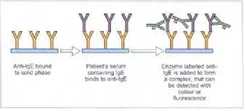
Serum IgE levels range between 0–0.00001 g/L and approximately 50% of the IgE is localized to the extra-vascular space. The concentration of IgE is age-dependent with cord blood levels being very low. However, atopic infants have a steep rise in their serum IgE during their early years. Extreme elevations of IgE are seen in parasitic infections and hyper-IgE syndrome. A normal or low level of IgE in an asthmatic individual suggests non-IgE mechanisms are playing a role in asthma pathogenesis. In atopic dermatitis, IgE levels are elevated in over 90% of the patients. However, total IgE is not very helpful in the management of allergic diseases. Several immunoassays are available for the measurement of serum total IgE and these include radioimmunoassay (RIA), enzyme linked immunosorbent assay (ELISA), and paper radioimmunosorbent assay (PRIST) (2.1).
Allergen specific IgE
The presence of allergen-specific IgE is highly indicative of an individual’s susceptibility to mount an allergic response upon re-exposure to that allergen. Allergen-specific IgE are assayed using an in vitro assay (radio allergen sorbent test, RAST) in serum (2.2). RAST measures the amount of IgE that is directed to a specific allergen. Skin tests are generally considered to be more sensitive than RAST assay and it is rare, though not unknown, for a patient to be skin test-negative and RAST-positive. RAST is more expensive than the skin test and results are not available immediately in the clinic. However, RAST may be appropriate in certain situations (Table 2.2).
2.1
Total IgE in blood is often
measured using radioimmunoassay.
2.2
Serum-specific IgE can be
measured
in vitro
by RAST. Several
kits are available for measurement of
specific IgE to aeroallergens, foods,
insect venom, and drugs.
Table 2.2
Indication for radio allergen sorbent
test (RAST)
RAST test is indicated:
• Where facilities and/or expertise for a skin test are not available
• When the skin test result is unexpected (e.g. in patients who present with a good history of sensitivity to a particular allergen, and yet produce equivocal or negative skin test results) • In patients with extensive atopic dermatitis where a large enough area of uninvolved skin is not
available
systemic reaction
• In patients with dermographism • In epidemiological and clinical research
composition of venoms for patients with hymenoptera sensitivity. These patients, who have potential cross-sensitivities, have opted for immunotherapy.
Mast cell tryptase (MCT)
MCT is a protease that is found in mast cell granules. It is secreted upon stimulation of the cell, together with histamine and many other mediators. Measurement of tryptase in the blood is indicated where there is doubt regarding the nature of a systemic reaction. Tryptase levels are usually, but not always, elevated following an allergic reaction. The assay of tryptase offers significant advantages over other methods of monitoring mast cell activation: it can be performed on serum samples; it is more specific than histamine; and it may be elevated several hours after an allergic reaction.
Table 2.3 Things to remember when performing
skin prick test (SPT)
• Always include negative (saline) and positive (histamine) controls • The prick should be gentle so as not to draw blood
• Care is taken to space individual tests at a sufficient distance from each other so as not to produce overlapping erythema
• Antihistamines should be discontinued 4 days prior to SPT
• Epinephrine and antihistamine should be at hand, but full resuscitation facilities may not be necessary
• Dermatographism can produce false-positive reactions
In vivotests
Skin prick tests(SPT)
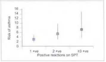
The skin prick or puncture test remains the ‘gold standard’ in the diagnosis of allergic diseases. It is a biological test for the presence of specific IgE antibodies to the allergen that is being tested (2.3–2.6).
2.3
Extracts are commercially
available for a large number of food
and aeroallergens.
2.4–2.6
SPTs involve placing a drop of
allergen extract on the skin of the
forearm or back, and introduction of
the allergen into the epidermis with a
needle puncture. After the prick, the
allergen is removed by blotting with a
tissue paper. An immediate reaction is
read at 15–20 minutes. The longest
diameters of the wheal and one
Table 2.4 SPT could be graded for
semi-quantitative assessment
Grade Wheal
0 <25% of histamine area 1+ 25–50% of histamine area 2+ 50–100% of histamine area 3+ 100–200% of histamine area 4+ >200% of histamine area
reaction denotes sensitivity of the subject to the specific allergen tested (Table 2.4). Sensitization on SPT carries a significant risk for the development of asthma and allergic diseases. The risk increases with increasing number of positive reactions (2.7–2.9).
Variation of these tests includes intra-dermal testing which is more sensitive than the standard puncture test. This test requires 0.02 ml of a serial dilution (10–1000-fold) of allergen extract to be injected intra-cutaneously through a 26–27 gauge needle to produce a superficial bleb. Like the skin test, the readings are performed in 15–20 minutes. False-negative reactions can occur when the allergen is injected subcutaneously. Both the wheal and the erythema are measured with a millimetre ruler or calipers. With some dark-skinned people, it is difficult to assess the erythema. Although the reproducibility of the intra-dermal test is better than SPT, this requires greater technical skill. There is also a greater risk of a systemic reaction.
The prick-to-prick test is also used occasionally when allergen extract is either not available or is not reliable, such as when testing for certain fruits. The lancet is pricked in the substance, and the skin is pricked in the same manner as in SPT. The skin reaction is observed and measured.
2.8
The risk of atopic dermatitis (odds
ratio and their 95% confidence
intervals) in 4-year-old children
sensitized to one, two, or three or more
allergens on SPT.
2.9
The risk of allergic rhinitis (odds
ratio and their 95% confidence
intervals) in 4-year-old children
sensitized to one, two, or three or more
allergens on SPT.
placed in direct contact with the skin,
usually on the upper back, within small
aluminium discs. Adhesive tape is used
to fix them in place, and the test sites
are marked. The patches are left in
place for 48 hours and are usually read
at 48 and 72 hours after placement of
the allergens.
Patch testing
Patch testing is a way of identifying whether a substance that comes in contact with the skin is causing inflammation of the skin. Patch tests are used for the diagnosis of allergic contact dermatitis. They are indicated in recurrent episodes of contact dermatitis, when needed to identify the offending allergen, and in patients who are not responding to conventional therapy. Patch tests document the presence of delayed-type hypersensitivity
(2.10).
The test is interpreted as: • Erythema 1+.
• Oedema or vesicle <50% of patch area 2+. • Oedema or vesicles >50% of the patch area 3+.
Systemic corticosteroids and immunosuppressants can suppress the responses. False-positive reactions are due to irritant effect, while false-negatives can occur because the patch tests do not reproduce the exact environment in which the allergen is encountered. Another limitation is the availability of a finite number of allergens.
A range of substances can be used for patch testing. Common substances causing allergic contact dermatitis are included in the European Series Standard Battery (or similar) which is applied to most patients (Table 2.5). In addition, other substances can be tested appropriate to the individual circumstances. Each substance has been tested to find the best concentration to demonstrate an allergic reaction without causing irritation to those who are not allergic to the material. Allergens for patch tests are available both in ready-to-use format (2.11) and as test material mixed in a vehicle (usually white petroleum).
Bronchial provocation tests
Table 2.5
Common chemicals included in the
Histamine and methacholine are the most commonly used substances. Nebulized histamine or methacholine dissolved in saline is administered through a dosimeter in doubling dilutions. The procedure is stopped if the forced expiratory volume in 1 second (FEV1) drops by at least 20%, or if the patient becomes symptomatic and is unable to
continue the procedure. The degree of airways responsiveness is expressed as PC20 the
concentration producing a 20% fall in FEV1(2.12).
sensitivity to a particular allergen is first demonstrated on a skin test before that particular allergen is used for allergen BPT. Spirometry is recorded to establish a baseline FEV1
which should be >70% predicted. After ensuring that the patient is suitable to continue, the subject inhales saline through a dosimeter. The FEV1 is measured and this value is
used to calculate the percentage fall from the post-saline baseline. Following the saline inhalation, the subject inhales varying concentrations of allergen, starting with the least concentration. After each concentration of allergen, a four-fold increment is administered, providing the FEV1 has not fallen by >10% from the post-saline value. If
the fall in FEV1 is between 10–15%, a two-fold increment is administered. If
2.11
‘True test’ is one of the
commercially available patch tests that
can be easily applied in an office
setting.
2.12
Methacholine challenge test in
normal and asthmatic airways
calculates the PC
20. Note there is very
minimal drop in the FEV
1in normal
20% drop occurred at 0.25 mg/ml of
methacholine.
the fall is >15% but <25% from the post-saline baseline, a dose of the same concentration is administered. The challenge is terminated when a fall in FEV1 of >25% from the
post-saline baseline FEV1 measurement is achieved. After the final concentration of allergen,
FEV1 measurements are taken at 5, 10, 15, 30, 45, and 60 minutes, and thereafter every
30 minutes up to 10 hours (2.13).
Foodchallenge
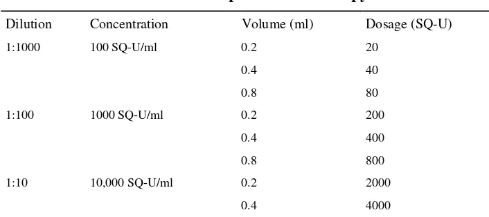
A food challenge is performed to confirm or refute the causative relationship of a presumed adverse reaction to a food. It is independent of mechanism, but immediate reactions are suggestive of IgE-mediated food allergy. A food challenge can be open, single blind, or double blind. An open food challenge is easiest to perform, but is subject to bias and is usually not helpful. During a blind food challenge, a patient is given the food to be tested or a placebo, masked in another food or drink to which the patient is not allergic or intolerant. A double blind placebo controlled food challenge (DBPCFC) is considered the ‘gold standard’ in diagnosing food allergy or intolerance. A strict elimination diet of the suspect foods for 7–14 days before the challenge is essential for all types of food challenges. During blind challenges, an equal number of randomly alternating food allergen and placebo challenges are given. The starting dose depends on the sensitivity of the patient (from the history) and the type of food. For example, subjects highly sensitive to nuts may have to have a labial challenge (2.14). The dose is gradually increased every 30–60 minutes. Clinical reactivity is ruled out once a substantial amount is given, which is usually the dose that the patient has previously reacted to.
There is no need to undertake a food challenge if a clear-cut history is supplemented by a positive SPT or RAST. Some precautions are recommended, such as avoiding antihistamines, beta 2 agonists, and cromolyn sodium 12 hours prior to the challenge.
Other tests
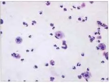
Sputum induction
2.13
FEV
1following an allergen
challenge in a patient with allergic
asthma shows an early asthmatic
response defined as a fall in FEV
1of at
least 25% within 15 minutes of
allergen inhalation, and the late
asthmatic response defined as a fall in
FEV
1of at least 15% between 3 and 10
hours. The result is expressed as
cumulative PD
20(provocation dose to
cause a 20% fall in FEV
1). Note the
early asthmatic response is associated
with an increase in the serum
Fibreoptic bronchoscopy
A bronchoscopy is not a routine test employed in the assessment of allergic airway disease, but serves as a tool to assess the inflammation in airways, and to evaluate the effects of treatment on the inflammatory cell population in the airways. Cytokine levels can also be assayed from bronchoalveolar lavage and bronchial biopsies, to provide clues to the pathophysiologic mechanisms underlying asthma (2.17).
2.14A, B
A labial challenge may be
advisable, as an initial procedure, for
those with a suspected reaction to nuts.
2.15
Sputum induction is carried out
using inhaled aerosolized hypertonic
saline (4.5%). Subjects are seated in an
induction chamber, 400 µg of inhaled
salbutamol is administered via a spacer
device, and the peak expiratory flow
(PEF) is recorded. Patients with a
post-bronchodilator FEV
1of <50% of
administered via a nebulizer. After
every 5 minutes, the nebulization is
stopped and PEF measured. The
procedure is discontinued when there
is a fall in PEF of >15%, or if there are
troublesome symptoms. Sputum is
expectorated into a Petri dish. The
induction period is for a maximum of
20 minutes, or until a satisfactory
quality of the sample is obtained.
2.16
Unselected sputum is processed
by adding an equal weight of 0.01 M
dithioerythritol. The contents are then
filtered through a 70 micron filter to
remove mucus. The filtrate is
to contribute to the
immunopathogenesis of asthma and
rhinitis.
Endobronchial ultrasound
Endobronchial ultrasound is a technique used in research to evaluate the thickness of the airway mucosa in asthma. It is useful in improving the diagnostic yield with transbronchial needle aspiration and transbronchial lung biopsies. However, its use as a research tool in asthma is still at a very early stage (2.18).
Nitric oxide (NO)
Nitric oxide is an inorganic, gaseous free radical that carries a variety of messages between cells. Vasorelaxation, neurotransmission, and cytotoxicity can all be potentiated through cellular response to NO. The formation of NO is catalysed by inducible NO synthase (iNOS). Cytokines, such as INF-γ, TNF, IL-1 and IL-2, and lipopolysaccharides, cause an increase in iNOS messenger ribonucleic acid (mRNA), protein, and activity levels. Protein kinase C stimulating agents exhibit the same effect on iNOS activity. After cytokine induction, iNOS exhibits a delayed activity response, which is then followed by a significant increase in NO production over a long period of time. NO, per se, can exert damaging effects on cells or could react with superoxide to form peroxynitrite, a powerful oxidant capable of exerting cytotoxic effects. NO also plays a role in regulating the immune response and can inhibit lymphocyte proliferation.
2.18
An endobronchial ultrasound
showing cross-section of a bronchus.
(1, first hyperechoic band: marginal
echo from mucosa/submucosa; 2,
second hypoechoic band:
Southampton General Hospital,
Southampton, UK.)
2.19
NO in exhaled breath is a simple,
quick, and non-invasive test that may
find a place in the clinical management
of asthma.
2.20
Forced oscillation technique
offers simple, quick, non-invasive, and
patient-friendly measurement of
airflow obstruction.
Forced oscillation technique
Forced oscillation technique is a relatively new technique used to asses the lung function. It measures the airway resistance which indicates the degree of airflow obstruction. During the test, patients have tidal breathing against the oscillatory wave (4–30 Hz) produced by a Pseudo Random Noise Oscillometer, and the impedance of the airway is calculated (2.20).
Potentially, there are several advantages of the forced oscillation technique. As it requires only passive co-operation, it could be done in infants, children, severe asthmatics, and elderly patients for whom traditional spirometry might be difficult. Furthermore, as this test is done during tidal breathing, it is more physiological and eliminates the problems associated with forced expiratory manoeuvres. Thus, it may provide a more reliable means of measuring lung function. In addition, this technique offers the potential to study resistance in large and small airways independently of each other.
Assessment of environmental allergens
Indoor allergens
Allergen avoidance forms an important aspect of management of allergic diseases. The assessment of exposure to a specific allergen may indicate the level of risk and the need to implement avoidance measures. Predominant indoor allergens include house dust mites, cockroaches, animal dander, and moulds. These allergens can be measured in the house dust collected with a dust collector.
2.21
Repeated application of
Acarosan
®, an antidust mite spray
(based on benzyl benzoate) led to the
reduction in the level of dust mite
allergens in the treated group.
(Adapted from Arshad HS
et al.
[1992]. Effect of food and house-dust
mite allergen avoidance on
development of allergic disorders in
infancy.
Lancet,
339:
1493–97.)
Outdoor allergens
2.22
Pollen trap. Air is sucked through
a narrow slit on the side of the pollen
trap and lands on a sticky tape or glass
slide. Pollen and other particles in the
air stick to the tape or glass surface.
The tape/slide is taken off and pollen
counting is done under the light
microscope.
2.23
Pollen calendar for the UK.
(Adapted from data from The National
Pollen and Aerobiology Research Unit,
University College Worcester,
Asthma
Epidemiology
The prevalence of asthma has increased to epidemic proportions, and the current health care expenditure for asthma in the industrialized countries is enormous. In Western Europe, asthma has doubled in the last 10 years, and in the US it has increased by over 60% since the 1980s. The human and economic burden associated with asthma is large and the economic cost is estimated to exceed that of tuberculosis and HIV/AIDS combined together. It is an irony that 43% of the cost of asthma care is related to the use of emergency departments in hospitals. The International Study of Asthma and Allergies in Childhood (ISAAC) looked at the prevalence and severity of asthma, rhinitis, and eczema in children living in different centres to help make comparisons within and between countries. The mean prevalence of any wheeze in the past 12 months ranged from under 5% in Albania, China, Greece, Georgia, Indonesia, Romania, and Russia to 29–32% in Australia, New Zealand, Republic of Ireland, and the UK (3.1). The prevalence of four or more attacks, which is a more specific indicator of clinically important asthma, ranged from <1% in Albania, Indonesia, Uzbekistan, Russia, Romania, Greece, Georgia, and China to over 9% in Australia, New Zealand, UK, and Canada. These rates of prevalence tended to correlate with those of less frequent wheezing. This suggests that higher prevalences of wheezing in these English-speaking countries are unlikely to be explained solely by over-reporting of mild symptoms. Studies in adults show similar variation (3.2).
diagnosed asthma, recent wheeze,
bronchial hyper-responsiveness
(BHR), and atopy in children. (Data
adapted from the ISAAC study.
Asthma, GIF [2002].
Global Strategy
for Asthma Management and
Prevention,
National Institute of
Health: a1–1176.)
3.2
The prevalence of asthma
symptoms in adults. The figure shows
the data on prevalence of diagnosed
asthma, recent wheeze, bronchial
hyper-responsiveness (BHR), and
atopy in adults. (Data adapted from
studies in the Australian population
and the European Community
Respiratory Health Survey (ECRHS).
Asthma, GIF [2002].
Global Strategy
for Asthma Management and
Prevention,
National Institute of
Health: a1–1176.)
Development and risk factors
who are non-atopic. The level of IgE is associated with the prevalence and severity of bronchial hyper-responsiveness (BHR) and asthma. However, asthma is non-atopic in nearly half of older children and adults, and important susceptibility genes may be inherent to the airway wall (3.4).
Most asthma originates in the early stages of life (3.5). Childhood asthma tends to be a predominantly male disease. After 20 years of age, the prevalence remains approximately equal until age 40, when the disease becomes more common in females. Abnormal BHR is a central feature of asthma. However, not all patients with BHR have symptoms of asthma. BHR frequently precedes, and is associated with, an elevated risk for wheeze onset and recurrent asthma in adolescents.
Exposure to dust mites within the first year of life is associated with later development of asthma, and possibly atopy (3.6). Inhalation of cigarette smoke during pregnancy has been linked with abnormal lung functions, BHR, and allergy in the newborn. Tobacco smoke is also an important trigger factor for asthma attacks. Some recent studies have shown a relationship between passive smoking and the development of asthma. In addition, air pollution plays a well-documented role in asthma attacks. Ozone is a key player in the development and aggravation of asthma. The ‘hygiene hypothesis’ suggests that lack of exposure to a childhood infection, endotoxin, and bacterial products are important determinants regarding development of atopic disease. Environmental exposure to microbial antigens and bacterial compounds helps the development of immune tolerance through the stimulation of Th1 cells and/or suppression of Th2 cells, preventing the development of allergic disorders in children.