EMSA Eritin Blocks Nuclear Factor-Kappa B in Diabetes Mellitus
Mice Model
Maic Audo Lin Sihombing
1, Sutiman B. Sumitro
2and Muhaimin Rifa’i
2*1
Master Program of Biology, Faculty of Mathematics and Natural Sciences, University of Brawijaya, Malang, Indonesia 2
Department of Biology, Faculty of Mathematics and Natural Sciences, University of Brawijaya, Malang, Indonesia
Abstract
Diabetes mellitus is one of the common metabolic disorders with increasing prevalence during recent years with hyperglycemia as its characteristic. DM has been shown to be a state of free radicals over production resulted from hyperglycemia that can activate cellular signaling pathways transcription of factor NF-κB which stimulates the production of several inflammatory mediators andl lead to chronic inflammation. Chronic inflammation is implicated in
β-cell damage and function and promotes apoptosis. EMSA Eritin is a polyherbal consisting of soy bean extracts, coconut water extract and red rice extract that assumed to be antidiabetic and anti-inflammatory. This study will assess the effectiveness of EMSA Eritin against inflammation in diabetes mellitus by measuring levels of NF-κB produced by immunocompetent cells in DM mice model. Streptozotocin 100 mg.kg-1 BW is used to induce diabetes mellitus in mice. Oral administration of EMSA Eritin was given for 14 days with dose of 0.3125 mg.g-1 BW, 3.125 mg.g-1 BW and 31.25 mg.g-1 BW. Data were analyzed using One-way ANOVA (p<0.05) and Duncan test using SPSS 16.0 for Windows. The results showed that EMSA Eritin can be used as an alternative therapy for the treatment of DM. The level of NF-κB in diabetic mice significantly decreased when the mice received EMSA Eritin.
Keywords:Diabetes Mellitus, EMSA Eritin, NF-κB, ROS
INTRODUCTION
Diabetes mellitus is one of the most common metabolic disorders with increasing prevalence during recent years. Changes in lifestyle, diet and environment becomes the causes of the increasing prevalence of this disease. Prevalence occurred in Asia and Africa, as a result of urbanization trend and lifestyle changes [1]. Statistical data confirmed that diabetes has reached epidemic proportions. More than 346 million people world wide have diabetes and it is predicted to be the seventh leading cause of death in the world by 2030 [2]. Meanwhile, the number of patients with DM in Indonesia reached 8.6 million in 2000 and will increase to 21.3 million by 2030. Due to the high number of deaths, Indonesia was ranked 4th in the world after United States, India and China [3].
Diabetes mellitus characterized by hyper-glycemia that occurs because the body is unable to secrete insulin in sufficient amounts or is unable to use insulin effectively or both [4,5,6]. DM can be a consequence of the reduction in the number of insulin receptors or failure in the
Correspondence author:
Muhaimin Rifa’i
Email : [email protected]
Address : Laboratory of Animal Physiology, University of Brawijaya, Jl. Veteran Malang, 65145
insulin-receptor binding, and failure in the transport of glucose into cells by the protein glucose transporter (GLUT) [7]. Previous research suggested that oxidative stress and inflammation play an important role in the development of DM [8].
Among various factors that are being involved in the progression of DM, oxidative stress could be typically induced by excessive nutritional factors like glucose and free fatty acids (FFA) which increasing formation of free radicals, auto oxidation of glucose and lipid peroxidation. In the process of oxidative stress, excessive reactive oxygen species (ROS) such as superoxide anion, hydroxyl radicals and hydrogen peroxide were produced by mitochondria and plasma mem-brane [9]. ROS can activate cellular signaling pathways such as nuclear factor-κB (NF-κB), which stimulates the production of several inflammatory mediators. Recent finding suggest that proinflammatory cytokines such as interleukin-1 (IL-1), interleukin-6 (IL-6) and
TNF-α, play important roles in the pathophysiology of
induction of ROS-mediated NF-κB activation, may be important in etiology of DM and related complications [11].
Appropriately prescribed drugs such as sulfonylureas or thiazolidinediones as monothe-rapy or add-on treatment are essential to cure the disease. However, many patients still cannot achieve satisfactory glycemic goals and even experienced serious side effects. Therefore, searching for alternative protective strategies becomes great interest. Recently, interests have been growing in the application of natural com-ponents as antidiabetic agents whicth have fewer side effects, cheap and easy to obtain [12,13]. EMSA Eritin is a dietary supplement that consist-ing of soy bean, coconut water and brown rice extract. Polyphenol compounds such as isofla-vones and flavonoids, found in soy and red rice and mneral from coconut water known have antioxidant activity, inflammatory, and
anti-diabetic to prevent β-cell apoptosis, increasing β -cell proliferation, secretion and activity of insulin [13,14,15]. Based on these references, poly herbal EMSA Eritin is believed to be useful in the process of DM treatments. Therefore, this study will assess the effectiveness of EMSA Eritin towards inflammation in DM by measuring levels of the transcrip-tion factor of inflammatory,
NF-κB produced by immunocompetent cells in diabetes mellitus mice model with streptozotocin induced.
MATERIALS AND METHODS
Animal and research protocol approval
Healthy Balb/c mice (25-35 g) were purchased from the Central Animal Room, Cellular and Molecular Biology Laboratory, University of Brawijaya. The mice were maintained in a temperature and humidity-regulated room with controlled lighting (12-h light/dark cycle). The animals were given standard pellets and tap water was provided ad libitum.
Experimental design
Total of 25 Balb/c female mice, between the ages of 4-6 weeks were used and divided into 5 groups of treatment include negative control (healthy mice and without EMSA Eritin administration) and positive control (diabetic mice with EMSA Eritin administration). EMSA Eritin was grouped into three doses: Low dose (0.3125 mg.g-1 BW), optimum dose (3.125 mg.g-1 BW) and high dose (31.25 mg.g-1 BW). Each treatment was repeated 5 times.
Drugs solution mice [4]. The mice were grown, fed and water by ad libitum. Mice were weaned at 4 weeks old and selected for screening by glucometer glucose tolerance test. Mice with glucose level ≥ 200 mg.dl-1 were considered to be diabetic and included in the research.
Oral administration of EMSA Eritin
Positive diabetic mice were given EMSA Eritin orally at a dose of 0 mg.g-1 BW (N); 0.3125 mg.g-1 BW (D1); 3.125 mg.g-1 BW (D2); and 31,25 mg.g-1 BW (D3). The dose is obtained from the conversion of the human dose that is 250 mg.kg-1 BW. EMSA Eritin of all doses was freshly prepared twice a day before administration and given orally by syringe at 9:00 a.m. and 03.00 p.m. Administration was conducted for 14 days. Blood glucose levels were measured every 3 days during the administration of EMSA Eritin to determine the change of blood glucose level.
Lymphocyte isolation
Mice were sectioned after 14 days treatment. Spleen were isolated and crushed in petri dish containing PBS. Lymphocyte homogenates were filtered using BD nylon cell strainerTM (100μm) and transferred into a new propylene tubes. Homogenates were centrifuged at 2500 rpm, 4°C for 5 min. Supernatant was removed and pellet resuspended in 1 mL PBS.
Antibody staining and flowcytometry analysis Pellet stained with antibodies with extra-cellular and intraextra-cellular staining. The following antibodies were used for extracellular staining are fluorescein isothiocyanate (FITC)-conjugated anti-mouse CD4, phycoerythrin (PE)-conjugated anti-mouse CD8, and fluorescein isothiocyanate (FITC)-conjugated anti-mouse CD68. Antibodies used for intracellular staining is PE-Cy5 conjuga-ted anti-mouse NF-κB.
For extracelluler staining, cells were stained
with 1μL of extracellular antibody that had been diluted with 50 μL of PBS, and then incubated in
20 minutes. After that, cells were added with 50
μL fixative solution (cytofix/cytosperm) and
incubated again in ice box (4°C) for 20 minutes.
Cells were added with 500 μL washperm to
removes the fixative solution and centrifuged at 2500 rpm, 4°C for 5 min. Pellet was stained with intracellular antibodies and then incubated in ice box (4°C) for 20 minutes. Suspension then was
added with 300 μL PBS, transferred to flow
cytometry cuvet and ready for analyzed with flow cytometry.
Statistical analysis
The data were evaluated by one-way ANOVA using the SPSS 16.0 program for Windows. The differences between the means assessed using Duncan's multiple range test. Statistical signifi-cance was considered at p<0.05.
RESULTS
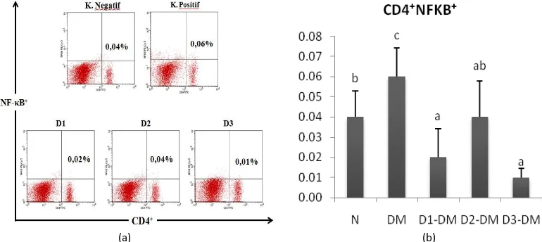
EMSA Eritin suppressed NF-κB in T lymphocyte Nuclear translocation of NF-κB in response to inflammatory insults is the primary regulatory step for its activation. NF-κB showed high expression in diabetic mice compared with normal mice (Fig. 1). STZ injection in the diabetic group (DM) can significantly increase the relative number of CD4+ T cells NF-κB+ 0.06 ± 0.01%
compared to the healthy group (N) 0.04%. EMSA Eritin which administered in diabetic mice showed immunomodulatory activity as immune-supresant of NF-κB which produced by CD4 lymphocyte T cells. The relative number of CD4+NF-κB+ T cells in EMSA Eritin treatment group (D1-DM, D2-DM, and D3-DM) was significantly decrease (p<0.05) compared with control group (N). The low dose, normal dose and high dose of EMSA Eritin can suppress the level of CD4+NF-κB+ with the relative number 0.02%, 0.04% and 0.01%, respectively.
EMSA Eritin also showed suppression activity of NF-κB that produced by CD8 T cells (Fig. 2). The relative number of NF-κB in negative control (DM) is 0.04% and increased after STZ injection 0.11%. Relative number of CD8+NF-κB+ T cells in all of dose of EMSA Eritin treatment group was lower than negative control group (DM), with the value of each are 0.05%, 0.05% and 0.08% (p>0.05). NF-κB that produced by CD4 and CD8 T cells was decreased in all doses of EMSA Eritin treatments. The relative number of NF-κB in treatment groups did not show a significant difference with healthy group. This result showed that the treatment group of diabetic mice had returned to normal condition or became healthier.
(a) (b)
Figure 1. EMSA Eritin decrease relative number of NF-κB on CD4+ T cells. Description:
N: healthy mice group,
DM: diabetic mice group were treated with EMSA Eritin 0 mg.g-1 BW;
D1-DM = diabetic mice group were treated with Emsa Eritin low dose 0.3125 mg.g-1 BW; D2-DM = diabetic mice group were treated with Emsa Eritin normal dose 3.125 mg.g-1 BW; D3-DM = diabetic mice group were treated with Emsa Eritin high dose 31.25 mg.g-1 BW. (a) The percentage of CD4+ T cells expressing positive NF-κB using flow cytometry.
(a) (b)
Figure 2. EMSA Eritin decrease relative number of NF-κB on CD8+ T cells Description:
N: healthy mice group
DM: diabetic mice group were treated with EMSA Eritin 0 mg.g-1 BW;
D1-DM = diabetic mice group were treated with Emsa Eritin low dose 0.3125 mg.g-1 BW;
D2-DM = diabetic mice group were treated with Emsa Eritin normal dose 3.125 mg/gr BW; D3-DM = diabetic mice group were treated with Emsa Eritin high dose 31.25 mg.g-1 BW. (a) The percentage of CD8+ T cells expressing positive NF-κB using flow cytometry.
(b) The bars are the number of CD8+ T cells expressing positive NF-κB on the spleen cells of mice after EMSA Eritin treatment.
Data are mean±SD in each group with p value < 0.05.
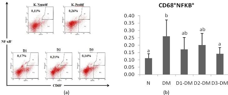
EMSA Eritin suppressed NF-κB in macrophage cells (CD68+)
We also found that administered EMSA Eritin decreased the level of NF-κB that is produced by CD68 macrophage cells (Fig. 3). NF-κB was increased significantly (p<0.05) after mice got induction of STZ (0.26%) compared to healthy mice (0.11%) in Fig.3. The relative number of
NF-κB in all dose treatment showed significant
difference compared to negative control group (DM) with the value of 0.17%, 0.20% and 0.14%, respectively. It showed that the treatment of EMSA Eritin improve the diabetic condition to be better. Increased levels of NF-κB would lead to proliferation of CD4 T cells, CD8 T cells and CD68 macrophage cells to produce proinflammatory cytokines that essential in the development of diabetes mellitus.
(a) (b)
Figure 3. EMSA Eritin decrease relative number of NF-κB on CD68+ macrophage cells Description:
N: healthy mice group,
DM: diabetic mice group were treated with EMSA Eritin 0 mg.g-1 BW;
D1-DM = diabetic mice group were treated with Emsa Eritin low dose 0.3125 mg.g-1 BW; D2-DM = diabetic mice group were treated with Emsa Eritin normal dose 3.125 mg.g-1 BW; D3-DM = diabetic mice group were treated with Emsa Eritin high dose 31.25 mg.g-1 BW).
(a) Bars are number of CD68+ macrophage cells expressing positive NF-κB on spleen cells of mice after EMSA Eritin treatment. (b) The percentage of CD68+ macrophage cells expressing positive NF-κB using flow cytometry.
DISCUSSIONS
Diabetes mellitus has been shown to be a state of free radicals over production resulting from hyperglycemia and FFA. Factors that attributed to the formation of free radicals may include not only elevated non-enzymatic and autooxidative glycosylation, but also metabolic stress as a result of alterations in energy metabolism, inflammatory mediators levels and the status of antioxidant defense [16]. In the process of oxidative stress, ROS primarily produced by mitochondria excessively. ROS activates cellular signaling pathways nuclear factor-κB (NF-κB) which stimulates the as diabetes mellitus. NF-κB, or nuclear factorκB, is a nuclear transcription factor found in all cell types. It is involved in cellular responses to stimuli such as stress, cytokines, free radicals, ultraviolet irradiation, bacterial or viral antigens andactivation of the adaptive immune receptors like TLR4 in response to FFA [10,17]. NF-κB promotes immunity by controlling the expression of genes involved in inflammation. NF-κB drives expression of target genes that mediate cell proliferation and release of cytokines to activate the immune response [17].
NF-κB is a transcription factor composed of p50 and p65 subunits (heterodimer) and
expressed in the cytoplasm of virtually all cell types. Its activity is controlled by a family of regulatory proteins, called inhibitors of NF-κB
(IκB). In the non-active condition, NF-κB in the cytoplasm constitute as heterodimer that binds
to Iκβ which responsible for the activation of NF
-κB. NF-kB activity is under the control of signaling from extracellular stimuli. Cytokine or PAMP ligation of cell surface receptors initiates signaling cascades that converge on the activation of the inhibitor of kB kinase (IKK) complex. The IKK complex consists of three subunits: the catalytic subunits IKKα and IKKβ,
and the regulatory subunit IKKγ. IKK phosphorylation of IkB molecules promotes their degradation and releases heterodimers p65 and p50 NF-kB dissociation from IKKα [17]. NF-κB binds to NF-κB binding domain on gene promoter and regulate the transcription of genes involved in innate immunity and inflammation. [18]. NF-κB signaling pathway can be activated by many proinflammatory stimuli such as TNF-α which is
not only being the activator of NF-kB but also a regulation product of NF-κB [7].
EMSA Eritin showed anti-inflammatory active-ty when administered to the lymphocytes in mice model diabetes mellitus. EMSA Eritin reduced the level of NF-κB that produced by CD4 T cells, CD8 T cells and CD68 macrophage cells. EMSA Eritin is a polyherbal that consists of soy bean extract, coconut water extract and red rice extract. This composition seem to be effective to suppress the transcription factor of inflammation, NF-κB. Soy isoflavone genistein is one of the active compound on EMSA Eritin and has been reported to be protective agent for diabetes by regulation of oxidative stress and inflammation as it decreases lipid peroxidation, inhibits cyclooxy-genase expression and myeloperoxidase activity as well as reacts with free radicals and neutralizes their effects [16]. Anthocyanin, especially C3G (cyanide-3-O-β-glucoside) is red rice pigment had antioxidative and anti-inflammatory activity [19]. Meanwhile, coconut water extract contains L-arginine, ascorbic acid, minerals such as calcium, magnesium and potassium which have hypolipidemic and antidiabetic activity [15].
Previous researches reported that genistein and anthocyanin can interfere NF-κB signaling by blocking translocation NF-κB p65 to nucleus,
reduce JNK activation and reduce IκBα
phosphorylation, thus interfere transcription genes that role in the inflammation [20,21]. Biological activity of genistein and anthocyanins directly inhibit p50-p65 NF-κB binding to the target of gene promoter NF-κB, stabilization of
IκBα by inhibit the phosphorylation of IκBα by
IKK-β. Thus the translocation of NF-κB into the nucleus can be blocked and transcription process will not occur [18]. In addition, L-arginine can inhibit the reaction of non-enzymatic glycosy-lation of proteins and the formation of AGEs by inhibiting the covalent modification proteins or AGEs formation [22]. AGE will increases ROS and triggers the MAPK and NF-κB signaling pathway. MAPK activation will trigger macrophage activity to synthesizes cytokine pro inflammation [23]. Inhibition of AGEs by L-arginine will reduce NF-κB activation.
CONCLUSION
suggest that NF-κB might be an important molecule contributing to diabetes mellitus and its complication.
Acknowledgement
The author would like to thank to Mr. Mansur Ibrahim for funding this research.
Ethics
This study has received ethical eligibility certificate (Ethical Clearance) from The Research Ethics Committee (Animal Care and Use Committee) Brawijaya University No. 384-KEP-UB.
REFERENCES
[1] American Diabetes Association. 2010. Diag-nosis and classification of diabetes mellitus.
Diabetes Care. 33.
[2] World Health Organization. 2014. Diabetes fact sheet No. 312. http://www.who.int. Accessed in December 15th 2014.
[3] Setiawan, A., Suciati, E. Yulinah, I.K. Adnyana, H. Permana, P. Sudjana. 2011. Efek Antidiabetes kombinasi ekstrak bawang putih (Allium sativum Linn.) dan rimpang kunyit (Curcumma domestica Val.) dengan pembanding glibenklamid pada penderita diabetes melitus tipe 2. MKB. 43 (1).
[4] Arulmozhi, D.K., A. Veeranjaneyulu, S.L. Bodhankar. 2004. Neonatal Streptozotocin-induced rat model of type 2 diabetes producing cells from clucotoxicity under high glucose conditions In-Vitro and ameliorates drug induced diabetes in rats. J. Translation. Med. 11. 115.
[6] Pedicino, D., G. Liuzzo, F. Trotta, A.F. Giglio, S. Giubilato, F. Martini, F. Zacardi, G. Scavone, M. Previtero, G. Massaro, P. Cialdella, M.T. Cardillo, D. Pitocco, G. Ghirlanda, F. Crea. 2013. Adaptive immunity, inflamation, and cardiovascular complications in type 1 and type 2 diabetes mellitus. J. Diabetes Res. Article ID 184258. [7] Cruz, N.G., L.P. Sousa, M.O. Sousa, N.T.
Pietrani, A.P. Fernandes, K.B. Gomes. 2013. The linkage between inflmation and type 2 diabetes mellitus. Diabetes Res. Clin. Pr. 99. 85-92.
[8] Donath, M.Y., E. Dalmas, N.S. Sauter, M. Boni-Schnetzler. 2013. Inflammation in obesity and diabetes: islet dysfunction and therapeutic opportunity. Cell Metabolism.
17 (6). 860-872.
[9] Li, Z, Y.N. Geng, J.D. Jiang, W.J. Kong. 2014. Antioxidant and anti-inflammatory activities of Berberine in the treatment of diabetes mellitus. Evidence-Based Complementary Altern. Med. Article ID 289264.
[10] Patel, S., D. Santani. 2009. Role of NF-κB in the pathogenesis of diabetes and its asso-ciated complications. Pharmacol. Reports
61. 595-603.
[11] Mariappan, N., C.M. Elks, S. Sriramula, A. Guggilam, Z. Liu, O. Borkhsenious, J. Francis. 2010. NF-kB-induced oxidative stress contri-butes to mitochondrial and cardiac
dysfunc-tion in type II diabetes. Cardiovasc. Res. 85.
473-483.
[12] Taha, H., A. Arya, M. Paydar, C.Y. Looi, W.F. Wong, M.C.R. Vasudeva, M.I. Boordin, H.M. ali, A.M. Mustafa, A.H. Hadi. 2014. Upregulation of insulin secretion and down regulation of proinflammatory cytokines, oxidative stress and hyperglycemia in STZ-nicotinamide-induced type 2 diabetic rats by Pseuduvaria monticola bark extract. Food Chem. Toxicol. 66. 295–306.
[13] Coman, C., O.D. Rugina, C. Socaciu. 2012. Plants and natural compounds with antidiabetic action. Not. Bot. Horti. Agrobo.
40 (1). 314-325.
[14] Bhattamisra, S.K., L. Mohapatra, S. Parida. 2013. Effect of Isoflavone rich soya seed extract on glucose utilization and endurance capacity in diabetic rat. Diabetalogia Croatica. 42 (2). 42-52.
[15] Sandhya, V.G., T. Rajamohan. 2008. Compa-rative evaluation of the hypolipidemic effects of coconut water and lovastatin in rats fed fat-cholesterol enriched diet. Food Chem. Toxicol. 46 (12). 3586-3592.
[16] El-Kordy, E., A.M. Alshahrani. 2015. Effect of genistein, a natural soy isoflavone, on pan-creatic β-Cells of streptozotocin-induced diabetic rats: histological and immunohisto-chemical study. J. Microsc. Ultrastruct. 63 (12).
[17] Baker, R.G., M.S. Hayden, S. Ghosh. 2011.
NFκB, inflamation, and metabolic disease.
Cell Metabolism. 13. 11-22.
tissue inflammation and diabetes mellitus.
Vasc. Pharmacol. 58. 3-20.
[19] Takikawa, M., S. Inoue, F. Horio, T. Tsuda. 2010. Dietary anthocyanin-rich bilberry extract ameliorates hypergycemia and insulin sensitivity via activation of AMP-activated protein kinase in diabetic mice. J. Nutr. 140. 527-533.
[20] Behloul, N., G. Wu. 2013. Genistein: a promising therapeutic agent for obesity and diabetes treatment. Eur. J. Pharmacol. 698. 31-38.
[21] Lee, S.G., B. Kim, Y. Yang, T.X. Pham, Y.K. Park, J. Manatou, S.I. Koo, O.K. Chun, J.Y. Lee. 2014. Berry anthocyanins suppress the expression and secretion of proinflamma-tory mediators in macophages by inhibiting nuclear translocation of NF-κB independent of NRF2-mediated mechanism. J. Nutr. Biochem. 25. 404-411.
[22] Mandal, M.D., S. Mandal. 2011. Coconut (Cocos nucifera L.: Araceae): in health promotion and disease prevention. Asian Pacific J. Trop. Med. 241-247.
[23] Maghfiroh, K., M. Rifa’I, S. Widyawarti.
2014. Adaptive immune response