Schizophrenic Subjects Show Aberrant fMRI Activation
of Dorsolateral Prefrontal Cortex and Basal Ganglia
during Working Memory Performance
Dara S. Manoach, Randy L. Gollub, Etienne S. Benson, Meghan M. Searl,
Donald C. Goff, Elkan Halpern, Clifford B. Saper, and Scott L. Rauch
Background: Working memory (WM) deficits in
schizo-phrenia have been associated with dorsolateral prefrontal cortex (DLPFC) dysfunction in neuroimaging studies. We previously found increased DLPFC activation in schizo-phrenic versus normal subjects during WM performance (Manoach et al 1999b). We now have investigated whether schizophrenic subjects recruit different brain regions, particularly the basal ganglia and thalamus, components of frontostriatal circuitry thought to mediate WM.
Methods: We examined regional brain activation in nine
normal and nine schizophrenic subjects during WM per-formance using functional magnetic resonance imaging. Subjects performed a modified version of the Sternberg Item Recognition Paradigm that included a monetary reward for correct responses. We compared high and low WM load conditions to each other and to a non-WM baseline condition. We examined activation in both indi-vidual subjects and averaged group data.
Results: Relative to normal subjects, schizophrenic
sub-jects exhibited deficient WM performance, at least an equal magnitude of right DLPFC activation, significantly greater left DLPFC activation, and increased spatial heterogeneity of DLPFC activation. Furthermore, only the schizophrenic group activated the basal ganglia and thalamus, even when matched for task performance with the normal group.
Conclusions: Aberrant WM performance and brain
acti-vation in schizophrenia may reflect dysfunction of fron-tostriatal circuitry that subserves WM. Future studies will elucidate the contribution of the anatomical components of this circuitry to WM deficits. Biol Psychiatry 2000;48: 99 –109 © 2000 Society of Biological Psychiatry
Key Words: Working memory, prefrontal cortex,
schizo-phrenia, basal ganglia, functional magnetic resonance imaging, functional brain mapping
Introduction
W
orking memory (WM) is the process of actively holding information “on-line” and manipulating it in the service of guiding behavior (Baddeley 1992). It is a temporary store whose contents are continually updated, scanned, and manipulated in response to immediate infor-mation-processing demands. WM is a critical building block of cognition, and it is impaired in schizophrenia (Park and Holzman 1992). WM deficits have been dem-onstrated in medicated and unmedicated schizophrenic patients (Carter et al 1996), persist throughout the course of illness (Park et al 1999), and are relatively resistant to pharmacotherapy (Goldberg and Weinberger 1996).The participation of the dorsolateral prefrontal cortex (DLPFC) in WM is well established (Friedman and Goldman-Rakic 1994; Petrides et al 1993b) and most neuroimaging studies of WM in schizophrenia demon-strate aberrant DLPFC activation (Manoach et al 1999b; Weinberger and Berman 1996). However, the neural circuitry underlying WM deficits in schizophrenia is not well understood. Working memory deficits may arise from primary DLPFC dysfunction or from a dysregulation of the DLPFC by other cortical or subcortical structures. The DLPFC projects to the striatum and receives projections back from the basal ganglia via the thalamus (Alexander et al 1986). This frontostriatal circuitry is thought to partic-ipate in WM (D’Esposito and Grossman 1996; Houk and Wise 1995). Like the DLPFC, the striatum shows meta-bolic activation during WM performance in nonhuman primates (Levy et al 1997). In addition, during the delay period of delayed-response tasks, striatal neurons exhibit sustained activity that closely resembles that of the DLPFC (Apicella et al 1992). Finally, lesions and dys-function of the basal ganglia in both human and nonhuman primates produce impairments on delayed response tasks (Battig et al 1960; Partiot et al 1996). In schizophrenia, aberrant prefrontal cortex activation has been associated with decreased metabolic rate in the basal ganglia (Buchs-baum et al 1992; Siegel et al 1993) and with a failure to suppress blood flow to the striatum during WM perfor-mance (Rubin et al 1991). These findings suggest that
From Beth Israel Deaconess Medical Center (DSM, MMS, CBS) and Massachu-setts General Hospital (RLG, ESB, DCG, EH, SLR), Harvard Medical School, Boston, Massachusetts.
Address reprint requests to Dara S. Manoach, Ph.D., Beth Israel Deaconess Medical Center, Behavioral Neurology Unit, 330 Brookline Avenue, Boston MA 02215. Received June 18, 1999; revised December 8, 1999; accepted December 9, 1999.
© 2000 Society of Biological Psychiatry 0006-3223/00/$20.00
dysfunction of frontostriatal circuitry may underlie WM deficits.
The primary goal of our study was to investigate whether schizophrenic subjects show aberrant activation of the subcortical components of frontostriatal neural circuitry, specifically the basal ganglia and thalamus, during WM performance. We also expected to replicate our previous findings of increased DLPFC activation and a relation between better WM performance and increased DLPFC activation in schizophrenic subjects (Manoach et al 1999b). Finally, based on their association with WM performance in our study of the SIRP in normal subjects (Manoach et al 1997) and numerous other WM studies (Cohen et al 1997; Jonides et al 1998; Smith et al 1998), we expected both groups to activate the supplementary motor area, lateral premotor and motor areas, and the intraparietal sulcus.
We used the Sternberg Item Recognition Paradigm (SIRP; Sternberg 1966) and fMRI to examine task-related differences in regional brain activity in normal and schizo-phrenic subjects. The SIRP is a continuous performance, choice reaction time (RT) task that reliably activates the DLPFC in both normal and schizophrenic subjects (Mano-ach et al 1997, 1999a, 1999b; Rypma et al 1999). RT is a linear function of the number of items held in WM (WM load; Sternberg 1966), and accurate responses are predi-cated upon the internal representation of these items. We compared a high WM load condition to a baseline task to identify group differences in regional activation associated with WM. We also compared the high WM load condition to a low WM load condition to insure that our findings in the first comparison could not be attributed to qualitative differences in the baseline task. Finally, because DLPFC activation is found to be related to WM performance
(Braver et al 1997; Callicott et al 1999), we examined group differences in activation when task performance was comparable. Because individuals with schizophrenia have WM deficits, matching groups by either selecting normal subjects for deficient performance or schizo-phrenic subjects for normal performance results in unrep-resentative samples. Instead, we matched groups for per-formance by comparing them at different levels of WM load. The performance of schizophrenic subjects in the low WM load condition was comparable to that of normal subjects in the high WM load condition. For our matched performance group comparison, we contrasted the regional activation of schizophrenic subjects in the low WM load versus the baseline comparison to that of normal subjects in the high WM load versus the baseline comparison.
Methods and Materials
Subjects
Nine schizophrenic outpatients (seven men and two women) were recruited from an urban mental health center (Table 1). Diagnoses were confirmed with Structured Clinical Interviews for DSM-III-R (Spitzer et al 1992). With the exception of one unmedicated subject, all of the schizophrenic subjects had been maintained on stable doses of antipsychotic medications for at least 6 weeks before scanning, one on atypical and seven on conventional agents. Symptomatology was characterized with the Brief Psychiatric Rating Scale (BPRS; Overall and Gorham 1962) and the Positive and Negative Syndrome Scale (PANSS; Kay et al 1987). Movement abnormalities were characterized with the Abnormal Involuntary Movement Scale (NIMH 1974) and the Simpson–Angus Rating Scale (Simpson and Angus 1970). Nine normal subjects (seven men, two women), without a history of psychiatric illness were recruited from the hospital community. All subjects were screened to exclude substance Table 1. Means, Standard Deviations, and Group Comparisons of Demographic Data and Rating
Scale Scores
Subject characteristics
Normal subjects (n59)
Schizophrenic subjects
(n59) t p
Level of severity Age 38.7610.6 42.467.8 1.33 .20
Laterality score (handedness) 67.2643.4 78.9630.7 0.66 .52 Education (years) 19.762.6 10.661.0 9.80 ,.0001a
Estimated verbal IQ 125.967.01 101.8611.8 5.28 ,.0001a
Parental socioeconomic statusb 1.8
61.1 3.761.0 z52.78 .004a
Age of onset 20.763.6 Length of illness (years) 25.065.6
BPRS 20.166.7 Minimal
PANSS negative 21.664.4 Mild to moderate PANSS positive 14.464.8 Minimal to mild
AIMS 4.465.2 Minimal
Simpson–Angus 2.262.2 Minimal
The z value is the result of a nonparametric Mann–Whitney U comparison.
aSignificant at p
#.05.
abuse or dependence within the past 6 months and any indepen-dent conditions that might affect brain function. Seven schizo-phrenic and six normal subjects were strongly right-handed as determined by a laterality score of 70 or above on the modified Edinburgh Handedness Inventory (White and Ashton 1976). Subject groups were matched for age and laterality score. The normal subjects had more years of education, higher estimated verbal IQs (American National Adult Reading Test; Blair and Spreen 1989), and higher parental socioeconomic status as determined by the Hollingshead Index (Hollingshead 1965) than the schizophrenic subjects. All subjects gave written informed consent after the experimental procedures had been fully explained.
Procedures
TASKS. Experimental tasks were controlled by a Macintosh PowerPC using Macintosh stimulus presentation software (Mac-Stim). Before scanning, subjects practiced until they understood the tasks. They were instructed to respond as quickly and accurately as possible and informed that they would be paid a 5¢ bonus for each correct response. Stimuli were projected onto a screen positioned on the head coil. Subjects responded by pressing a keypad with their thumbs on either hand. Response RT and side (right or left) were recorded.
Each WM task condition began with the instruction, “Learn these” followed by the presentation of a set of digits (targets) for 5000 msec. In each of the 14 WM trials that followed, subjects were presented with a single digit. They responded with a right-trigger press if the digit was a target (a member of the memorized set) and a left-trigger press if the digit was a foil (not a member of the memorized set). We varied the number of targets to produce high (five targets) and low (two targets) WM load conditions. In our baseline condition (Arrows), each trial consisted of the display of an arrow pointing right or left, and subjects responded by pressing the corresponding trigger. Each trial lasted 2600 msec, including a random interstimulus interval ranging from 150 to 1000 msec. Within each condition (5t, 2t, Arrows), half the trials required a right-trigger press, and half required a left-trigger press. Blocks of the three conditions were alternated within each run (Figure 1A includes a graphic depic-tion of a run). Subjects performed four runs of 4 min 32 sec each. Each run contained 28 trials of each condition. The total experiment time was approximately 25 min.
IMAGE ACQUISITION. Functional magnetic resonance im-ages were collected with a General Electric Signa 1.5 Tesla high-speed imaging device (modified by Advanced NMR Sys-tems, Wilmington, MA) using a quadrature head coil. Head stabilization was achieved with a plastic bite bar molded to each subject’s dentition or, for edentulous subjects, head cushioning and a forehead strap. The structural scan was a sagittal localizer (spoiled gradient recall acquisition in a steady state (SPGR), 60 slices, resolution 0.90 3 0.90 3 2.80 mm). An automated shimming program maximized field homogeneity. Blood oxygen level– dependent (BOLD) imaging was performed with a gradi-ent echo T2*-weighted sequence (TR/TE/Flip 5 2000msec/ 50msec/70°) to measure variations in blood flow and
oxygen-ation. Fifteen contiguous horizontal 8-mm slices parallel to the intercommissural plane (voxel size 3.1333.1338 mm) were acquired interleaved.
fMRI DATA ANALYSIS. The T2*-weighted images were corrected for motion using an algorithm (Jiang et al 1995), based on Woods et al (1992). Motion was estimated for each subject as the average maximal displacement of subsequent images from the reference image across the four functional scans correspond-ing to the four runs of the task (Jiang et al 1995). The functional scans were normalized by scaling the whole brain signal intensity to a set number. The four functional scans of each subject were vertically averaged. Both the functional and structural scans were then transformed into Talairach space using anatomical land-marks and resliced in the coronal orientation over 57 slices (voxel dimensions x, y, and z: 3.1333.1333 mm; Talairach and Tournoux 1988). Functional scans also were vertically averaged across subjects within each group to produce averaged group data.
We identified voxels with significant positive task-related signal changes (“activation”) in each subject’s data and in the averaged group data using pairwise t tests of the task conditions. Images collected during instructional prompts and presentation of targets were excluded from analyses. Drift correction and spatial smoothing (0.7 pixel gaussian Hanning filter) were incorporated into statistical mapping. A cluster-growing algo-rithm was used to identify local maxima in the statistical maps and to define and visually display the surrounding voxels that met our activation threshold of p,131024
(Bush et al 1998). This threshold provides an overall p value of .05, corrected for multiple comparisons based on the approximately 500 voxels in the DLPFC of each hemisphere. Voxels in non a priori regions were considered to be activated if they met the more stringent threshold of p,131026
, which corrects for the approximately 16,000 voxels in the entire brain. Activated voxels were exam-ined to confirm that they were in the brain and did not overlie areas of susceptibility artifact. The location of activation clusters was determined according to the Talairach coordinates of the voxel with the maximum t statistic (max voxel).
DLPFC DEFINITION AND ANALYSIS. Unlike other
pri-mates, the human prefrontal cortex is not bounded by definitive sulcal landmarks. The term DLPFC frequently is used to refer to Brodmann’s Areas 9 and 46, both of which are activated during WM performance (Petrides et al 1993a; 1993b). In our study, we defined the DLPFC to include portions of these areas using conservative Talairach coordinates (Rajkowska and Goldman-Rakic 1995; Area 9: A/P153 to126, D/V150 to125; Area 46: A/P150 to129, D/V136 to114). An activation cluster was considered to be within the DLPFC if the max voxel was within the lateral cortical ribbon and if both the max voxel and the majority of voxels were within 2 mm of these criteria (within the limits of our spatial resolution).
in the voxel with the peak task-related signal change, scaled by the error variance. Two additional indices were derived to determine the consistency of the findings with the max voxel
index because excluding subjects with the least activation biases group comparisons by inflating the mean activation of the group. Finally, we measured the number of activated voxels (# voxel index). Both the magnitude and spatial extent of activation influence this index.
ANALYSIS OF BEHAVIORAL MEASURES. Behavioral measures, RT and response accuracy, were subject to repeated measures analyses of variance. RTs from incorrect trials were excluded. Group comparisons of performance and activation were evaluated with pairwise t tests. We used analyses of covariance with the interaction of the group and the covariate (max voxel) to compare the relation of activation with task performance. Pearson correlations were used to describe the relationships in each group. A statistic was considered to be significant if its exact two-tailed probability value was#.05.
Results
Task Performance
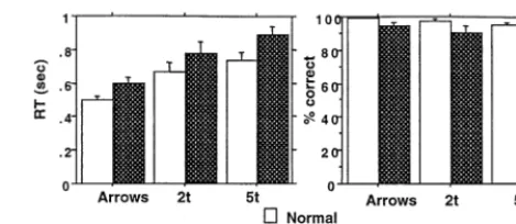
All of the normal subjects and eight of nine schizophrenic subjects performed significantly above chance in all three conditions. One schizophrenic subject performed below chance in the 5t condition only. She was not excluded because her 5t errors were primarily omissions, probably reflecting slow RT rather than disengagement from the task as was suggested by her performance in the other conditions. Schizophrenic subjects showed a trend to have longer RTs [F(1,16)5 3.84, p5 .07], and there was no interaction of diagnosis with condition (Figure 2). Schizo-phrenic subjects made significantly more errors [F(1,16)57.33, p5.02] than did normal subjects. There was a significant interaction of diagnosis by condition for errors [F(2,32) 5 4.21, p 5 .02; Figure 2]. Normal subjects were less variable in error performance than were schizophrenic subjects and performed near ceiling level across conditions.
Group Comparisons of DLPFC Activation: Data from Individuals
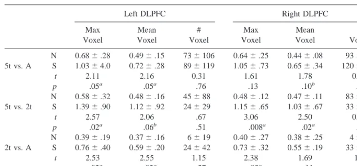
All of the normal subjects and eight of nine schizophrenic subjects exhibited DLPFC activation in the 5t versus Arrows comparison (Figure 1A depicts bilateral DLPFC activation in a schizophrenic subject). (Analyses of acti-vation use the max voxel index unless otherwise speci-fied.) Schizophrenic subjects showed a significantly greater magnitude of activation than did normal subjects in the left but not the right DLPFC (Table 2). In the 5t versus 2t comparison, schizophrenic subjects showed signifi-cantly greater activation in both the right and left DLPFC, demonstrating that group differences were not attributable to the Arrows baseline condition.
We also compared DLPFC activation when the groups
were matched for WM performance. When we compared the schizophrenic group’s performance in the 2t condition with the normal group’s performance in the 5t condition, the groups did not differ in either RT (t50.55, p5.59) or errors (t 51.20, p5 .25; Figure 2). The activation of schizophrenic subjects in the 2t versus Arrows comparison did not differ from that of the normal subjects in the 5t versus Arrows comparison (left DLPFC: t 5 0.53, p 5
.60; right DLPFC: t50.70, p 5.49).
The groups were not different in the lateralization of DLPFC activation (quantified by the equation (right 2
left)/(right 1 left)) in the 5t versus Arrows comparison (t 50.26, p5 .80). Within each group, activation of the right and left DLPFC was comparable (schizophrenia: t5
0.10, p 5.92; normal: t5 0.37, p5 .72).
The findings using the mean voxel index were generally consistent with those of the max voxel index (Table 2). The groups did not differ in the # voxels index, however. As in our previous study (Manoach et al 1999b), this index showed a high degree of intersubject variability and for this reason may be relatively insensitive to group differences.
Relation of DLPFC Activation to WM Performance
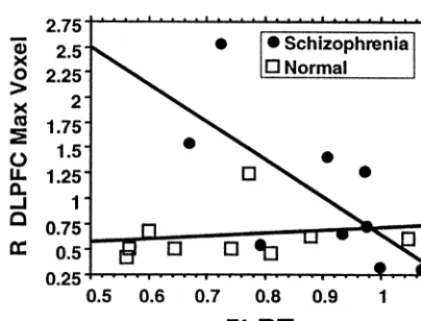
Within the schizophrenic group, better performance (fewer errors and shorter RTs) was consistently related to in-creased activation (5t vs. Arrows comparison) and several of these relations met or approached significance for the right DLPFC (Table 3 and Figure 3). In the normal group, DLPFC activation was not significantly related to RT or errors, but the interpretation of the correlations for errors is limited by the severely restricted range of errors in normal subjects. Analyses of covariance with an interac-tion of group with the covariate did not reveal significant group differences in the relations of activation to performance.
Group Comparisons of Regional Brain Activation: Averaged Group Data
DLPFC ACTIVATION. The averaged group data was analyzed to identify and map significantly activated vox-els. Both groups exhibited bilateral DLPFC activation in the 5t versus Arrows comparison; however, the groups activated different regions of the DLPFC. Only 3.5% of the voxels activated by the schizophrenic group over-lapped with those activated by the normal group (Table 4, Figure 1B, a). Within each group, the location of DLPFC activation was consistent across each of the three compar-isons of task conditions. This suggests that the group difference in location of DLPFC activation was not an artifact of the Arrows baseline condition (5t vs. 2t) or of task performance differences (Figure 1B illustrates DLPFC activation in the matched performance group comparison).
In addition, the normal group activated more DLPFC voxels than did the schizophrenic group (normal group: 310; schizophrenic group: 172). This finding from the
averaged group data is discrepant with the data derived from individual subjects in which the schizophrenic group activated more voxels (though not significantly more). Within each group, we examined the overlap of activation clusters within the DLPFC for each individual with those of the averaged group data. In the schizophrenic group, only 24% of the individual clusters overlapped with the group clusters, while in the normal group, 71% of the individual clusters overlapped. These findings indicate that the schizophrenic group was more heterogeneous in the spatial distribution of activated voxels within the DLPFC.
BASAL GANGLIA AND THALAMIC ACTIVATION. The most striking group difference in activation is that only the schizophrenic group activated the thalamus and basal ganglia including the head of the caudate and the lentiform nucleus (Figure 1B, a). Inspection of the indi-vidual data revealed that seven out of nine schizophrenic subjects (including the unmedicated subject and the sub-Table 2. Means, Standard Deviations, and t Tests of Group Differences in Dorsolateral Prefrontal
Cortex (DLPFC) Activation
Left DLPFC Right DLPFC Max
Voxel
Mean Voxel
# Voxel
Max Voxel
Mean Voxel
# Voxel N 0.686.28 0.496.15 736106 0.646.25 0.446.08 936158 5t vs. A S 1.0364.0 0.726.28 896119 1.056.73 0.656.34 1206216
t 2.11 2.16 0.31 1.61 1.78 0.30
p .05a .05a .76 .13 .10b .77
N 0.586.32 0.486.16 45688 0.486.12 0.476.11 836193 5t vs. 2t S 1.396.90 1.126.92 24629 1.156.65 1.036.67 33662
t 2.57 2.06 .67 3.06 2.50 0.75
p .02a .06b .51 .008a .02a .47
N 0.396.19 0.376.16 6619 0.406.27 0.386.25 467 2t vs. A S 0.766.40 0.596.20 24642 0.736.32 0.556.19 33647
t 2.53 2.55 1.15 2.38 1.69 1.8
p .02a .02a .27 .03a .11 .09b
The findings are presented by hemisphere for each activation index for the 5t vs. Arrows, 5t vs. 2t, and 2t vs. Arrows comparisons. N, normal group; S, schizophrenic group.
aSignificant at p
#.05.
bTrend at p
#.10.
Table 3. Relations of Dorsolateral Prefrontal Cortex (DLPFC) Activation in the 5t vs. Arrows Comparison to Working Memory Performance as Measured by Reaction Time (RT) and Errors in the Normal (N) and Schizophrenic (S) Groups
Group Condition
Left DLPFC Right DLPFC
RT Errors RT Errors
N 5t r5.38, p5.33 r5.51, p5.17 r5.19, p5.64 r5.60, p5.09a
2t r5.42, p5.27 r5 2.52, p5.16 r5.04, p5.92 r5 2.43, p5.26 S 5t r5 2.32, p5.41 r5 2.35, p5.37 r5 2.69, p5.04b r
5 2.62, p5.08a
2t r5 2.40, p5.30 r5 2.43, p5.26 r5 2.75, p5.02b r
5 2.59, p5.10a
aTrend at p
#.10.
bSignificant at p
ject on an atypical antipsychotic medication) and none of the normal subjects activated these regions. The schizo-phrenic group also activated these regions in the 2t versus 5t (not shown) and 2t versus Arrows comparisons (Figure 1B), but to a lesser extent. Again, this suggests that the differential activation of these regions is not a function of either the baseline condition or task performance differences.
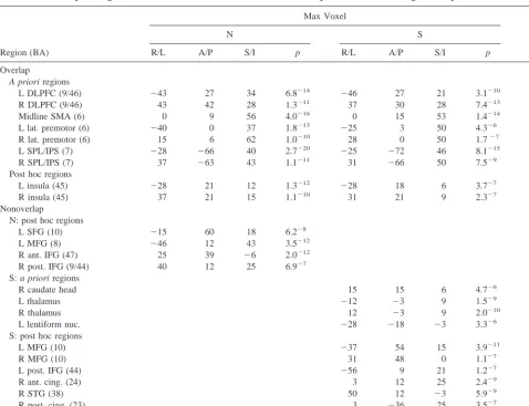
OTHER REGIONS. As predicted, the normal and schizophrenic groups showed overlapping activation in lateral premotor and motor areas, the supplementary motor area, and the intraparietal sulcus. Both groups also acti-vated the insula. The amount of overlap in these regions was variable, but exceeded the amount of overlap in the DLPFC. The groups also showed nonoverlapping activa-tion in a number of regions (Table 4).
Data Quality Considerations
Although eight out of nine normal subjects used a bite bar for head stabilization, only two out of nine schizophrenic subjects could because of poor dentition. This likely contributed to their significantly increased motion relative to normal subjects (average maximal displacement within a scan: normal: 0.29 mm 6 0.16; schizophrenia: 1.73 mm 6 1.2; t 5 3.57, p 5 .003). Group differences in motion represent a potential confound in our comparisons of activation. Motion can increase the variance of the fMRI signal and is usually associated with decreased power to detect differences between conditions; however, task-related motion may also artifactually lead to activa-tion (Hajnal et al 1994). Moactiva-tion was not correlated with
either right or left DLPFC activation (max voxel index; 5t vs. Arrows comparison) in either group. In addition, the groups were not different in the variability of signal intensity in either the right or left DLPFC as defined according to the a priori Talairach coordinates (variability was measured by taking the mean of the standard devia-tions of voxel signal intensity for every DLPFC voxel across all of the scans and within each of the three conditions). These findings suggest that the group differ-ences in DLPFC activation are not a function of increased motion or variance in the schizophrenic group. In addition, our DLPFC findings replicate those of our previous study in which the groups were not different with regard to motion (Manoach et al 1999b).
Analysis of Control Variables
Years of education, estimated verbal IQ, and parental socioeconomic status were not correlated with either DLPFC activation (5t vs. Arrows comparison) or task performance in either group. Within the schizophrenic group, DLPFC activation and task performance were not correlated with general psychopathology (BPRS) or rat-ings of positive or negative symptoms. Although the power to detect real relationships is low in nine subjects, none of the relations computed approached statistical significance, and there was no consistency in the direction of the relations of the control variables to DLPFC activa-tion and task performance.
Discussion
Relative to normal subjects, schizophrenic subjects showed at least an equal magnitude of right DLPFC activation and significantly greater left DLPFC activation during WM performance. This replicates our previous study using the same paradigm but different subjects, scanners, imaging methodology, and analysis techniques (Manoach et al 1999b). The schizophrenic group also activated the basal ganglia and thalamus, even when matched for performance with the normal group. These findings suggest that schizophrenic subjects recruit differ-ent neural circuitry for WM performance.
These findings contrast with the literature that demon-strates “task-related hypofrontality” in schizophrenia. Findings of hypofrontality have been challenged as a possible artifact of poor task performance. Several factors may have contributed to our finding of increased DLPFC activation. We rewarded correct responses, subjects were able to perform the task accurately, and, because accurate responding is predicated on the internal representation of items, we ensured that subjects used WM rather than an alternate strategy for task performance. We hypothesize
that reward may have enhanced motivation, task perfor-mance, and activation. This is consistent with studies of single-unit recordings in the principal sulcus of primates that demonstrate increased firing of WM neurons during WM delays in anticipation of a preferred reward (Wa-tanabe 1996). With regard to the relation of activation to task performance, recent findings suggest that although DLPFC activation increases with WM load (Braver et al 1997), when WM capacity is exceeded, DLPFC activation decreases (Callicott et al 1999; Goldberg et al 1998). Although the schizophrenic subjects performed signifi-cantly worse than the normal subjects did, the WM load did not exceed their capacity. Their increased DLFPC activation and poorer performance may reflect that given identical WM load, performance was more effortful for them (Frith et al 1995). The groups did not differ in
DLPFC activation when they were matched for perfor-mance by reducing the WM load for the schizophrenic group. Previous studies that employed tasks with greater WM demands may have exceeded the WM capacity of schizophrenic subjects and consequently found hypofron-tality. Finally, our findings may reflect that the SIRP differs from many other WM tasks (e.g., n-back, Tower of London, Wisconsin Card Sort Test) in that it constrains strategy and emphasizes the maintenance component of WM rather than manipulative processes such as the updating and temporal tagging of the contents of WM.
Another potential contributor to the different findings is that, in addition to examining averaged group data, we measured activation in individual subjects. Many, but certainly not all (e.g., Callicott et al 1998) previous studies that demonstrated task-related hypofrontality relied on Table 4. Summary of Regional Brain Activation in the 5t vs. Arrows Comparison for the Averaged Group Data
Region (BA)
Max Voxel
% Overlap
N S
R/L A/P S/I p R/L A/P S/I p
Overlap
A priori regions
L DLPFC (9/46) 243 27 34 6.8214
246 27 21 3.1210 3.5
R DLPFC (9/46) 43 42 28 1.3211 37 30 28 7.4213 3.5
Midline SMA (6) 0 9 56 4.0216 0 15 53 1.4214 54.1
L lat. premotor (6) 240 0 37 1.8213
225 3 50 4.326 20.0
R lat. premotor (6) 15 6 62 1.0210 28 0 50 1.727 12.5
L SPL/IPS (7) 228 266 40 2.7220
225 272 46 8.1215 93.0
R SPL/IPS (7) 37 263 43 1.1211 31
266 50 7.529 72.6
Post hoc regions
L insula (45) 228 21 12 1.3212
228 18 6 3.727 61.1
R insula (45) 37 21 15 1.1210 31 21 9 2.327 76.9
Nonoverlap
N: post hoc regions
L SFG (10) 215 60 18 6.228
L MFG (8) 246 12 43 3.5212
R ant. IFG (47) 25 39 26 2.0212
R post. IFG (9/44) 40 12 25 6.927
S: a priori regions
R caudate head 15 15 6 4.726
L thalamus 212 23 9 1.529
R thalamus 12 23 9 2.0210
L lentiform nuc. 228 218 23 3.326
S: post hoc regions
L MFG (10) 237 54 15 3.9211
R MFG (10) 31 48 0 1.127
L post. IFG (44) 256 9 21 1.227
R ant. cing. (24) 3 12 25 2.429
R STG (38) 50 12 23 5.929
R post. cing. (23) 3 236 25 3.527
L ITG (37) 250 257 29 7.8211
group averaging. Such methods may underestimate DLPFC activation in schizophrenia because of increased heterogeneity of the location of activation within the DLPFC. We found that the schizophrenic and normal groups activated different subterritories of the DLPFC and that as individuals, schizophrenic subjects were more variable than normal subjects were in the location of DLPFC activation. Increased spatial heterogeneity of ac-tivation in schizophrenia has also been reported in motor regions during performance of a sensorimotor task (Holt et al 1998) and in the DLPFC during performance of the n-back WM task (Holt et al 1999). There are several possible explanations for the increased spatial heterogene-ity and different location of DLPFC activation in the schizophrenic group. There is substantial structural vari-ability of the DLPFC in normal subjects (Rajkowska and Goldman-Rakic 1995). In imaging studies, this is compen-sated for, in part, by spatial normalization and image smoothing. Schizophrenic subjects may be even more variable than normal subjects are in the gross morphology of the DLPFC, its functional organization, or both. The current study cannot distinguish between these possibili-ties. They may also be more variable and less efficient in their use of strategies to accomplish the task. In this way, their differential activation may represent a compensatory response to dysfunction of WM neural circuitry. Although increased motion may have contributed to these findings, it is unlikely a complete explanation because, in contrast to the DLPFC, the schizophrenic and normal groups showed substantial overlap of activation in several cortical regions that are repeatedly associated with WM performance (Cohen et al 1997; Jonides et al 1998; Manoach et al 1997; Smith et al 1998). These a priori regions were the supplementary motor area, lateral premotor and motor areas, and the intraparietal sulcus (Figure 1B).
Although we did replicate the major findings of our previous studies (Manoach et al 1997, 1999b), findings regarding the laterality of DLPFC activation were not entirely consistent. We did not replicate our previous post hoc findings of differences in the laterality of DLPC activation between the schizophrenic and normal groups (Manoach et al 1999b). In addition, although both studies predicted and found relationships between better task performance and increased DLPFC activation in the schizophrenic group, the findings were not identical. In the present study, better performance (RT and errors) was related to increased right DLPFC activation, whereas in the previous study, RT was unrelated to activation, but increased response accuracy (errors) was related to in-creased left DLPFC activation. These inconsistent findings may be a consequence of insufficient power, and larger samples will be required to evaluate them.
The most striking finding is that only the schizophrenic
group activated the thalamus and basal ganglia. Neurolep-tic exposure may have contributed to this differential activation. Conventional neuroleptics can alter both the resting perfusion (Miller et al 1997) and volume of basal ganglia structures (Chakos et al 1994). Although it is unclear how such changes might relate to the task-related differences observed here, we cannot rule out a medication effect.
Accumulating evidence from single neuron recording and lesion studies in animals (Apicella et al 1992; Battig et al 1960) and lesion and dysfunction studies in humans (Owen et al 1997; Partiot et al 1996) suggests that frontostriatal neural circuitry subserves WM. Several neu-roimaging studies of normal WM report basal ganglia and thalamic activation under conditions of increased WM load (Barch et al 1997; Callicott et al 1999; Goldberg et al 1998; Rypma et al 1999). Activation of these regions in the schizophrenic group only may reflect diminished WM capacity; however, even when the groups were matched for performance, only the schizophrenic group activated the basal ganglia and thalamus.
anatomic components of this circuitry to WM deficits in schizophrenia.
This study was supported by the Scottish Rite Schizophrenia Research Program (DSM), The G. Harold and Leila Y. Mathers Charitable Foundation (CBS, DSM), the National Institute on Drug Abuse, K21-DA00275 (RLG), the National Alliance for Research on Schizophrenia and Depression (SLR), and the National Institute of Mental Health, MH01215 (SLR). The authors gratefully acknowledge the contributions of Robert Weisskoff, Edward Amico, Mary Foley, Daniel Z. Press, Alvaro Pascual-Leone, and Todd Kramer.
References
Alexander GE, Delong MR, Strick PL (1986): Parallel organi-zation of functionally segregated circuits linking basal gan-glia and cortex. Annu Rev Neurosci 9:357–381.
Apicella P, Scarnati E, Ljungberg T, Schultz W (1992): Neuronal activity in monkey striatum related to the expectation of predictable environmental events. J Neurophysiol 68:945– 960.
Baddeley A (1992): Working memory. Science 255:556 –559. Barch DM, Braver TS, Nystrom LE, et al (1997): Dissociating
working memory from task difficulty in human prefrontal cortex. Neuropsychologia 35:1373–1380.
Battig K, Rosvold HE, Mishkin M (1960): Comparison of the effect of frontal and caudate lesions on delayed response and alternation in monkeys. J Comp Physiol Psychol 53:400 – 404.
Blair JR, Spreen O (1989): Predicting premorbid IQ: A revision of the National Adult Reading Test. Clin Neuropsychol 3:129 –136.
Braver TS, Cohen JD, Nystrom LE, et al (1997): A parametric study of prefrontal cortex involvement in human working memory. Neuroimage 5:49 – 62.
Buchsbaum MS, Haier RJ, Potkin SG, et al (1992): Frontostriatal disorder of cerebral metabolism in never-medicated schizo-phrenics. Arch Gen Psychiatry 49:935–942.
Bush G, Whalen PJ, Rosen BR, et al (1998): The counting stroop: An interference task specialized for functional neuro-imaging—validation study with functional MRI. Hum Brain
Mapping 6:270 –282.
Callicott JH, Mattay VS, Bertolino A, et al (1999): Physiological characteristics of capacity constraints in working memory as revealed by functional MRI. Cereb Cortex 9:20 –26. Callicott JH, Ramsey NF, Tallent K, et al (1998): Functional
magnetic resonance imaging brain mapping in psychiatry: Methodological issues illustrated in a study of working memory in schizophrenia. Neuropsychopharmacology 18: 186 –196.
Carter C, Robertson L, Nordahl T, et al (1996): Spatial working memory deficits and their relationship to negative symptoms in unmedicated schizophrenia patients. Biol Psychiatry 40: 930 –932.
Chakos MH, Lieberman JA, Bilder RM, et al (1994): Increase in caudate nuclei volumes of first-episode schizophrenic pa-tients taking antipsychotic drugs. Am J Psychiatry 151:1430 – 1436.
Cohen JD, Perlstein WM, Braver TS, et al (1997): Temporal dynamics of brain activation during a working memory task.
Nature 386:604 – 608.
D’Esposito M, Grossman M (1996): The physiological basis of executive function and working memory. Neuroscientist 2:345–352.
Friedman HR, Goldman-Rakic PS (1994): Coactivation of pre-frontal cortex and inferior parietal cortex in working memory tasks revealed by 2DG functional mapping in the rhesus monkey. J Neurosci 14:2775–2788.
Frith CD, Friston KJ, Herold S, et al (1995): Regional brain activity in chronic schizophrenic patients during the perfor-mance of a verbal fluency task. Br J Psychiatry 167:343–349. Goldberg TE, Berman KF, Fleming K, et al (1998): Uncoupling cognitive workload and prefrontal cortical physiology: A PET rCBF study. Neuroimage 7:296 –303.
Goldberg TE, Weinberger DR (1996): Effects of neuroleptic medications on the cognition of patients with schizophrenia: A review of recent studies. J Clin Psychiatry 57(suppl 9):62– 65.
Hajnal JV, Myers R, Oatridge A, et al (1994): Artifacts due to stimulus correlated motion in functional imaging of the brain.
Magn Reson Med 31:283–291.
Hollingshead AB (1965): Two Factor Index of Social Position. New Haven, CT: Yale University Press.
Holt JL, Van Horn JD, Esposito G, et al (1998, November): Variability in functional neuroanatomy in schizophrenia: Group vs. single-subject PET activation data. Abstract pre-sented at the 28th annual meeting of the Society for Neuro-science, Los Angeles.
Holt JL, Van Horn JD, Meyer-Lindenberg A, et al (1999, October): Multiple sources of signal abnormality underlying prefrontal hypofunction and increased variability in the sites of activation within BA 9/46 in individual medication free schizophrenic patients. Abstract presented at the 29th annual meeting of the Society for Neuroscience, Miami Beach, FL. Houk JC, Davis JL, Beiser DG (1995): Models of Information
Processing in the Basal Ganglia. Cambridge, MA: MIT
Press.
Houk JC, Wise SP (1995): Distributed modular architectures linking basal ganglia, cerebellum, and cerebral cortex: Their role in planning and controlling action. Cereb Cortex 5:95– 110.
Jiang A, Kennedy D, Baker J, et al (1995): Motion detection and correction in functional MR imaging. Hum Brain Mapping 3:1–12.
Jonides J, Schumacher EH, Smith EE, et al (1998): The role of parietal cortex in verbal working memory. J Neurosci 18: 5026 –5034.
Jueptner M, Weiller C (1998): A review of differences between basal ganglia and cerebellar control of movements as revealed by functional imaging studies. Brain 121:1437–1449. Kay SR, Fiszbein A, Opler LA (1987): The positive and negative
syndrome scale (PANSS) for schizophrenia. Schizophr Bull 13:261–276.
Knight RA, Manoach DS, Elliott DS, Hershenson M (in press): Perceptual organization in schizophrenia: the processing of symmetrical configurations. J Abnorm Psychol.
Differential activation of the caudate nucleus in primates performing spatial and nonspatial working memory tasks.
J Neurosci 17:3870 –3882.
Manoach DS, Benson ES, Chang Y, et al (1999a, October): Test-retest reliability of a fMRI working memory paradigm in normal and schizophrenia subjects. Abstract presented at the 29th annual meeting of the Society for Neuroscience, Miami Beach, FL.
Manoach DS, Press DZ, Thangaraj V, et al (1999b): Schizo-phrenic subjects activate dorsolateral prefrontal cortex during a working memory task as measured by fMRI. Biol
Psychi-atry 45:1128 –1137.
Manoach DS, Schlaug G, Siewert B, et al (1997): Prefrontal cortex fMRI signal changes are correlated with working memory load. Neuroreport 8:545–549.
Mattay VS, Callicott JH, Bertolino A, et al (1997): Abnormal functional lateralization of the sensorimotor cortex in patients with schizophrenia. Neuroreport 8:2977–2984.
Miller DD, Rezai K, Alliger R, Andreasen NC (1997): The effect of antipsychotic medication on relative cerebral blood perfu-sion in schizophrenia: assessment with technetium-99m hexa-methyl-propyleneamine oxime single photon emission com-puted tomography. Biol Psychiatry 41:550 –559.
NIMH (1974): Abnormal Involuntary Movement Scale (AIMS), U.S. Public Health Service Publication No. MH-9-17. Wash-ington, DC: U.S. Government Printing Office.
Overall JE, Gorham DR (1962): The brief psychiatric rating scale. Psychol Rep 10:799 – 812.
Owen AM, Iddon JL, Hodges JR, et al (1997): Spatial and non-spatial working memory at different stages of Parkin-son’s disease. Neuropsychologia 35:519 –532.
Park S, Holzman PS (1992): Schizophrenics show spatial work-ing memory deficits. Arch Gen Psychiatry 49:975–982. Park S, Puschel J, Sauter BH, et al (1999): Spatial working
memory deficits and clinical symptoms in schizophrenia: A 4-months follow-up study. Biol Psychiatry 46:392– 400. Partiot A, Verin M, Pillon B, et al (1996): Delayed response tasks
in basal ganglia lesions in man: further evidence for a striato-frontal cooperation in behavioural adaptation.
Neuro-psychologia 34:709 –721.
Petrides M, Alivisatos B, Evans AC, Meyer E (1993a): Dissoci-ation of human mid-dorsolateral from posterior dorsolateral frontal cortex in memory processing. Proc Natl Acad Sci
U S A 90:873– 877.
Petrides M, Alivisatos B, Meyer E, Evans AC (1993b):
Func-tional activation of the human prefrontal cortex during the performance of verbal working memory tasks. Proc Natl
Acad Sci U S A 90:878 – 882.
Rajkowska G, Goldman-Rakic PS (1995): Cytoarchitectonic definition of prefrontal areas in the normal human cortex: II. Variability in locations of areas 9 and 46 and relationship to the Talairach coordinate system. Cereb Cortex 5:323–337. Rubin P, Holm S, Friberg L, et al (1991): Altered modulation of
prefrontal and subcortical brain activity in newly diagnosed schizophrenia and schizophreniform disorder. Arch Gen
Psy-chiatry 48:987–995.
Rypma B, Prabhakaran V, Desmond JE, et al (1999): Load-dependent roles of frontal brain regions in the maintenance of working memory. Neuroimage 9:216 –226.
Siegel B, Buchsbaum M, Bunney W, et al (1993): Cortical-striatal-thalamic circuits and brain glucose metabolism activ-ity in 70 unmedicated male schizophrenic patients. Am J
Psychiatry 150:1325–1336.
Silverstein SM, Bakshi S, Chapman RM, Nowlis G (1998): Perceptual organization of configural and nonconfigural ef-fects of repeated exposure. Cogn Neuropsychiatry 3:209 – 223.
Simpson GM, Angus JWS (1970): A rating scale for extrapyra-midal side effects. Acta Psychiatr Scand Suppl 212:11–19. Smith EE, Jonides J, Marshuetz C, Koeppe RA (1998):
Compo-nents of verbal working memory: Evidence from neuroimag-ing. Proc Natl Acad Sci U S A 95:876 – 882.
Spitzer RL, Williams JB, Gibbon M, First MB (1992): The Structured Clinical Interview for DSM-III-R (SCID). I: His-tory, rationale, and description. Arch Gen Psychiatry 49:624 – 629.
Sternberg S (1966): High-speed scanning in human memory.
Science 153:652– 654.
Talairach J, Tournoux P (1988): Co-Planar Stereotaxic Atlas of
the Human Brain. New York: Thieme Medical Publishers.
Watanabe M (1996): Reward expectancy in primate prefrontal neurons. Nature 382:629 – 632.
Weinberger DR, Berman KF (1996): Prefrontal function in schizophrenia: Confounds and controversies. Philos Trans R
Soc Lond B Biol Sci 351:1495–1503.
White K, Ashton R (1976): Handedness assessment inventory.
Neuropsychologia 14:261–264.
Woods RP, Cherry SR, Mazziotta JC (1992): Rapid automated algorithm for aligning and reslicing PET images. J Comput
![Figure 1. (A) Left:schizophrenic subject during WM performance (five targets [5t] vs. Arrows comparison)](https://thumb-ap.123doks.com/thumbv2/123dok/3144205.1383684/4.612.68.543.78.547/figure-left-schizophrenic-subject-performance-targets-arrows-comparison.webp)