International Journal of Integrated Health Sciences. 2015;3(1) 11 Correspondence:
Tjahhjodjati, Urology Division, Department of Surgery, Faculty of Medicine, Universitas Padjadjaran-Dr. Hasan Sadikin General Hospital
Jl. Pasteur No. 38, Bandung, Indonesia e-mail: [email protected]
Ultrasound Guided Fine Needle Aspiration Biopsy in Renal Tumors
Tjahjodjati, Joko PitoyoUrology Division, Department of Surgery, Faculty of Medicine, Universitas Padjadjaran-Dr . Hasan Sadikin General Hospital
Abstract Objective: To assess the sensitivity and specificity of ultrasound guided fine
needle aspiration biopsy (FNAB) in renal tumors.
Methods: A diagnostic study was performed on 23 patients with renal tumors who visited the Urology Division, Department of Surgery, Dr. Hasan Sadikin General Hospital from January 2011 to August 2012. First time ultrasound guided was performed to the patients before nephrectomy. Renal tissues that were obtained from both procedures were examined histopathologically, and the result from nephrectomy was used as the gold standard. Analysis was
conducted by measuring the sensitivity, specificity, positive predictive value
(PPV), and negative predictive value (NPV) of the ultrasound guided FNAB.
Results: The results showed that the sensitivity value of ultrasound guided
FNAB was 85.71%, with 50% specificity, 94.74% PPV and 25% NPV. From 23
patients, only 2 patients had hematoma.
Conclusions: Ultrasound guided FNAB is sufficiently sensitive with a fair
specificity for diagnosing renal tumors and is safe for patients.
Keywords:Renal tumor, sensitivity, specificity, ultasound guided FNAB
IJIHS. 2015;3(1):11–3
Introduction
Renal cell carcinoma (RCC) represents about 2–3% of all cancers with the highest incidence occurring in western countries. During the last two decades, there has been an increase in the incidence of approximately 2% per year in Europe and throughout the world. Renal cell carcinoma is a solid tumor and accounts for
approximately 90% of all malignancies found
in kidney.1 Nowadays in western countries, most renal tumors (60%) are found in early
stage (less than 4 cm).2 Kidney tumors can be
primary tumors or secondary tumors such as lymphoma.3
In determining the histopathology of the tumor, a biopsy procedure is performed. Biopsy procedure comes in various types, including percutaneous biopsy and open biopsy. The percutaneous renal biopsy has been used for
a long time, but it has not been used anymore in the last decade because of the low accuracy in retrieving tumor tissues.4–6 With further development of imaging equipments, such as ultrasound, Computerized Tomography scan (CT scan) and Magnetic Resonance Imaging (MRI), percutaneous biopsy is reused, albeit in combination with those equipments.2,3,5,7–9 Remzi and Marberger6 reported that biopsy
using CT-scan guided fine-needle aspiration
method in kidney tumors has the ability to
predict the presence of 92–96% malignancy, 67–70% grading of tumors, and 78–92%
tumor’s subtype determination.A report from Reichelt et al.10 stated that ultrasound guided Fine Needle Aspiration Biopsi (FNAB) has an accuracy of 83.3% in predicting renal tumors. The use of ultrasound guided FNAB has many advantages when compared to CT-scan or MRI. In addition, ultrasound is cheaper and it does not involve radiation exposure. This ultrasound can be performed in real-time and is more readily available in hospitals, especially in Indonesia. The study was conducted to
measure the sensitivity and specificity of the
ultrasound guided FNAB for renal mass tumor.
Original Article
Received: June 17, 2014
Revised:
September 18, 2014
Accepted: February 4, 2015
12 International Journal of Integrated Health Sciences. 2015;3(1)
Methods
A diagnostic study on 23 patients with renal tumors who visited the Urology Division of the Department of Surgery, Dr. Hasan Sadikin General Hospital was performed in the period of January 2011 to August 2012. Patients underwent ultrasound guided FNAB in the operating room shortly before nephrectomy.
The procedure used 25G or 23G biopsy fine
needle and the biopsy was performed once
or twice until sufficient tumor tissues were
obtained for histopathologic examination. The histopathologic examination was performed by two different pathologists. One examined the tumor tissues from the FNAB and the other examined the tumor tissues from the nephrectomy. The gold standard used was the histopathology results from the nephrectomy. The histopathology results that were obtained from both specimens were then compared and analyzed using 2x2 table diagnostic test
consisting of sensitivity, specificity, positive
predictive value (PPV) and negative predictive value (NPV) values.
Results
This study found that from 23 samples, 14 of them were renal tumors with T4, followed by
6 with T3 and 3 with T2 (Table 1).
Table 1 Staging of Renal Tumors
Stage Frequency
Fine-needle biopsy can cause complications such as hematoma. In this study, there were 2 cases of hematoma out of 23 samples.
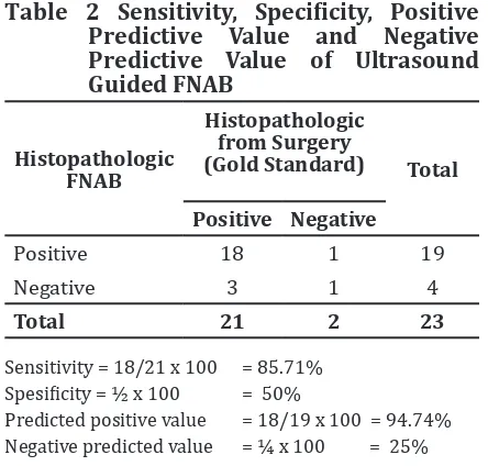
The sensitivity was 85.71%, specificity was 50%, PPV was 94.74% and NPV was 25% for
ultrasound guided FNAB in this study (Table 2).
Two cases with T2 and one case with T3 that were found negative for malignancy based on the ultrasound guided FNAB histopathologic results were positive based
on the results of the surgery histopathologic
results. On the contrary, one case with T4 that
received positive result for malignancy based on the histopathologic results from ultrasound guided FNAB was negative based on the results of the surgery.
Table 2 Sensitivity, Specificity, Positive Predictive Value and Negative Predictive Value of Ultrasound Guided FNAB
Positive 18 1 19
Negative 3 1 4
Total 21 2 23
Sensitivity = 18/21 x 100 = 85.71% Spesificity = ½ x 100 = 50%
Predicted positive value = 18/19 x 100 = 94.74% Negative predicted value = ¼ x 100 = 25%
Discussion
Fine-needle biopsy can be used to avoid open-biopsy surgery for patients with high risk factors for surgery.2,4,6 This study found that the accuracy of the renal tumor diagnosis using ultrasound guided FNAB has a sensitivity of 85.71%. This means that the histopathologic results from ultrasound guided FNAB is
sufficiently sensitive to diagnose malignancy.
This is supported by the fact that this approach
has a PPV of 94.74%. This is similar to the
result found by Reichelt et al.10 that reported the accuracy of ultrasound guided FNAB for renal tumor of 83.3%, mainly for a tumor
of less than 4 cm. Furthermore, the study found that the specificity of the ultrasound
guided FNAB was 50% and the NPV was 25%, meaning that the ability of ultrasound guided FNAB to detect a negative results was only 50% and the percentage of true negative cases was 25%. Remzi and Marberger6 reported that
CT-scan guided FNAB was able to predict 92– 96% malignancy, 67–70% grading the tumors, and 78–92% tumor’s subtype determination.
Most patients came to the hospital at T3 stage
or above. This situation influences the value of sensitivity and specificity of ultrasound guided
FNAB. Based on this information, further
Ultrasound Guided Fine Needle Aspiration Biopsy in Renal Tumors
International Journal of Integrated Health Sciences. 2015;3(1) 13 studies should be conducted on patients with
smaller tumors (less than T2).
Fine needle aspiration biopsy can cause complications such as hematoma with an incidence of less than 5%.6,8,10 There are also other complications, such as pneumothorax, tumor seeding, arterial-venous malformation (AVM), and death.2,3,6 Malformation of the arterial venous occurred in less than 2% of the cases and the mortality rate is 0.031%. Only 2 cases of hematoma were found in this study.
This indicates that the use of the ultrasound guided FNAB for renal tumors is relatively safe.
The ultrasound guided FNAB can be used to diagnose malignant kidney tumors. It can
be used to obtain sufficient tumor tissues
with no radiation effects. This approach is also affordable and a minimally invasive. Hence,it does not need general anesthesia and hospitalization.
Tjahjodjati, Joko Pitoyo
:11–3
References
1. Ljungberg B, Cowan N, Hanbury DC, Hora M, Kuczyk MA, Merseburger AS, et al. European
Association of Urology guidelines on renal cell carcinoma: the 2010 update. Eur Urol.
2010;58(3):398–406.
2. Volpe A, Finelli A, Gill IS, Jewett MA, Martignoni G, Polascik TJ, et al. Rationale for percutaneous biopsy and histologic characteristic or renal
tumours. Eur Urol. 2012;62(3):491–504.
3. Somani BK, Nabi G, Thorpe P, N’Dow J, Swami S, McClinton S, et al. Image-guided
biopsy-diagnosed renal cell carcinoma: Critical appraisal of technique and long-term
follow-up. Eur Urol. 2007;51(5):1289–95.
4. Schmidbauer J, Remzi M, Memarsadeghi M, Haitel A, Klingler HC, Katzanbeisser D, et al.
Diagnostic accuracy of computed tomography-guided percutaneous biopsy of renal masses. Eur Urol. 2008:53(5):1003–11.
5. Sahni VA, Silverman SG. Biopsy of renal masses:
when and why. Cancer Imaging. 2009;9:44–55.
6. Remzi M, Marberger M. Renal tumor biopsies for evaluation of small renal tumors: why, in
whom and how? Eur Urol. 2009;55(2):359–67.
7. Leveridge MJ, Finelli A, Kachura JR, Evans A, Chung H, Shiff DA, et al. Outcomes of small
renal mass needle core biopsy, nondiagnostuc percutaneous biopsy, and the role of repeat
biopsy. Eur Urol. 2011;60(3):578–84.
8. Crispen PL, Blute ML. Do percutaneous renal tumor biopsies at initial presentation affect treatment strategies? Eur Urol.
2009;55(2):307–9.
9. Kummerlin IP, Smedts F, Kate FJt, Horn T, Algaba F, Trias I, et al. Cytological punctures in the diagnosis of renal tumours: a study on accuracy and reproducibility. Eur Urol.
2009;55(1):187–95.
10. Reichelt O, Gajda M, Chyhrai A, Wunderlich H, Junker K, Schubert J. Ultrasound-guided biopsy of homogenous solid renal masses. Eur Urol.