www.elsevier.com/locate/jinsphys
Immunolocalization and possible effect of a moth allatotropin-like
substance in a fly, Phormia regina (Diptera: Calliphoridae)
Meng-Ping Tu
a, Rong Kou
b, Zong-Shun Wang
a, John G. Stoffolano Jr.
a,
Chih-Ming Yin
a,*aDepartment of Entomology, University of Massachusetts, Amherst, MA 01003, USA bInstitute of Zoology, Academia Sinica, Nankang 11259, Taipei, Taiwan, ROC
Received 15 December 1999; accepted 15 June 2000
Abstract
Insect allatotropin upregulates the biosynthesis of juvenile hormones by the corpus allatum. We raised two rabbit antisera against the allatotropin of Manduca sexta (Mas AT) using a synthetic, multiple-antigenic-peptide that contains a branching heptalysine core and eight Mas AT molecules. Both antisera recognized specifically the same neurons in the larval brain, frontal ganglion and terminal abdominal ganglion of M. sexta as previously reported by others. Immunoassay showed reactivity specific to the Mas AT. Very low or nearly no cross-reactivity was found for two Mas AT-like peptides, a myotropin from Locusta migratoria and a Mas AT-like peptide deduced from the DNA sequence of Aedes aegypti, respectively. Immunopositive neurons also were identified in adult Phormia regina, Dacus dorsalis, Oncopeltus fasciatus, and Mythimna loreyi, and in larval M. loreyi, Bombyx mori, and
Andraca bipunctata. At 20 pmol per 25µl incubation medium (i.e. 8×1027
M), synthetic Mas AT significantly stimulated in vitro juvenile hormone biosynthesis by the corpus allatum of adult, sugar-fed females of P. regina to 2.64-fold that of controls. Thus, this study provides the first demonstration that at the higher end of the physiological concentration range, the Mas AT has allatotropic effect in vitro to CA of non-lepidopterans. However, in vivo functions of Mas AT and/or Mas AT-like peptide in P. regina remain to be defined. 2001 Elsevier Science Ltd. All rights reserved.
Keywords: Aedes aegypti; Bombyx mori; Dacus dorsalis; Manduca sexta; Oncopeltus fasciatus; Juvenile hormone
1. Introduction
Anautogenous species, like Phormia regina, require a protein meal before oo¨genesis occurs (Fraenkel, 1940; Stoffolano et al., 1992; Yin and Stoffolano, 1990; Yin et al., 1993), while adults of autogenous species begin oo¨genesis without the need of a protein meal. This pro-tein meal serves at least two major functions (Yin and Stoffolano 1994, 1997). It activates the brain, which then triggers the entire neuroendocrine cascade [including the activation of corpus allatum (CA)] involved in the hor-monal regulation at each stages of oo¨genesis. It also vides raw materials needed for the formation of the
pro-* Corresponding author. Tel.:+1-413-545-1060; fax:+ 1-413-545-2115.
E-mail address: [email protected] (C.-M. Yin).
0022-1910/01/$ - see front matter2001 Elsevier Science Ltd. All rights reserved. PII: S 0 0 2 2 - 1 9 1 0 ( 0 0 ) 0 0 1 1 0 - 4
teins, carbohydrates, and lipids to be deposited in the yolk of developing oo¨cytes.
Experimentation with treatments like allatectomy, pre-cocene application, and hormone therapy showed that formation (including vitellogenin biosynthesis/release by the fat body) and uptake of vitellogenin by the developing oo¨cytes are regulated by juvenile hormones (JHs). A specific blend of JHs consisting of juvenile
hor-mone III bisepoxide (JHB3), juvenile hormone III (JH
connected to the brain via nerve fibers. In certain species, including the queen blow fly, Phormia regina, the CA is unpaired. The CA activity can be regulated by neural and/or endocrine stimulation or inhibition (Feyereisen, 1985). Two groups of neuropeptides, allatotropins (ATs)
and allatostatins (ASTs), secreted from the
brain/suboesophageal ganglion (SOG) neurosecretory cells, can up or downregulate the JH synthesis (Khan, 1988; Hoffmann et al., 1999), respectively. ASTs have been isolated from a criket, Gryllus bimaculatus, a moth,
Manduca sexta, a honey bee, Apis mellifera, and
cock-roaches, Blatella germanica, Diploptera punctata, and
Periplaneta americana. In vitro study shows that some
allatostatic factors may exist in the Drosophila brain (Moshitzky and Applebaum, 1995). Peptides that are structurally homologous with the cockroach allatostatins have been identified from the fly, Calliphora vomitoria and named callatostatins. However, callatostatins are myoinhibitory rather than allatostatic in this fly (Duve et al., 1994). To date, AT (Mas AT) has been isolated only from the heads of pharate adult M. sexta (Kataoka et al., 1989). Similar mode of CA control of JH synthesis has also been observed in larvae of the larger wax moth,
Galleria mellonella, and adults of locust, Locusta migratoria, cricket, G. bimaculatus, true bug, Pyrrhoc-oris apterus, and honey bee, A. mellifera (Ga¨de et al.,
1997). In blow flies, results show that brain median neu-rosecretory cells (MNCs) may release hormones to acti-vate the CA (Thomsen 1942, 1952; Thomsen and Moller, 1960; Applin 1979, 1981), but the chemical nat-ure of these hormones remains unknown. A thorough understanding of oo¨cyte development in P. regina, as well as in any other insect, should include a thorough understanding of how CA activity and JH biosynthesis are governed by these hormones.
To help better understand these governing mech-anisms, this paper reported the production of two rabbit anti-Mas AT antisera using a synthetic Mas AT immun-ogen designed following a multiple-antigenic-peptide (MAP) strategy of Tam (1988). Neurons positive to these anti-Mas AT antisera were revealed in the brain, frontal ganglion, and terminal abdominal ganglion of day-2, 5th instar M. sexta larvae. The cell number and location were exactly those previously reported by Taylor et al. (1996). Our antisera also revealed Mas AT-immunoreactive cells in the brain of both sugar- and liver-fed, adult females of P. regina. Likewise, the cor-pus cardiacum (CC) of liver-fed, adult females also con-tained Mas AT-immunoreactive substances. Similar immunostaining was found in the brain and/or frontal ganglion of the following insects: the adult large milk-weed bug, Oncopeltus fasciatus; the adult oriental fruit fly, Dacus dorsalis; the larval and adult loreyi leafworm,
Mythimna loreyi; and the larval tea silk moth, Andraca bipunctata. In contrast, no immunopositive cells appeared in the adult brain of the honey bee, A. mellifera
(foraging workers); the carpenter ant, Camponotus sp. (foraging workers); the Colorado potato beetle,
Leptinot-arsa decemlineata (adults); and the walking stick, Megacrania tsudai (adults).
Since immunopositive cells for Mas AT were ident-ified in P. regina, we were interested in knowing if Mas AT would stimulate JH biosynthesis by this fly. In vitro assay results showed that synthetic Mas AT significantly increases, in a dose-dependent manner, the JH pro-duction by the CA of sugar-fed, adult females of P.
regina. The presence of free/circulating Mas AT and/or
Mas AT-like substance, as well as their possible in vivo function, remains to be determined for this fly. Thus, we have demonstrated the presence and possible function of Mas AT and/or Mas AT-like substance in P. regina. Many questions remain. Is this substance the authentic Mas AT or just a homolog? Is there a fly-specific allato-tropin that is functionally related but structurally unre-lated to Mas AT in P. regina? Are there non-allatotropic biological functions for the Mas AT-like substance in P.
regina? These questions remain to be answered.
2. Materials and methods
2.1. Animals
P. regina was reared and maintained as previously
described (Stoffolano, 1974; Zou et al., 1988; Yin et al., 1994). Mature larvae were allowed to crawl out of the medium into sand to pupariate. Puparia were collected daily and allowed to emerge in screened cages. Flies emerging within 12 h were sexed and each sex placed in the same age group, or cohort (hour 0, day 1). During the first 3 days after emergence, a 4.3% sucrose solution was provided to all flies. The same sugar solution was also provided to all flies, ad libitum, following a feeding bout on beef liver. All flies were kept at 28±2°C under a 16 h light, 8 h dark photoregime. Beef liver was used to provide dietary protein, ad libitum, at 72 h of adult-hood for 1.5 h to obtain liver-fed flies. To better synch-ronize the flies’ physiology, specific procedures and pre-cautions in fly selection were followed as described by Yin et al. (1994).
Larvae of the tobacco hornworm, M. sexta, were obtained regularly as a generous gift from Dr E.S. Bow-dan, Departement of Biology, University of
Massachu-setts. They were reared at 28±2°C under a photoregime
of 16 h light and 8 h dark on an artificial diet (Carolina Biological Supply) as a group prior to the 3rd instar, and then kept individually until pupation. Day-2, 5th instar larvae were used in this study.
Honey bees and carpenter ants were collected at the University of Massachusetts, Amherst campus. D.
experimental colonies either at the Department of Ento-mology, University of Massachusetts, or at the Institute of Zoology, Academia Sinica, Taipei, Taiwan.
2.2. Design and synthesis of a Mas AT-MAP immunogen
The MAP immunogen consists of a core of branching heptalysine conjugated in a specific way to permit coval-ent linking of eight idcoval-entical peptides through their C-termini (Tam, 1988). In the present case, eight Mas AT molecules were linked to this core with eight free N-termini. This synthetic immunogen was made by Ms C.C. Lin of the Protein/Nucleic Acid Sequencer Core Facility, Institute of Zoology, Academia Sinica, using a model 432A Peptide Synthesizer from Applied Biosys-tems (Foster City, CA). The crude MAP immunogen was purified by HPLC on a 140B Solvent Delivery System equipped with an Aquapore RP-300 C 8 column (4.6×200 mm, 7µm) using a linear gradient solvent sys-tem (where solvent A was 0.1% aqueous TFA and B
was 0.08% TFA in CH3CN) at 0.3 ml/min flow rate.
Absorbance was monitored at 220 nm.
2.3. Immunization scheme and collection of antisera
Two female, New Zealand white rabbits (CY 16 and CY 17) were each immunized initially with the above described synthetic Mas AT immunogen injected (200
µg in 0.5 ml phosphate buffered saline) into the ear vein without any adjuvant. One month later, each rabbit received a subcutaneous booster injection, into the dorsal
neck fold, of 200 µg of immunogen emulsified in 250
µl of phosphate buffered saline and 250 µl of Freund’s
complete adjuvant. After three more weeks, each animal
received a second booster injection containing 400 µg
of immunogen in 250 µl of phosphate buffered saline
and 250 µl of Freund’s incomplete adjuvant delivered
intramuscularly into the thigh. One week later, anti-Mas AT antiserum was collected and tested for the presence of specific antibodies from each rabbit. Subsequent col-lections were carried out biweekly with or without prior booster injection until sufficient quantity of antisera was accumulated.
2.4. Characterization of the antisera
The specificity of our Mas AT antisera was determ-ined by a number of experiments. In conventionally designed immunogens for small peptides, molecules of a small peptide are linked to a large carrier, thus the resulting antiserum may contain considerable antibodies that are reactive, not to the small peptide, but to the car-rier. This undesirable side effect is less likely to occur to a MAP immunogen because the bulk of it is made of the multiple copies of the small peptide with a carrier
portion made of only seven lysines. To ascertain that our anti-Mas AT antisera have low or no reactivity to the lysine carrier, checker board experiments were perfor-med using ELISA wells coated with a different MAP immunogen (i.e. MylATr MAP consisted of eight copies of Ile–Tyr–Leu–Glu–Ile–Lys conjugated to the heptalys-ine core). Strips of ELISA wells were coated with 40
ng/well of the MylATr MAP in 100 µl of
carbonate/bicarbonate buffer (pH 9.6). After washing off the unadsorbed MylATr MAP, unoccupied well surfaces
were blocked with 200µl of a blocking solution
contain-ing gelatin and carbowax (Mr 20,000) for 30 min. The
wells were then thoroughly washed with TBST washing solution [TBST, 20 mM Tris, 500 mM NaCl, 0.01% (W/V) thimerosal, pH 7.5 plus 0.05% (V/V) Tween-20]. Antisera CY16 and CY17, at 1:1250: 1:2500; 1:5000; 1:10,000, 1:20,000; or 1:40,000 dilutions, in an antibody buffer [TBST plus 1.0% (W/V) bovine serum albumin], were added to the wells, respectively, after each well
was rehydrated by adding 200 µl of distilled water for
30 min. The strips were placed in a moist chamber to
reduce evaporation during the 3 h incubation at 37°C.
After this, the wells were rinsed seven times before
adding 100 µl/well of a peroxidase-conjugated affinity
purified goat anti-rabbit IgG (Jackson ImmunoResearch Laboratories, Inc., Plymouth Meeting, PA) diluted 1:4000 in the same antibody buffer. After loading the enzyme-linked secondary antibodies, the wells were
placed back into the moist chamber for 80 min at 37°C.
After this incubation, the strips were rinsed five times and developed for 60 min at room temperature with 100
µl of 3,39,5,59-tetramethylbenzidine and urea peroxide. The color development was stopped and color intensified
by adding 50µl/well of 0.2 M H2SO4. Absorbance was
recorded at 450 nm using a ThermoMax microtiter plate reader from Molecular Devices, Palo Alto, CA.
Following the protocol used before in our laboratory, a competitive enzyme-linked immunosorbent assay (cELISA) for Mas AT was developed using the above mentioned anti-Mas AT antisera. This cELISA was used to establish the reactivity between our antisera and the synthetic Mas AT (H–Gly–Phe–Lys–Asn–Val–Glu–
Met–Met–Thr–Ala–Arg–Gly–Phe–NH2). It was also
used to compare the possible cross-reactivity of our anti-sera to two structurally similar peptides. One of the two peptides was [Ala6, Leu7, Ser8]-allatotropin, a myotropin
to the cDNA sequence as an amidated peptide with 14
amino acids (Ala–Pro–Phe–Arg–Asn–Ser–Glu–Met–
Met–Thr–Ala–Arg–Gly–Phe). The Mas AT and the two Mas AT-related peptides, used in this study, were syn-thesized at the Protein/Nucleic Acid Sequencer Core Facility, Institute of Zoology, Academia Sinica, Taiwan. To set up the cELISA, each microtiter plate well was
coated with 40 ng of the Mas AT MAP in 100 µl of
carbonate/bicarbonate buffer. After adsorption of the coating immunogen and washing off the excessive Mas AT MAP, unoccupied well surfaces were blocked with
200 µl of the same blocking solution mentioned above
for 30 min at room temperature. Then, the wells were thoroughly washed with TBST, patted dry, and stored in a desiccator until the time of assay. It has been demon-strated that to set up an ELISA one may try a coating antigen that is structurally different from the immunizing immunogen to achieve a certain level of sensitivity and specificity (Wie and Hammock, 1984). However, for this present study, we reached our goal in sensitivity and specificity for our ELISA when using the same immuniz-ing immunogen as the coatimmuniz-ing antigen. Consequently, we decided not to invest money and time to make a different coating antigen.
Standard solutions of synthetic Mas AT, synthetic Lom MT, and synthetic deduced mosquito AT-like pep-tide were prepared. To begin each assay, the coating antigen on the well was first rehydrated with distilled
H2O for 45 min. Standard solutions (2.5 µl/well) were
added followed by 100µl of our anti-Mas AT antiserum
(CY16) at 1:400,000 dilution (CY17 was applied at 1:200,000 dilution) in the antibody buffer [i.e. TBST plus 1.0% (W/V) bovine serum albumin]. The incu-bation, washing, color development, stopping, and absorbance recording were the same as described above. From a different angle, the specificity of our antisera was determined by showing that certain identifiable AT neurons of larval M. sexta could be also recognized by our antisera. These neurons have been identified already by other investigators (Taylor et al., 1996) using in situ hybridization and an antiserum raised against Mas AT with an immunogen synthesized through a conjugation design quite different from the current MAP design. Day-2, 5th instar M. sexta larvae were anesthetized by
CO2and kept immobilized by chilling on ice. The brains
and ventral nerve cords were then dissected in ice-cold 4% (W/V) paraformaldehyde in sodium phosphate buffer (0.1 M, pH 7.4), after the insects had been injected with the same ice-cold paraformaldehyde solution. The neural tissues were removed from the larvae and kept in the fixative for approximately 18 h at 4°C.
Whole-mount preparations were processed using an immunofluorescence method adapted from Davis (1987). Briefly, the fixed tissues were washed thoroughly 4–6 times (1 h each) in phosphate buffered saline plus 0.5% Triton X-100 (PBST). The tissues were then blocked in
10% normal goat serum (NGS) in PBST for 1 h before incubating them in the anti-Mas AT antisera (CY17 diluted 1: 1000 in 10% NGS/PBST or CY16 at 1:500 dilution) for 48 h at 4°C (without agitation). After being treated with primary antiserum, tissues were washed five times (1 h each) in PBST, blocked a second time with 10% NGS in PBST for 1 h. Tissues were then treated with rhodamine (TRITC)-conjugated goat anti-rabbit IgG (1:200 dilution in 10% NGS/PBST) in the dark overnight (ca. 18 h) at room temperature. Tissues were then washed three times with PBST (20 min each) in the dark before clearing in a glycerol series (40, 60, and 80% glycerol in carbonate/bicarbonate buffer, pH 9.5, 30 min in each glycerol solution). Preparations were
stored in 80% glycerol at 4°C in the dark for later use
or mounted on a microscopic slide with Vectashield mounting medium. The edges of the coverglass were sealed with clear nail polish to extend the storage life of the preparations. The slides were observed using an epi-fluorescent microscope (Zeiss) equipped with a filter for rhodamine and photographed using an automatic Zeiss MC 63/M35F Photomicro Gaphic camera. For some specimens, digitized images were prepared with a con-focal microscope (MRC600 Krypton-Argon, Bio-Rad, using an exciter wavelength of 568 nm and GR2 emis-sion filter block) located at the Central Microscope Facility of the University of Massachusetts, Amherst. Digitized images were recorded using the computer software COMOS from Bio-Rad and processed for pub-lication using Photoshop 5.0 from Adobe. A comparable procedure was used also to study the neural tissues of
P. regina and other insects.
As a control, a different series of P. regina brain and ventral nerve cord were probed with antisera that were incubated first with synthetic Mas AT. Such pretreated antisera should have reduced or zero capability to react with Mas AT and/or Mas AT-like materials
immunoch-emically. For each pretreatment, each 100 µl of CY17
at 1:1000 dilution (or CY16 at 1:500 dilution) was added to an 1.5 ml siliconized microcentrifuge tube containing
20, 40, or 80 µg of synthetic Mas AT. After thorough
mixing, the antisera were placed in a refrigerator for ca. 18 h (overnight) before used next day.
2.5. Mas AT-stimulated JH biosynthesis, in vitro, by the CA of P. regina
The influence of the Mas AT on the in vitro JH biosynthesis by the CA of sugar-fed, adult female P.
regina was examined using a method based on that of
Tobe and Pratt (1974) with some modification (Liu et al., 1988; Zou et al., 1989). Two CAs from sugar-fed
females at 72 h of adulthood were incubated in 25µl of
a custom TC199 tissue culture medium from Gibco [i.e. regular TC199 minus methionine, which was then spiked
with 1 µCi of [H3
activity 200 mCi/mmol)]. We used CAs from sugar-fed flies because they showed less background activity than liver-fed flies. Before the CAs were introduced into the medium, 2, 20, or 200 pmol of synthetic Mas AT were added. The CAs were incubated for 4 h before JH biosynthesis was estimated using a scintillation counter
to monitor the incorporation of [H3]-methionine into
the JHs.
3. Results
3.1. Generation of the antisera
Each rabbit produced over 100 ml of high titer anti-serum designated as CY16 and CY17. CY16 and CY17 (at different dilution of 1:500 and 1:1000, respectively) yielded comparable results when used immunochem-ically in our whole-mount preparations of insect neural tissues. This suggests that antisera might have some slight differences in affinity and/or antibody titer towards tissue-bound Mas AT or Mas AT-like substance. Anti-sera from both rabbits were also used to develop com-petitive enzyme-linked immunosorbent assays (cELISA) for testing their cross-reactivity to Mas AT, Lom MT,
and a deduced mosquito AT-like peptide at
nanogram/test level. Under our cELISA conditions, comparable results were obtained if antisera CY16 and CY17 were used at 400,000- and 200,000-fold dilutions, respectively. Again, this suggests that there were slight differences in our antisera in terms of their affinity and/or titer towards Mas AT.
3.2. Characterization of anti-Mas AT antisera
3.2.1. Localization of Mas AT-immunoreactive cells in M. sexta larvae
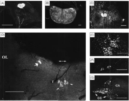
When probed with the CY17 antiserum, immunoposi-tive neurons were identified from day-2, last (5th) instar larval M. sexta in their frontal ganglion [Fig. 1(A)], brain [Fig. 1(B)], and terminal abdominal ganglion [Fig. 1(C)]. The number and the location of these neurons were identical to those reported by Taylor et al. (1996). As they have reported, there are two neurons each in the frontal and the terminal abdominal ganglia, as well as numerous immunoreactive neurons in the brain. CY16 antiserum yielded the same results.
3.2.2. Lack of cross-reactivity to other Mas AT-like peptides
The cELISA for Mas AT was used to check possible
cross-reactivity to other peptides that somewhat
resemble Mas AT. Two peptides were synthesized and tested. The first is the [Ala6, Leu7, Ser8]-allatotropin,
which is originally discovered from the male accessory gland of L. migratoria. The second is a peptide deduced
from the DNA of A. aegypti showing a high degree of homology to Mas AT. Results from experiments with CY16 (Fig. 2) showed that under the stated conditions, the cELISA had a linear detection range of 25–2500 ng of synthetic Mas AT. It took ca. 110 ng of Mas AT, or greater than 2000 ng of [Ala6, Leu7, Ser8]-allatotropin,
to reach the point of 50% inhibition. The difference was about 20-fold. Interestingly, the affinity of CY16 was so low towards the deduced mosquito AT-like peptide, competitive inhibition never exceeded 20% in this assay. CY17 antiserum yielded similar results.
3.2.3. Lack of reactivity to MylATr MAP
Our results showed practically no reactivity between our antisera and the MylATr MAP. Regardless of the concentration of antisera tested, the reactivity to Mas AT MAP was at least 17 times (ranged from 17 to 46 times) higher than that to the MylATr MAP. It appeared that our antisera contained little or no antibodies that recog-nize the heptalysine core, which is the only feature shared by both MylATr MAP and Mas AT MAP. Other-wise, we would see considerable reactivity of the anti-sera towards both MylATr MAP and Mas AT MAP. We conclude that our antisera were specific to the Mas AT portion of the MAP.
3.3. Localization of Mas AT-immunoreactive cells in P. regina and other insects
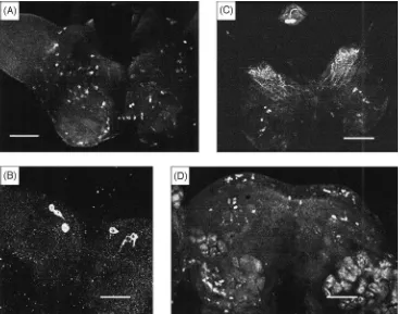
When probed with CY17 antiserum, immunopositive neurons were identified in the cephalic nervous tissues of P. regina [Fig. 1(D–G)] but not in the thoracico-abdominal ganglion. In each brain hemisphere of sugar-and liver-fed adult P. regina females, two immunoposi-tive neurons were found in the region where lateral neu-rosecretory cells would be expected [Fig. 1(D,F)]. Also, 9–11 immunopositive cells were found in a region located in between where median neurosecretory cells would be expected and the pharyngeal aperture [Fig. 1(D,E)]. This pattern of distribution of Mas AT immuno-positive neurons differed significantly to the distribution pattern of such cells in the larval brain of M. sexta [Fig. 1(B)]. It was interesting to note that immunoreactive material was present in the CC of liver-fed P. regina (6 h after the onset of the feeding). The CA of both sugar-and liver-fed flies, as well as the CC of sugar-fed flies, was void of Mas AT-like substance. In addition, no Mas AT immunoreactivity was observed in the anterior midgut of this fly. CY16 antiserum yielded similar results. When CY17 antiserum was used against the brain and/or frontal ganglion of other insects, immunore-active materials were identified in: (1) the adult brain of a hemipteran, O. fasciatus [Fig. 3(A)]. In this bug, immunopositive cells were found scattered all over the brain; (2) the adult brain of the oriental fruit fly, D.
Fig. 1. Manduca sexta allatotropin (Mas AT) immunoreactivity in the central nervous system of 2-day old, 5th instar larva of M. sexta. (A)
Dorsal view of frontal ganglion of larval M. sexta. Two distinct neurons contained strong immunoreactivity in the perikaryons and axons descending into the recurrent nerve (not shown). Scale bar=105µm. (B) Dorsal view of the brain. Many immunoreactive neurons are present in the entire brain. Scale bar=105µm. (C) Dorsal view of the terminal abdominal ganglion. Two distinct neurons containing large quantities of immunoreactive materials. In (B) and (C), no Mas AT immunoreactivity was observed in any nerve fibers. Scale=105µm. (D)–(E) Mas AT-like immunoreactivity in the central nervous system of liver-fed, adult female, P. regina. (D) Posterior view of the brain showing the left optical lobe (OL), position of the median furrow of the protocerebrum (double-headed arrow pointing to each hemisphere), trachea (T), two clusters of immunoreactive neurons located in the region in between where the median neurosecretory cells may be expected and the foramen for the pharynx or pharyngeal aperture (thin arrow points to either left or right cluster of neurons), and two distinctively reactive neurons (thick arrow) in the left hemisphere in the region where lateral neurosecretory cells may be expected. Scale bar=140µm. (E) Immunoreactive neurons at the median region. It was clear that the nuclei are not immunopositive. Scale bar=50µm. (F) Two immunoreactive neurons at the lateral region. Again, nuclei are not immunoreactive. Scale bar=50µm. (G) Immunoreactivity in the CC–CA complex. Strong immunopositive responses were restricted to the CC portion of the complex. The overall appearance of the immunoresponses in the CC were quite different from that found in the brain neurons. The immunoreactivity did not appear to be associated with the somata of innate CC neurons. Scale bar=50µm. All micrographs, except (D), were obtained using a confocal microscope, (D) was obtained by using a fluorescent microscope.
showed up clearly in three pairs of neurons; and (3) the brain and frontal ganglion from the last larval instar lor-eyi leafworm, M. lorlor-eyi. Two immunopositive cells were identified in the frontal ganglion [Fig. 3(C)], with dozens of cells found scattered around the entire brain [Fig. 3(C)]; and (4) brains of both adult female [Fig. 3(D)] and male (results not shown) M. loreyi contained immu-nopositive cells, which were scattered around the two hemispheres. Results (not shown), from two more larval lepidopterans, B. mori and A. bipunctata, were very similar to those observed for the larval loreyi leafworm. No immunopositive Mas AT materials were found in the adult brain neurons of A. mellifera (honey bees),
Camponotus sp. (carpenter ants), L. decemlineata
(Colorado potato beetles), and M. tsudai (walking sticks).
Also, no immunopositive Mas AT neurons were found in the female adult brains of sugar- and liver-fed P. regina if the brains were probed with antisera that had been pretreated with synthetic Mas AT. This
indicated that at the three doses (20, 40, and 80 µg
Fig. 2. Relative affinities of Mas AT, Lom MT, and deduced Aee AT-like peptide toward our anti-Mas AT antiserum. Results of a com-petitive enzyme-linked immunosorbent assay (cELISA) for Mas AT showed that the affinity of Lom MT was 20-fold less than that of Mas AT towards the antiserum. Very little inhibition was detected for the Aee AT-like peptide up to 2000 ng level suggesting very low affinity to the antiserum.
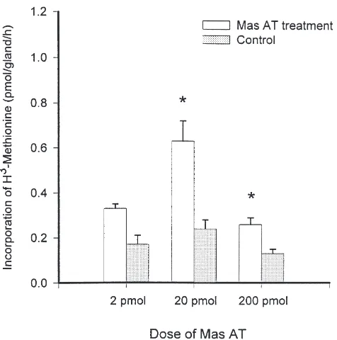
3.4. Stimulation of JH biosynthesis by the CA of P. regina with synthetic Mas AT
Three replicates were conducted to examine the effect of three doses of Mas AT on the in vitro JH biosynthesis of the CA of P. regina. In each replication, 3–5 incu-bations were conducted. Results (Fig. 4) showed that the synthetic Mas AT stimulated JH biosynthesis by the CA from sugar-fed females in a dose-dependent manner at
2, 20, and 200 pmol of Mas AT per 25 µl of TC199 (or
80, 800, and 8000 nM). Stimulated CAs produced 1.96, 2.64, and 2.00 times more JHs than the respective con-trols.
4. Discussion
We demonstrated Mas AT-immunoreactive sub-stances in the nervous system of P. regina and a number of other insects for the first time using rabbit antisera that we raised against a synthetic Mas AT MAP immun-ogen. Such MAP immunogens [(synthesized according to the design of Tam (1988)] have been used in many different studies. These studies successfully developed
antisera against the influenza virus hemagglutinin (Toth et al., 1993), S1 glycoprotein of infectious bronchitis virus (Jackwood and Hilt, 1995), N-terminal human parathyroid hormone 1-37 (Magerlein et al., 1998), and to structurally related Drosophila melanogaster peptides (Nichols et al., 1997). Likewise, our cELISA results indicated that our antisera show different degrees of reactivity to three structural related peptides belonging to the Mas AT family varying from high, low to nearly none.
This MAP strategy is chemically defined and provides an efficient way to make an immunogen for small pep-tides that are not immunogenic by themselves. The MAP immunogen compares well with the more conventional hapten-carrier protein type when the specificity of the antibodies generated by it is evaluated. Our results sup-port the above idea because our present antisera recog-nized the same Mas AT containing neurons in M. sexta as previously published (Taylor et al., 1996). Both anti-sera revealed that the frontal and the terminal abdominal ganglia each contained two strongly immunoreactive neurons. The larval moth frontal ganglia from A.
bipunctata, B. mori, and M. loreyi also contained two
such cells. The presence of AT or AT-like substance in the frontal ganglion of lepidopterous larvae of various species calls for a new critical look into its function in CA regulation. Separately, frontal ganglion and JH have been both invoked in the regulation of diapause of the southwestern corn borer, Diatraea grandiosella (Yin and Chippendale 1973, 1974). It would be very interesting to determine if this immunoreactivity is indeed allato-tropic in diapausing southwestern corn borers because a recent study of the larval noctuid moth, Helicoverpa
armigera has shown that the Mas AT acts as a
Fig. 3. Mas AT-like immunoreactivity in the nervous tissue of several test insects. (A) Dorsal view of the brain of adult, Oncopeltus fasciatus. Dozens of neurons were positive. Scale bar=170µm. (B) Dorsal-posterior view of an adult brain of Dacus dorsalis. Three distinct pairs of neurons were found immunoreactive. All neurons at the median region were negative. Scale bar=53µm. (C) Dorsal view of the frontal ganglion and the brain of larval (last instar) Mythimna loreyi. The frontal ganglion was flipped over unintentionally during mounting thus the intended dorsal view had become a ventral view with the ventral-posterior portion of the ganglion superficially presented as dorsal-anterior portion. Like the result of larval M. sexta, two distinct immunopositive cells were evident. Dozens of cells of varying immunoreactivity were present all over the brain. Scale bar=180µm. (D) Frontal view of an adult brain of female M. loreyi. Many protocerebral neurons are immunoreactive. Scale bar=180µm.
When one works with small peptides (or a small sec-tion of a larger peptide or protein), there is another advantage for using a MAP immunogen over the more conventional hapten-carrier protein design. The carrier protein of choice (i.e. HSA, BSA or keyhole limpet hemocyanin) is typically much larger than the haptens and most likely is antigenic itself, thus resulting in the production of antibodies against the carrier. By the MAP design, the actual peptide fragment is replicated eight times and collectively contributes to the bulk of the mass and structure of the immunogen. The ‘carrier’ portion contains only seven lysines. Such structural design, put-ting more emphasis on the heptan, can result in anti-serum that is highly specific to the heptan and shows little, if any, background affinity to the carrier. Our result that neither CY16 nor CY17 recognized MylATr MAP is consistent with the above idea.
Several lines of evidence supported the notion that our antisera are sufficiently specific to Mas AT. (1) when they were used immunochemically to characterize the larval nervous system of M. sexta, only those cells that have been previously identified as Mas AT producing neurons (Taylor et al., 1996) showed positive reactions. (2) pretreatment (i.e. preincubation) of Mas AT
anti-sera with synthetic Mas AT totally abolished the ability of the antisera to recognize any immunoreactive sub-stances in the nervous tissues. And (3), when the antisera were used in a cELISA, cross-reactivities to two other homologous peptides (e.g. Lom MT and Aee Mas AT-like peptide) were either low or nearly absent. Nonethe-less, we recognize that antiserum specificity is seldom absolute.
Fig. 4. Synthetic Mas AT stimulated JH synthesis by the CA of sugar-fed adult female P. regina. Two CAs were incubated in 25 µl of TC199 spiked with 2, 20, or 200 pmol of Mas AT in the radiochem-ical assay for estimating the stimulation. JH synthesis was expressed as incorporation of 3H-methyl moiety of the tritiated-methionine.
Asterisks marked the treatments where the differences between test and control were statistically significant (two samples t-test, P,0.05). Among all three Mas AT doses, the highest activation occurred at 20 pmol [Tukey (HSD) test, P,0.05].
Table 1
The amino acid sequences of three structurally related peptidesa
Lom MTb Gly–Phe–Lys–Asn–Val–Ala–Leu–Ser–Thr–Ala–Arg–Gly–Phe–NH 2
Mas ATb Gly–Phe–Lys–Asn–Val–Glu–Met–Met–Thr–Ala–Arg–Gly–Phe–NH 2
Aee AT-likeb - - - Phe–Arg–Asn–Ser–Glu–Met–Met–Thr–Ala–Arg–Gly–Phe–OH
aAmino acid sequences were compared to that of Mas AT. Different and missing acids are underlined.
b Lom MT=Locusta migratoria myotropin, Mas AT=Manduca sexta allatotropin, Aee AT-like=Aedes aegypti allatotropin-like peptide deduced
from cDNA.
reduction in cross-reactivity to the antiserum. At this time, we do not know whether the remaining 20% cross-reactivity of Lom MT comes from its N-terminus or C-terminus, or both. Thus, it appears that the affinity of our antiserum towards Mas AT is associated primarily with either or both termini and to a lesser degree with the middle section. The cross-reactivity pattern of our antiserum complements the antiserum raised using a conventional immunogen design by Veenstra and Hage-dorn (1993). For the specificity of their antiserum, it appears that the 5 N-terminal amino acids are not as important, the 3 amino acids at positions 6, 7, and 8 are somewhat important, and the 5 C-terminal amino acids are of major importance. In other words, when both con-ventional and MAP immunogen design are used, one might be able to raise a panel of antisera that can
differ-entially recognize the structural differences in either the N-terminus, middle section, or C-terminus of homologs from the same peptide family.
The cELISA we reported here has a linear detection range of 25–2500 ng of synthetic Mas AT. We picked this range to show that even at higher than normal physiological doses there is little or no cross-reactivity. The quanitity of Mas AT or Mas AT-like substance presented in M. sexta and P. american a ranged from 0.01 to 3.3 pmol/tissue/insect (Veenstra and Hagedorn, 1993). To determine Mas AT in this physiological range requires a cELISA with a linear range of 10–1000 pg. We are presently working on such a cELISA.
AT is a neuropeptide first isolated and identified from adult heads of M. sexta (Kataoka et al., 1989) and has the ability to stimulate JH biosynthesis by the CA of adult moths only. Peptides with similar biological activi-ties have only been partially characterized in A. mellifera
carnica (Rachinsky, 1996), G. mellonella (Bogus and
Scheller 1994, 1996), G. bimaculatus and Achaeta
dom-esticus (Lorenz and Hoffmann, 1995), L. migratoria
(Gadot and Applebaum, 1985; Rembold et al., 1986; Lehmberg et al., 1992), and P. apterus (Hodkova et al., 1996). Although the chemical structures of these pep-tides are not defined, it is clear that the spectrum of occurrence of AT-like peptides may be much wider among insects than previously recognized but somewhat expected. This idea is supported by various data from immunochemical and immunoassay studies. Mas-AT
immunoreactive cells are found in the nervous system of M. sexta throughout its development (Veenstra and Hagedorn, 1993; Veenstra et al., 1994). These cells also exist in both larval and adult Drosophila in the central nervous system (Zitnan et al. 1993, 1995). Further, Veenstra and Hagedorn (1993) have estimated the Mas AT levels using a sensitive ELISA in the brain, ventral nerve cord and retrocerebral complex of adult M. sexta, and reported an AT-immunoreactive substance in the nervous system of adult P. americana. In this study, we have found immunopositive neurons in the brain of both sugar- and liver-fed, adult female, P. regina; the adult large milkweed bug, O. fasciatus; the adult oriental fruit fly, D. dorsalis; the larval and adult loreyi leafworm,
M. loreyi and the larval tea silk moth, A. bipunctata.
adult female P. regina also contained Mas AT-like sub-stances, but the CA did not. The difference between sug-ar- and liver-fed flies may suggest that the Mas AT-like substances is transported from the brain to the CC in sufficient quantity in liver- but not sugar-fed female flies. The difference between CC and CA in liver-fed female adults may suggest that the Mas AT-like material is of sufficient storage quantity in the neurohemal organ (e.g. CC) but of insufficient hormone-receptor quantity at the expected target organ (i.e. CA) to be detected by the current immunochemical method. In other words, a more sensitive method may reveal its presence on the CA. In contrast, no immunopositive cells appeared in the adult brain of the honey bee, A. mellifera (foraging workers); the carpenter ant, Camponotus sp. (foraging workers);
the Colorado potato beetle, L. decemlineata
(reproductively active males and females), and the walk-ing stick, M. tsudai (adults). The above findwalk-ings need to be independently confirmed to establish the spectrum of AT occurrence. More work remains also to confirm the allatotropic and/or myotropic function of these Mas AT-like immunopositive substances.
As already mentioned, Mas AT activates JH synthesis by the CA in adult, but not larval, M. sexta (Kataoka et al., 1989), yet it exists in both stages. Its presence in larval M. sexta is independently confirmed using immunological and/or molecular biological methods (Taylor et al., 1996; present study). Is there non-allato-tropic functions existing for the larvae? Indeed, Veenstra et al. (1994) reported a non-allatotropic excitatory effect of Mas AT on the heart of pharate adult M. sexta. Equ-ally interesting is the recent finding that Mas AT inhibits the short-circuit current (Isc) across the posterior midgut
of fourth instar feeding larvae, and day-2, fifth instar larvae of M. sexta (Lee et al., 1998). The Iscof midguts
from wandering or pharate fifth instar larvae is virtually unaffected by Mas AT. Likewise, posterior midguts of
Hyalophora cecropia and B. mori are not affected either.
Also, as mentioned earlier, Mas AT acts as a myostimul-atory agene in larval H. armigera (Duve et al., 1999). More non-allatotropic functions for Mas AT may remain to be discovered.
In the past, because Mas AT also stimulates JH syn-thesis by the CA of adult female Heliothis virescens, but not the CA of adult female Tenebrio molitor,
Schisto-cerca nitens, and P. americana, its activity is thought to
be restricted to the adult lepidopterans (Kataoka et al., 1989). Such view appears to be too narrow when recent data are considered. In honey bee, A. mellifera, Mas AT exhibits a stage-specific stimulation of JH release by the CA of feeding stage larvae in a dose-dependent fashion, but not by the prepupal CA (Rachinsky and Feldlaufer,
2000). They find that at a rather high level of 1024 M,
Mas AT can cause a 2.2-fold increase in JH release. This observation is the first report of stimulating effect of Mas AT on the CA of a non-lepidopteran, albeit the fact that
1024 M may be considered to be pharmacological. The
present study showed that at 8×1027 M, synthetic Mas
AT can increase the JH biosynthesis in vitro by the CA of 3-day-old, sugar-fed, female P. regina 2.64-fold. This represents the first case of an allatotropic effect of syn-thetic Mas AT in a non-lepidopteran at a physiological dose. With in vitro system, it is not uncommon to
observe hormonal effect at 1027 M. For example, the
early puffs from the giant chromosome 3 of the salivary glands of D. melanogaster are induced over a wide range of ecdysone concentrations, from 1 nM to 500 nM with a 50% response recorded at 100 nM (Russell and Ash-burner, 1996). However, the absolute stimulatory effect for the synthetic Mas AT is lesser in P. regina (264% of the control) than in M. sexta (500%) in the stages tested. Nevertheless, the idea deduced from immunoch-emical evidence that the structure of Mas AT has been sufficiently preserved through evolution to be present and is biologically active in other insects (Veenstra and Hagedorn, 1993) appears to receive support from our results. At least, the present data may provide new impetus for us to test the above idea more seriously. Further support for this broader preservation idea will come from reports demonstrating the allatotropin effect in vitro and/or in vivo in insects other than dipterans and lepidopterans.
Acknowledgements
The authors thank Dr N.T. Davis for his critical review and most helpful suggestions. This work was supported by the National Science Foundation (IBN-9306650, to C.-M. Yin and J.G. Stoffolano, Jr.) and the Massachusetts Agricultural Experiment Station (Hatch 632 to J.G. Stoffolano and Hatch 743 to C.-M. Yin) and published as Contribution No. 3244.
References
Applin, D.G., 1979. Effect of diet on the neuroendocrine system and egg development in the sheep blowfly, Lucilia sericata. Physiologi-cal Entomology 4, 291–299.
Applin, D.G., 1981. Long-term effects of diet on the neuroendocrine system of the sheep blowfly, Lucilia sericata. Physiological Ento-mology 6, 129–134.
Bogus, M., Scheller, K., 1994. Identification of allatotropin-secreting cells in the brain of an insect larva. Naturwissenschaften 81, 87–89. Bogus, M., Scheller, K., 1996. Allatotropin released by the brain con-trols larval molting in Galleria mellonella by affecting juvenile hormone synthesis. International Journal of Developmental Biology 40, 205–210.
Dai, J.-D., Stoffolano, J.G. Jr., Yin, C.-M., 1987. Median neurosecre-tory cells and associated nerves of the adult black blow fly,
Phor-mia regina (Diptera: Calliphoridae). Annals of the Entomological
Society of America 80, 339–345.
amer-icana (L.), and identification of mandibular and maxillary motor
neurons associated with this system. Journal of Comparative Neur-ology 259, 604–621.
Dethier, V.G., 1976. The Hungry Fly. Harvard University Press, Cam-bridge, MA.
Duve, H., East, P.D., Thorpe, A., 1999. Regulation of lepidopteran foregut movement by allatostatins and allatrtropin from the frontal ganglion. Journal of Comparative Neurology 413, 405–416. Duve, H., Johnson, A.H., Scott, A.G., East, P.D., Thorpe, A., 1994.
[Hyp3]Met-callatostatin. Identification and biological properties of a novel neuropeptide from the blowfly Calliphora vomitoria. Jour-nal of Biological Chemistry 269, 21059–21066.
Feyereisen, R., 1985. Regulation of juvenile hormone titer: synthesis. In: Kerkut, G.A., Gilbert, L.I. (Eds.). Comprehensive Insect Physi-ology, Biochemistry, and PharmacPhysi-ology, vol. 7. Pergamon Press, Oxford, pp. 391–429.
Fraenkel, G., 1940. Utilization and digestion of carbohydrates by the adult blowfly. Journal of Experimental Zoology 17, 18–29. Gadot, M., Applebaum, S.W., 1985. Rapid in vitro-activation of
corpora allata by extracted locust brain allatotropic factor. Archives of Insect Biochemistry and Physiology 2, 117–129.
Ga¨de, G., Hoffmann, K.H., Spring, J.H., 1997. Hormonal regulation in insects: facts, gaps, and future directions. Physiological Review 77, 963–1032.
Hodkova, M., Okuda, T., Wagner, R., 1996. Stimulation of corpora allata by extract from neuroendocrine complex; comparison of reproducing and diapausing Pyrrhocoris apterus (Heteroptera: Pyrrhocoridae). European Journal of Entomology 93, 535–543. Hoffmann, K.H., Meyering-Vos, M., Lorenz, M.W., 1999.
Allatostat-ins and allatotropAllatostat-ins: is the regulation of corpora allata activity their primary function? European Journal of Entomology 96, 255–266. Jackwood, M.W., Hilt, D.A., 1995. Production and immunogenicity of multiple antigenic peptide (MAP) constructs derived from the S1 glycoprotein of infectious bronchitis virus (IBV). Advances in Experimental Medicine and Biology 380, 213–219.
Kataoka, H., Toschi, A., Li, G.P., Carney, R.L., Schooley, D.A., Kramer, S.J., 1989. Identification of an allatotropin from adult
Manduca sexta. Science 243, 1481–1483.
Khan, M.A., 1988. Brain-controlled synthesis of juvenile hormone in adult insects. Entomologia Experimentalia and Applicata 46, 3–17. Langley, P.A., 1965. The neuroendocrine system and stomatogastric nervous system of the adult tsetse fly Glossina morsitans. Proceed-ings of the Zoological Society of London 144, 415–424. Lee, K.-Y., Horodyski, F.M., Chamberlin, M.E., 1998. Inhibition of
midgut ion transport by allatotropin (Mas-AT) and Manduca FLRFamides in the tobacco hornworm Manduca sexta. Journal of Experimental Biology 201, 3067–3074.
Lehmberg, E., Ferenz, H.J., Applebaum, S.W., 1992. Maturation and responsiveness to extracts of corpora allata from male Locusta
migratoria containing allatotropic factors. Zeischrift fur
Naturfor-schung 47C, 449–452.
Liu, M.-A., Jones, G.L., Stoffolano, J.G. Jr., Yin, C.-M., 1988. Con-ditions for estimation of corpus allatum activity in the blowfly
Phormia regina, in vitro. Physiological Entomology 13, 69–79.
Lorenz, M.W., Hoffmann, K.H., 1995. Allatotropic activity in the suboesophageal ganglia of crickets, Gryllus bimaculatus and
Achaeta domesticus (Ensifera Gryllidae). Journal of Insect
Physi-ology 41, 191–196.
Magerlein, M., Hock, D., Adermann, K., Neidlein, R., Forssmann, W.G., Strein, K., 1998. Production of sequence specific polyclonal antibodies to human parathyroid hormone 1-37 by immunization with multiple antigenic peptides. Arzneimittel-Forschung 48, 783–787.
Nichols, R., McCormick, J., Lim, I., 1997. Multiple antigenic peptides designed to structurally related Drosophila peptides. Peptides 18, 41–45.
Nijhout, H.F., 1994. Insect Hormones. Princetion University Press, Princeton, NJ.
Paemen, L., Tips, A., Schoofs, L., Proost, P., Van Damme, J., De Loof, A., 1991. Lom-AG-mytropic peptide from the male accessory glands of Locusta migratoria. Peptides 12, 7–10.
Rachinsky, A., 1996. Brain and suboesophageal ganglion extracts affect juvenile hormone biosynthesis in honey bee larvae (Apis
mel-lifera carnica). Zoology: Analysis of Complex Systems 99, 277–
284.
Rachinsky, A., Feldlaufer, M.F., 2000. Responsiveness of honey bee (Apis mellifera L.) corpora allata to allatoregulatory peptides from four insect species. Journal of Insect Physiology 46, 41–46. Rembold, H., Schlagintweit, B., Ulrich, G.M., 1986. Activation of
juv-enile hormone synthesis in vitro by a corpus cardiacum factor from
Locusta migratoria. Journal of Insect Physiology 32, 91–94.
Moshitzky, P., Applebaum, S.W., 1995. Pathway and regulation of JH III-bisepoxide biosynthesis in adult Drosophila melanogaster corpus allatum. Archives of Insect Biochemistry and Physiology 30, 225–237.
Russell, S., Ashburner, M., 1996. Ecdysone-regulated chromosome puffing in Drosophila melanogaster. In: Gilbert, L.I., Tata, J.R., Atkinson, B.G. (Eds.) Metamorphosis: Postembryonic Reprogram-ming of Gene Expression in Amphibian and Insect Cells. Academic Press, San Diego, CA, pp. 109–144.
Stoffolano, J.G. Jr., 1974. Influence of diapause and diet on the devel-opment of the gonads and accessory reproductive glands of the black blowfly Phormia regina (Meig.). Canadian Journal of Zoology 52, 981–988.
Stoffolano, J.G. Jr., Li, M.-F., Zou, B.-X., Yin, C.-M., 1992. Vitellog-enin uptake, not synthesis, is dependent on juvenile hormone in adults of Phormia regina (Meigen). Journal of Insect Physiology 38, 839–845.
Tam, J.P., 1988. Synthetic peptide vaccine design: Synthesis and properties of a high-density multiple antigenic peptide system. Pro-ceedings of the National Academy of Sciences USA 85, 5409– 5413.
Taylor, P.A. III, Bhatt, T.R., Horodyski, F.M., 1996. Molecular charac-terization and expression analysis of Manduca sexta allatotropin. European Journal of Biochemistry 239, 588–596.
Thomsen, E., 1942. An experimental and anatomical study of the cor-pus allatum in the blow-fly Calliphora erythrocephala Meig. Vid-ensh. Medd. Dan. Natuhist. Foren. 106, 319–405.
Thomsen, E., 1952. Functional significance of the neurosecretory brain cells and the corpus cardiacum in the female blow-fly Calliphora
erythrocephala Meig. Journal of Experimental Biology 29, 137–
172.
Thomsen, E., Moller I., 1960. Further studies on the function of the neurosecretory brain cellls of the adult Calliphora female. In: The Ontogeny of Insects, Acta symposii de evolutione insectorum, Praha, 1959.
Tobe, S.S., Pratt, G.E., 1974. The influence of substrate concentrations on the rate of juvenile hormone biosynthesis by corpora allata of the desert locust in vitro. Biochemical Journal 44, 107–113. Toth, G.K., Varadi, G., Nagy, Z., Monostroi, E., Penke, B., Hegedus,
Z., Ando, I., Fazekas, G., Kurucz, I., Mak, M., 1993. Branched polypeptides as antigens for influenza virus hemagglutinin and T-cell receptor subunits. Peptide Research 6, 272–280.
Veenstra, J.A., Costes, L., 1999. Isolation and identification of a pep-tide and its cDNA from the mosquito Aedes aegypti related to
Man-duca sexta allatotropin. Peptides 20, 1145–1151.
Veenstra, J.A., Hagedorn, H.H., 1993. Sensitive enzyme immunoassay for Manduca allatotropin and the existence of an allatotropin-immunoreactive peptide in Periplaneta americana. Archives of Insect Biochemistry and Physiology 23, 99–109.
Wie, S.I., Hammock, B.D., 1984. Comparison of coating and immuniz-ing antigen structure on the sensitivity and specificity of immunoas-says for benzoylphenylurea insecticides. Journal of Agricultural and Food Chemistry 32, 1294–1301.
Yin, C.-M., 1994. Juvenile hormone III bisepoxide: new member of the insect juvenile hormone family. Zoological Studies 33, 237–245. Yin, C.-M., Chippendale, G.M., 1973. Endocrine system of mature diapause and nondiapause larvae of the southwestern corn borer,
Diatraea grandiosella. Annals of the Entomological Society of
America 66, 943–947.
Yin, C.-M., Chippendale, G.M., 1974. Juvenile hormone and the induc-tion of larval polymorphism and diapause of the southwestern corn borer Diatraea grandiosella. Journal of Insect Physiology 20, 1833–1847.
Yin, C.-M., Chippendale, G.M., 1975. Insect frontal ganglion: fine structure of its neurosecretory cells in diapause and non-diapause larvae of Diatraea grandiosella. Canadian Journal of Zoology 53, 1093–1100.
Yin, C.-M., Chippendale, G.M., 1979. Diapause of the southwestern corn borer, Diatraea grandiosella: further evidence showing juven-ile hormone to be the regulator. Journal of Insect Physiology 25, 513–523.
Yin, C.-M., Stoffolano, J.G. Jr., 1990. The interactions among nutrition, endocrines and physiology on the reproductive develop-ment of the black blowfly, Phormia regina (Meigen). Bulletin of the Institute of Zoology Academia Sinica, Monograph 15, 87–108. Yin, C.-M., Stoffolano, J.G. Jr., 1994. Endocrinology of vitellogenesis in blow flies. In: Davey, K.G., Peter, R.E., Tobe, S.S. (Eds.), Per-spectives in Comparative Endocrinology. National Research Coun-cil of Canada, Ottowa, pp. 291–298.
Yin, C.-M., Stoffolano, J.G. Jr., 1997. Juvenile hormone regulation
of reproduction in the Cyclorrhaphous Diptera with emphasis on oogenesis. Archives of Insect Biochemistry and Physiology 35, 513–537.
Yin, C.-M., Duan, H., Stoffolano, J.G. Jr., 1993. Hormonal stimulation of the brain for its control of oogenesis in Phormia regina (Meign). Journal of Insect Physiology 39, 165–171.
Yin, C.-M., Zou, B.-X., Jiang, M., Li, M.-F., Qin, W., Potter, T.L., Stoffolano, J.G. Jr., 1995. Identification of juvenile hormone III bisepoxide (JHB3), juvenile hormone III and methyl farnesoate
secreted by the corpus allatum of Phormia regina (Meigen), in vitro and function of JHB3 either applied alone or as a part of a
juvenoid blend. Journal of Insect Physiology 41, 473–479. Yin, C.-M., Zou, B.-X., Li, M.-F., Stoffolano, J.G. Jr., 1994. Discovery
of a midgut peptide hormone which activates the endocrine cascade leading to oogenesis in Phormia regina (Meigen). Journal of Insect Physiology 40, 283–292.
Zitnan, D., Sehnal, F., Bryant, P.J., 1993. Neurons producing specific neuropeptides in the central nervous system of normal and pupari-ation-delayed Drosophila. Developmental Biology 156, 117–135. Zitnan, D., Kingan, T.G., Kramer, S.J., Beckage, N.E., 1995.
Accumu-lation of neuropeptides in the cerebral neurosecretory system of
Manduca sexta larvae parasitized by the braconid wasp Cotesia congregata. Journal of Comparative Neurology 356, 83–100.
Zou, B.-X., Stoffolano, J.G. Jr., Nordin, J.H., Yin, C.-M., 1988. Sub-unit composition of vitellin, and concentration profiles of vitellog-enin, and vitellin in Phormia regina (Meig.) following a protein meal. Comparative Biochemistry and Physiology 90B, 861–867. Zou, B.-X., Yin, C.-M., Stoffolano, J.G. Jr., Tobe, S.S., 1989. Juvenile
hormone biosynthesis and release during oocyte development in