This page
All rights reserved.
No part of this ebook may be reproduced in any form, by photostat, microfilm, xerography, or any other means, or incorporated into any information retrieval system, electronic or mechanical, without the written permission of the publisher. All inquiries should be emailed to [email protected]
PUBLISHINGFORONEWORLD
NEW AGE INTERNATIONAL (P) LIMITED, PUBLISHERS
4835/24, Ansari Road, Daryaganj, New Delhi - 110002 Visit us at www.newagepublishers.com
In the recent past, the world has witnessed the smooth transition
of the
‘quantum state’
of a trapped Ca
2+ion to another Ca
2+ion
via
meticulously teleported means in a critically controlled manner thereby
mustering enough hope that in the near future
‘teleportation’
of
individual atoms and molecules would pave the way to teleportation of
molecules and
‘microorganisms’.
‘‘Dreams will only help you actualize your goals. I started dreaming
when I was a child...
I became a scientist because I dreamt’’.
This page
PREFACE
The textbook of ‘Pharmaceutical Microbiology’ specifically aims at the ever demanding thoughtful need of an absolutely well-documented compilation of factual details related to : theoritical principles, classifications, diagramatic profiles, graphic presentations, critical explanation, latest examples for the Pharmacy Degree (B. Pharm.,) throughout the Indian Universities, SAARC-countries, and similar curricula adopted abroad.
Modern invigorative society, based on the overwhelming and overemphasized broad-spectrum importance vis-a-vis utilities of ‘Microbiology’ profusely gets benefited from the intricate species of scores of microorganisms in several ways and means, namely : antibiotics, vaccines, enzymes, vitamins
etc. Nevertheless, a quantum-leap-forward in the field of ‘Modern Biotechnology’ rests predominantly upon reasonably sound microbiological foundation. Besides, microorganisms do modulate a plethora of vital and critical functionalities, such as : (a) enable completion of cycles of C, O, N and S which essentially occur in both terrestrial and aquatic systems ; (b) provide absolutely indispensable components of prevailing ecosystem ; and (c) serve as a critical source of ‘nutrients’ occurring at the grass-root of practically a large segment of ecological food webs and chains.
The entire course-content presented in ‘Pharmaceutical Microbiology’ has been meticulously and painstakingly developed and expanded as per the AICTE-Approved Syllabus–2000. Each chapter has been duly expatiated in a simple, lucid, and crisp language easily comprehensible by its august readers. A unique largely acceptable style of presentation has been adopted, viz., brief introduction, principles, labeled figures, graphics, diagrams of equipments, descriptions, explanations, pharmaceutical applications, and selected classical examples. Each chapter is duly elaborated with adequate foot-notes, references, and ‘further reading references’ at the end.
An exhaustive ‘Glossary of Important Microbiological Terminologies’ has been duly annexed at the end of the textbook. A fairly up to date computer-generated ‘Index’ in the textbook will surely enlarge the vision of its readers in gaining an easy access of subject enriched well documented text materials.
Pharmaceutical Microbiology consists of Ten Chapters : (1) Introduction and Scope ; (2) Structure and Function : Bacterial Cells ; (3) Characterization, Classification and Taxonomy of Microbes ; (4) Identification of Microorganisms ; (5) Nutrition, Cultivation and Isolation : Bacteria-Actinomycetes-Fungi-Viruses ; (6) Microbial Genetics and Variations ; (7) Microbial Control by Physical and Chemical Methods ; (8) Sterility Testing : Pharmaceutical Products ; (9) Immune Systems ; and (10) Microbiological (Microbial) Assays : Antibiotics–Vitamins–Amino Acids.
The text material essentially embodies not only an ample emphasis on the vivid coverage of fundamental principles of microbiology as a scientific discipline but also maintains a manageable length for the apprehension of brilliant students.
sufficient experimental details to enable students, research scholars, and budding scientists to pursue their objectives in the field of Pharmaceutical Microbiology.
The author earnestly believes that ‘Pharmaceutical Microbiology’ may prove to be of paramount importance for B. Pharm. (Pharmacy Degree), M. Sc., (Food Microbiology), M. Sc., (Microbiology), and M. Sc., (Environmental Science) students as well.
I extend my sincere thanks to Shri Saumya Gupta, MD and his excellent production wing to have the project completed in a record time frame.
CONTENTS
1. Introduction and Scope ... 1
1.1 Introduction ... 1
1.2 Historical Development of Microbiology ... 3
1.2.1. The Microscope ... 3
1.2.2. Spontaneous Generation Vs Biogenesis ... 4
1.2.3. Fermentation ... 6
1.2.4. Germ Theory ... 6
1.2.5. Classical Laboratory Methods and Pure Cultures ... 7
1.2.6. Immunity ... 8
1.2.7. Medical Microbiology ... 9
1.2.8. Pharmaceutical Microbiology ... 10
1.2.9. Industrial Microbiology ... 14
1.2.10. Emergence of Molecular Biology ... 15
1.2.11. Emergence of Virology ... 17
1.2.12. Microorganisms as Geochemical Agents ... 19
1.2.13. Microbiology in the New Millennium ... 19.
2. Structure and Function : Bacterial Cells ... 23
2.1 Introduction ... 23
2.2 Characteristic Features ... 23
2.2.1. Shape ... 23
2.2.2. Size ... 23
2.2.3. Reproduction ... 24
2.2.4. Formation of Colony ... 24
2.2.5. Mutation ... 24
2.2.6. Motility ... 24
2.2.7. Food and Oxygen Requirements ... 24
2.2.8. Temperature Requirements ... 24
2.3 Activities ... 25
2.4 Organization of Microbial Cells ... 25
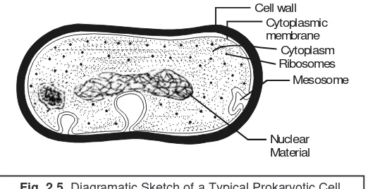
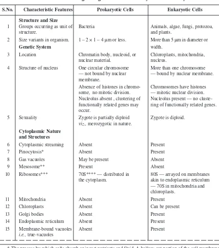
2.4.1. Type of Cells ... 26
2.4.1.1. Eukaryotic Cells ... 27
2.4.1.2. Prokaryotic Cells ... 33
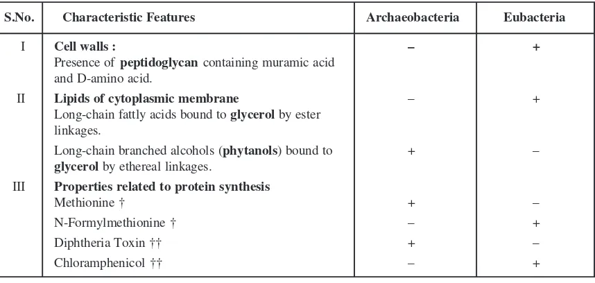
2.5 Archaeobacteria and Eubacteria ... 37
2.5.1. Methanogenic Bacteria [Methanogens] ... 38
2.5.2. Extreme Halophiles ... 40
2.5.3. Thermoacidophiles ... 41
2.5.3.1. Thermoplasma ... 41
2.5.3.2. Sulfolobus ... 41
2.6 The Bacterial Cells ... 42
2.6.1. Typical Bacterial Cells ... 43
2.6.2. Capsules and Slimes ... 44
2.6.3. Flagella and Fimbria ... 46
2.6.3.1. Flagella ... 46
2.6.3.2. Fimbria [or Pili] ... 48
2.6.4. Cell Envelope ... 49
2.6.5. Gram-Positive and Gram-Negative Bacteria ... 51
2.6.6. Significance of Teichoic Acids ... 53
2.6.7. The Cell Membrane ... 54
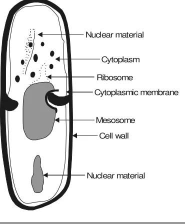
2.6.8. Bacterial Cytoplasm ... 55
2.6.9. Ribosomes ... 57
2.6.10. Cellular Reserve Materials ... 58
3. Characterization, Classification and Taxonomy of Microbes ... 62
3.1 Introduction ... 62
3.2 Characterization ... 62
3.2.1. Morphological Characteristics ... 63
3.2.2. Chemical Characteristics ... 64
3.2.3. Cultural Characteristics ... 64
3.2.4. Metabolic Characteristics ... 66
3.2.5. Antigenic Characteristics ... 66
3.2.6. Genetic Characteristics ... 67
3.2.6.1. DNA Base Composition ... 67
3.2.6.2. Sequence of Nucleotide Bases in DNA ... 68
3.2.7. Pathogenecity ... 69
3.2.8. Ecological Characteristics ... 69
3.3 Classificiation ... 70
3.3.1. Difficulties Encountered in Classification of Microorganisms ... 70
3.3.2. Objectives of Classification ... 70
3.3.3. Genetic Methods of Classifying Microbes ... 71
3.3.3.1. Genetic Relatedness ... 71
3.3.3.2. The Intuitive Method ... 72
3.3.3.3. Numerical Taxonomy ... 72
3.3.4. Systemetized Classification ... 75
3.3.4.1. Natural Classification ... 75
3.3.4.3. Linnear Binomial Scheme ... 76
3.3.4.4. Phenotypic Classification ... 77
3.3.4.5. Microscopic Examination ... 79
3.3.4.6. Cataloguing rRNA ... 80
3.3.4.7. Computer Aided Classification ... 81
3.3.4.8. Bacterial Classification ... 82
3.4 Taxonomy ... 87
3.5 The Kingdom Prokaryotae ... 88
3.5.1. Actinomyctes ... 89
3.5.1.1. General Characteristics ... 89
3.5.1.2. Significance of Actinomycetes ... 90
3.5.1.3. Classification ... 91
3.5.1.3.1. Whole Cell Carbohydrate Patterns of Aerobic
Actinomycetes ... 91
3.5.1.3.2. Major Constituents of Cell Wall Types of
Actinomycetes ... 91
3.5.1.3.3. Groups of Actinomycetes Based on Whole Cell Carbohydrate Pattern and Cell
Wall Type ... 92
3.5.1.3.4. Actinomycetes with Multiocular Sporangia ... 92
3.5.1.4. Actinomycetes and Related Organisms ... 93
3.5.1.4.1. Group ... 93
3.5.1.4.2. Genus ... 94
3.5.1.4.3. Order ... 97
3.5.1.4.4. Family ... 98
3.5.2. Bacteria ... 102
3.5.2.1. Salient Features ... 103
3.5.2.2. Structure and Form of the Bacterial Cell ... 104
3.5.2.2.1. Size and Shape ... 105
3.5.2.2.2. Structure ... 105
3.5.3. Rickettsia and Coxiella ... 107
3.5.4. Spirochaetes ... 108
4. Identification of Microorganisms ... 112
4.1 Introduction ... 112
4.2 Morphology ... 113
4.3 Selective and Diagnostic Media ... 113
4.3.1. Differential Media ... 116
4.3.1.1. Eosin Methylene Blue Agar [EMB-Agar] ... 116
4.3.1.2. MacConkey Agar ... 116
4.3.1.3. Hektoen Enteric Agar [HE-Agar] ... 116
4.3.2. Enrichment Media ... 116
4.3.2.1. Blood Agar ... 116
4.3.2.2. Chocolate Agar ... 117
4.3.3. Characteristic Media ... 117
4.3.3.1. Triple Sugar Iron Agar [TSI-Agar] ... 117
4.4 Cultural Characteristics ... 119
4.5 Biochemical Tests (or Properties) ... 120
4.5.1. Carbohydrate (Sugar) Fermentation ... 120
4.5.2. Litmus Milk ... 120
4.5.3. Indole Production ... 120
4.5.4. Methyl Red Test [MR-Test] ... 121
4.5.5. Voges-Proskauer Test [VP-Test] ... 121
4.5.6. Citrate Utilization ... 121
4.5.7. Nitrate Reduction ... 122
4.5.8. Ammonia Production ... 122
4.5.9. Urease Test ... 122
4.5.10. Production of Hydrogen Sulphide ... 123
4.5.11. Reduction of Methylene Blue ... 123
4.5.12. Production of Catalase [Tube Catalase Test] ... 123
4.5.13. Oxidase Reaction ... 123
4.5.14. Egg-Yolk Reaction ... 124
4.5.15. Growth in Presence of Potassium Cyanide ... 124
4.5.16. Composite Media ... 124
4.6 Profile of Microbial Stains ... 127
4.6.1. Preparation of Bacterial Specimens for Light Microscopy ... 128
4.6.1.1. Standard Preparations ... 128
4.6.1.2. Preparation of Smears for Staining ... 128
4.6.1.3. Gram Staining ... 129
4.6.1.4. Differential Staining ... 131
4.6.1.4.1. Gram’s Stain ... 131
4.6.1.4.2. Acid-Fast Stain ... 131
4.6.1.5. Miscellaneous Staining ... 131
4.6.1.5.1. Capsule Staining ... 132
4.6.1.5.2. Endospore Staining ... 132
4.6.2. Microscopy : The Differential Instruments ... 133
4.6.2.1. Concepts ... 133
4.6.2.2. Microscope Variants ... 134
4.6.2.2.1. Bright-Field Microscope ... 134
4.6.2.2.2. Dark-Field Microscope ... 136
4.6.2.2.3. Phase-Contrast Microscope ... 136
4.6.2.2.4. Differential Interference Contrast
(DIC) Microscope ... 139
4.6.2.2.5. Fluorescence Microscope ... 139
4.6.2.2.6. Electron Microscope ... 141
4.6.2.2.6.1. Transmission Electron
Microscope (TEM) ... 142
4.6.2.2.6.2. Scanning Electron
Microscope (SEM) ... 143
5. Nutrition, Cultivation and Isolation : Bacteria-Actinomycetes-Fungi-Viruses ... 146
5.1 Introduction ... 146
5.2 Bacteria ... 146
5.2.1. Nutrition of Microorganisms ... 146
5.2.2. Cultivation of Bacteria ... 147
5.2.2.1. Binary Fission ... 148
5.2.2.2. Normal Growth Curve of Microorganisms ... 149
5.2.2.3. The Lag Phase of Microbial Growth ... 150
5.2.2.4. Translational Periods Between Various Growth Phases ... 150
5.2.2.5. Synchronous Growth ... 151
5.2.2.6. Effect of Nutritional Concentration Vs Growth Rate of
Bacterial Culture ... 152
5.2.2.7. Growth Determining Techniques ... 152
5.2.3. Isolation of Bacteria ... 154
5.2.3.1. Selective and Diagnostic Media ... 154
5.2.3.2. Bismuth Sulphate Agar ... 154
5.2.3.3. Selective Media for Staphylococci ... 155
5.3 Actinomycetes ... 155
5.4 Fungi ... 156
5.4.1. Reproduction of Fungi ... 158
5.4.1.1. Asexual Reproduction ... 158
5.4.1.2. Sexual Reproduction ... 159
5.4.2. Industrial Importance of Fungi ... 159
5.4.2.1. Production of Wines and Beer ... 159
5.4.2.2. Production of Bakery Products ... 160
5.4.2.3. Production of Cheeses ... 160
5.5 Viruses ... 160
5.5.1. Bacteriophages ... 161
5.5.2. Growth of Bacteriophages in the Laboratory ... 164
5.5.3. Bacteriophage Lambda : The Lysogenic Cycle ... 164
6. Microbial Genetics and Variations ... 167
6.1 Introduction ... 167
6.2 Microbial Genetics ... 169
6.2.1. Structure and Function of Genetic Material ... 169
6.2.2. Genotype and Phenotype ... 170
6.2.3. Adaption and Mutation ... 170
6.2.4. DNA and Chromosomes ... 171
6.2.5. DNA Replication ... 172
6.2.6. Rate DNA Replication ... 174
6.2.7. Flow of Genetic Information ... 175
6.2.8. Bacterial Transformation ... 177
6.2.9. Bacterial Transcription ... 180
6.2.10. Bacterial Translation ... 182
6.2.11. Bacterial Conjugation ... 186
6.2.12. Bacterial Transduction ... 188
6.2.12.1. Generalized Transduction ... 189
6.2.12.2. Specialized Transduction ... 190
6.2.13. Bacterial Transfection ... 192
6.2.14. Phage Conversion ... 192
6.3 Microbial Variations [Genetic Manipulation in Microorganisms] ... 193
7. Microbial Control By Physical and Chemical Methods ... 198
7.1 Introduction ... 198
7.2 Physical Methods ... 198
7.2.1. Heat ... 199
7.2.2. Moist Heat ... 200
7.2.2.1. Boiling ... 201
7.2.2.2. Autoclaving ... 201
7.2.2.3. Pasteurization ... 204
7.2.2.4. Dry-Heat Sterilization ... 205
7.2.2.6. Cold ... 207
7.2.2.7. Desiccation ... 208
7.2.2.8. Osomotic Pressure ... 208
7.2.2.9. Radiation ... 209
7.2.2.9.1. Ionizing Radiation ... 209
7.2.2.9.2. Nonionizing Radiation ... 210
7.3 Chemical Methods ... 214
7.3.1. Effective Disinfection—Fundamentals ... 214
7.3.2. Disinfectant—Critical Evaluation ... 215
7.3.2.1. Use-Dilution Tests ... 215
7.3.2.2. Filter Paper Method ... 216
7.3.3. Disinfectant Variants ... 216
7.3.3.1. Alcohols ... 216
7.3.3.2. Aldehydes ... 217
7.3.3.3. Chlorohexidine ... 218
7.3.3.4. Gaseous Chemosterilizers ... 218
7.3.3.5. Heavy Metals and Derivatives ... 219
7.3.3.6. Halogens ... 219
7.3.3.7. Organic Acids and Derivatives ... 221
7.3.3.8. Oxidizing Agents ... 222
7.3.3.9. Phenol and Phenolics ... 223
7.3.3.10. Quaternary Ammonium Compounds [QUATS] ... 224
7.3.3.11. Surface-Active Agents ... 226
7.4 Experimental Parameters Influencing the Antimicrobial Agent Activity ... 228
7.4.1. Population Size ... 228
7.4.2. Population Composition ... 228
7.4.3. Concentration of Antimicrobial Agent ... 229
7.4.4. Duration of Exposure ... 229
7.4.5. Temperature ... 229
7.4.6. Local Environment ... 229
8. Sterility Testing : Pharmaceutical Products ... 231
8.1 Introduction ... 231
8.2 Test for Sterility : Pharmaceutical Products ... 233
8.2.1. Membrane Filtration ... 233
8.2.2. Direct Inoculation ... 239
8.2.2.1. Nutrient Broth ... 239
8.2.2.2. Cooked Meat Medium and Thioglycollate Medium ... 240
8.2.2.3. Sabouraud Medium ... 240
8.3 Sampling : Probability Profile ... 243
8.4 Overall Conclusions ... 245
9. Immune Systems ... 246
9.1 Introduction ... 246
9.1.1. Discrimination ... 247
9.1.2. Specificity ... 247
9.1.3. Anamnesis ... 247
9.1.4. Transferability by Living Cells ... 247
9.2 Types of Specific Immunity ... 248
9.2.1. Acquired Immunity ... 248
9.2.2. Active Immunity ... 249
9.2.3. Cell-Mediated Immunity ... 249
9.2.4. Congenital Immunity ... 249
9.2.5. Herd Immunity ... 249
9.2.6. Humoral Immuity [or B-Cell Mediated Immunity] ... 249
9.2.7. Local Immunity ... 250
9.2.8. Natural Immunity ... 250
9.2.9. Passive Immunity ... 250
9.3 Duality of Immune Systems ... 250
9.4 Immunological Memory ... 251
9.5 Natural Resistance and Nonspecific Defence Mechanisms ... 252
9.5.1. Natural Resistance ... 253
9.5.1.1. Species Resistance ... 253
9.5.1.2. Racial Resistance ... 253
9.5.1.3. Individual Resistance ... 254
9.5.1.4. External Defence Mechanisms ... 254
9.5.2. Internal Defense Mechanisms ... 256
9.5.3. Nonspecific Defense Mechanisms ... 256
9.5.3.1. Complement System ... 257
9.5.3.2. Phagocytosis ... 259
9.5.3.2.1. Functions of Phagocytes ... 260
9.5.3.2.2. Mechanism of Phagocytosis ... 260
9.5.3.3. Natural Killer Cells [NK Cells] ... 262
9.5.3.4. Interferons [IFNs] ... 263
9.5.3.4.1. Salient Features ... 263
(xvii)
9.5.3.4.3. Interferon Based on Recombinant
DNA Technology ... 264
9.5.3.4.4. Classical Recombinant Interferons [r IFNs] ... 265
10. Microbiological (Microbial) Assays : Antibiotics–Vitamins–Amino Acids ... 268
10.1 Introduction ... 268
10.1.1. Importance and Usefulness ... 268
10.1.2. Principle ... 269
10.1.3. Methodologies ... 269
10.1.3.1. Cylinder-Plate Method ... 269
10.1.3.2. Turbidimetric (or Tube Assay) Method ... 269
10.1.4. Present Status of Assay Methods ... 270
10.2 Variants in Assay Profile ... 270
10.2.1. Calibration of Assay ... 270
10.2.2. Precision of Assay ... 271
10.2.3. Accuracy of Assay ... 272
10.2.4. Evaluation of Assay Performance ... 272
10.3 Types of Microbiological (Microbial) Assays ... 273
10.3.1. Agar-Plate Diffusion Assays (Method A) ... 273
10.3.1.1. One-Dimensional Assay ... 273
10.3.1.2. 2D-or 3D-Assay ... 274
10.3.1.3. Dynamics of Zone Formation ... 274
10.3.1.4. Management and Control of Reproducibility ... 275
10.3.1.5. Measurement of Zone of Inhibition ... 277
10.3.1.6. Calibration ... 277
10.3.1.6.1. Standard Curves ... 277
10.3.1.6.2. 2-By-2-Assay ... 278
10.3.2. Rapid-Reliable-Reproducible Microbial Assay Methods ... 279
10.3.2.1. Urease Activity ... 279
10.3.2.2. Luciferase Assay ... 280
10.4 Radioenzymatic [Transferase] Assays ... 281
10.4.1. Calibration ... 282
10.4.2. Non-Isotopic Modification ... 283
10.5 Analytical Methods for Microbial Assays ... 283
10.5.1. High Performance Liquid Chromatography [HPLC] ... 283
10.5.2. Reverse-Phase Chromatography [RPC] ... 286
10.5.3. Ion-Pair (or Paired-Ion) Chromatography [IPC] ... 286
10.6.1.2. Standard Preparation and Units of Activity ... 287
10.6.1.2. Preparation of Standard Solution ... 289
10.6.1.3. Preparation of Sample Solution ... 289
10.6.1.4. Test Organisms ... 291
10.6.1.5. Preparation of Inoculum ... 294
10.6.1.5.1. For Method A ... 294
10.6.1.5.2. For Method B ... 294
10.6.1.6. Temperature Control ... 294
10.6.1.7. Spectrophotometer ... 295
10.6.1.8. Cylinder-Plate Assay Receptacles ... 295
10.6.1.9. Turbidimetric Assay Receptacles ... 295
10.6.1.10. Assay Designs ... 295
10.6.1.10.1. Methods ... 296
[A] Cylinder-Plate or Cup-Plate Method ... 296
A-1. One Level Assay with Standard Curve ... 297
A-2. Two Level Factorial Assay ... 298
A-3. Other Designs ... 298
[B] Turbidimetric or Tube Assay Method ... 298
10.7 Assay of Antibiotics by Turbidimetric (or Nephelometric) Methods ... 300
10.7.1. Assay of Chlorotetracycline ... 300
10.7.2. Cognate Assays ... 301
10.7.3. Assay of Vitamins ... 301
10.7.3.1. Calcium Pantothenate ... 302
10.7.3.2. Niacin (or Niacinamide) ... 304
10.7.3.2. Vitamin B12 [or Cyanocobalamin] ... 306
10.7.4. Assay of Amino Acids ... 307
Glossary ... 308
1
INTRODUCTION AND SCOPE
1.1.
INTRODUCTION
Microbiology is the — ‘scientific study of the microorganisms’.
In fact, microorganisminvariably refers to the minute living body not perceptible to the naked eyes, especially a bacterium or protozoon.
Importantly, microorganisms may be carried from one host to another as follows :
(a) Animal Sources. Certain organisms are pathogenic for humans as well as animals and may be communicated to humans via direct, indirect, or intermediary animal hosts.
(b) Airborne.Pathogenic microorganisms in the respiratory track may be discharged from the mouth or nose into the air and usually settle on food, dishes or clothing. They may carry infection if they resist drying.
(c) Contact Infections. Direct transmission of bacteria from one host to another viz., sexually transmitted diseases (STD).
(d) Foodborne. Food as well as water may contain pathogenic organisms usually acquired from the handling the food by infected persons or via fecal or insect contamination.
(e) Fomites. Inanimate objects e.g., books, cooking utensils, clothing or linens that can harbor microorganisms and could serve to transport them from one location to another.
(f) Human Carriers. Persons who have recovered from an infectious disease do remain carri-ers of the organism causing the infection and may transfer the organism to another host. (g) Insects. Insects may be the physical carriers, for instance : housefly (Musca domestica), or
act as intermediate hosts, such as : the Anopheles mosquito.
(h) Soilborne. Spore-forming organisms in the soil may enter the body via a cut or wound. Invariably fruits and vegetables, particularly root and tuber crops, need thorough cleansing before being eaten raw.
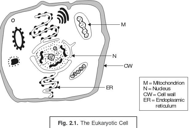
Microbiology is the specific branch of ‘biology’ that essentially deals with the elaborated inves-tigation of ‘microscopic organisms’ termed as microbes, that are composed of only one cell. These are typically either unicellular or multicellular microscopic organisms that are distributed abundantly both in the living bodies of plants and animals and also in the air, water, soil, and marine kingdom.
1
• Introduction
Interestingly, each and every microbe essentially bear both specific and special characteristic features that enable it to survive adequately in a wide spectrum of environments, such as : streams, ponds, lakes, rivers, oceans, ice, water-borne pipes, hot-springs, gastro-intestinal tract (GIT), roots of plants, and even in oil wells. In general, the microorganisms are usually characterized by very typical and extremely high degree of adaptability. Microbes are invariably distributed over the entire biosphere*, lithosphere, hydrosphere, and above all the atmosphere.
One may also define microbiology as — ‘the study of living organisms of microscopic size, that include essentially bacteria, fungi, algae, protozoa and the infectious agents at the very borderline of life which are broadly known as viruses.
It is mainly concerned with a variety of vital and important aspects, such as : typical form, inher-ent structure, reproduction, physiological characteristics, metabolic pathways (viz., anabolism, and ca-tabolism), and logical classification. Besides, it includes the study of their :
• Distribution in nature,
• Relationship to each other and to other living organisms, • Specific effects on humans, plants, and animals, and • Reactions to various physical and chemical agents.
The entire domain of microbiology may be judiciously sub-divided into a plethora of diversified, well-recognized, and broadly accepted fields, namely :
Bacteriology : the study of organism (bacteria),
Mycology : the study of fungi,
Phycology : the study of algae,
Protozoology : the study of protozoans, and
Virology : the study of viruses.
Advantages : The advantageous fields of microbiology are essentially the ones enumerated below : 1. Aero-Microbiology — helps in the overall preservation and preparation of food, food-prone
diseases, and their ultimate prevention.
2. Beverage Microbiology — making of beer, shandy, wine, and a variety of alcoholic bever-ages e.g., whisky, brandy, rum, gin, vodka. etc.
3. Exomicrobiology — to help in the exploration of life in the outerspace. 4. Food Microbiology — making of cheese, yogurt.
5. Geochemical Microbiology — to help in the study of coal, mineral deposits, and gas forma-tion ; prospecting the deposits of gas and oil, coal, recovery of minerals from low-grade ores. 6. Industrial Microbiology — making of ethanol, acetic acid, lactic acid, citric acid, glucose
syrup, high-fructose syrup.
7. Medical Microbiology — helps in the diagnostic protocol for identification of causative agents of various human ailments, and subsequent preventive measures.
8. Pharmaceutical Microbiology — making of life-saving drugs, ‘antibiotics’ e.g., penicillins, ampicillin, chloramphenicol, ciprofloxacin, tetracyclines, streptomycin.
INTRODUCTION AND SCOPE 3
9. Soil and Agricultural Microbiology — helps in the maintenance of a good farm land by keeping and sustaining a reasonable and regular presence of microbes in it.
10. Waste-Treatment Microbiology — treatment of domestic and industrial effluents or wastes by lowering the BOD*, and COD**.
Disadvantages : The apparently disadvantageous and detrimental manner whereby the microor-ganisms may exhibit their effects are, namely : disease-producing ormicroor-ganisms viz.,typhus fevercaused by Rickettsia prowazekii, malaria caused by Plasmodium falciparum ; food-spoilage microbes ; and a host of organisms that essentially deteriorate materials like optical lenses (in microscopes and spectrophotometers), iron-pipes, and wood filings.
1.2.
HISTORICAL DEVELOPMENT OF MICROBIOLOGY — MILESTONES
It is more or less a gospel truth that in science the ultimate credit, glory, and fame goes to the one who actually succeeds to convince the world, and not to the one who first had conceived the original concept and idea. Hence, in the development of microbiology the most popular and common names are invariably of those researchers/scientists who not only convinced the world in general, but also developed a tool or a specific technique or an idea (concept) which was virtually adopted or who expa-tiated their observations/findings rather vividly or astronomically that the science grew and prospered in particular.
Evidence from the literature reveals that Antony van Leeuwenhoek’s (1632-1723) lucid expla-nations with regard to the ubiquitous (i.e., found everywhere) nature of the microbes practically enabled Louis Pasteur (1822–1895) almost after two centuries to discover the involvement of these microorgan-isms in a variety of fermentation reaction procedures that eventually permitted Robert Koch (1843-1910), Theobald Smith, Pasteur and many others to establish and ascertain the intimate relationship of the various types of microbes with a wide range of dreadful human diseases. In fact, Robert Koch bagged the most prestigious Nobel prize in the year 1905 for his spectacular and wonderful discovery for the isolation and characterization of the bacteria that cause anthrax*** and tuberculosis.****
With the passage of time the ‘mankind’ has won several gruesome battles with dreadful micro-organisms quite successfully and have adequately mustered the knack not only to make them work in an useful and beneficial manner but also to control and prevent some of those that are rather dangerous and harmful in nature.
1.2.1. The Microscope
The evolution of microscope gathered momentum in the year 1674, when a Dutch cloth mer-chant Antony van Leeuwenhoek first of all had a glimpse at a drop of lake-water via a lens made of glass that he had ground himself. Through this simple device using a magnifying lens Leeuwenhoek first and foremost ever had an ‘amazing sight’ of the most fascinating world of the microbes.
*BOD : Biological oxidation demand. **COD : Chemical oxidation demand.
***Anthrax : Acute infectious disease caused by Bacillus anthracis, usually attacking cattle sheep, horses, and
goats. Humans contract it from contact with animal hair, hides or waste.
****Tuberculosis [TB]. An infectious disease caused by the tubercle bacillus, Mycobacterium tuberculosis, and
Later on, Leeuwenhoek critically and explicitly described the finer details of a plethora of micro-organisms viz.,protozoa, algae, yeast, and bacteria to the august Royal Society of London (UK) in a series of letters. It is worthwhile to mention here that the entire description was so precise and accurate that as to date it is now quite possible to assign them into each particular genera without any additional description whatsoever.
The earlier observations of microorganisms were made duly by several researchers chronologi-cally as given below :
Roger Bacon (1220–1292) : first ever postulated that a disease is caused by invisible living creatures.
Girolamo Fracastoro (1483–1553) and Anton von Plenciz (1762) : these two reseachers also made similar observations, assertions, and suggestions but without any experimental concrete evidences/ proofs.
Athanasius Kircher (1601–1680) : made reference of these ‘worms’ that are practically invis-ible to the naked eyes and found in decaying meat, milk, bodies, and diarrheal secretions. Kircher was, in fact, the pioneer in pronouncing the cognizance and significance of bacteria and other microbes in disease(s).
Antony van Leeuwenhoek (1632–1723) : initiated the herculian task of ‘microscope making’ through his inherent hobby of ‘lens making’. During his lifespan stretching over to 89 years he meticu-lously designed more than 250 microscopes ; of which the most powerful one could magnify about 200-300 times only. However, these microscopes do not have any resemblance to the present day ‘com-pound light microscope’ that has the ability to even magnify from 1,000-3,000 times.
1.2.2. Spontaneous Generation Vs Biogenesis
The wonderful discovery of microbes both generated and spurred enough interest not only in the fundamental origin of ‘living things’ but also augmented argument and speculation alike.
Based upon the various experimental evidences the following observations were duly made by scientists as enumerated below :
John Needham (1713-1781) : Precisely in the year 1749, while experimenting with raw meat being exposed to hot ashes, he observed meticulously the appearance of organisms that were not present at the initial stages; and, therefore, inferred that the bacteria virtually originated from the raw meat itself.
Lazaro Spallanzani (1729-1799) : actually boiled ‘beef broth’ for a duration of 60 minutes, and subsequently sealed the flasks tightly. After usual incubation for a certain length of time, practi-cally no microbes appeared. However, Needham never got convinced with Spallanzani’s findings, and vehemently insisted that ‘air’ happened to be an essential component to the process of spontaneous generation of the microbes, and that it had been adequately excluded from the flasks by sealing them precisely by the later.
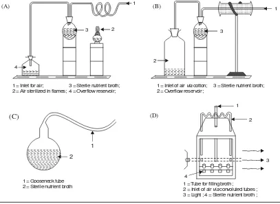
Franz Schulze (1815-1873) and Theodor Schwann (1810–1882) : these two scientists inde-pendently fully endorsed and justified the earlier findings of Spallanzani by allowing air to pass through strong acid solutions into the boiled infusions, and by passing air into the flasks via red-hot tubes respectively (Fig. 1.A). In neither instance did microorganisms appear.
INTRODUCTION AND SCOPE 5
H. Schröder and T. von Dusch (~ 1850) : carried out a more logical and convincing experimen-tal design by passing air via cotton fibers so as to prevent the bacterial growth ; and thus, it ultimately initiated and gave rise to a basic technique of ‘plugging’ bacterial culture tubes with ‘cotton plugs’
(stoppers), which technique being used still as to date (Fig. : 1.B).
Felix Archimede Pouchet (1800–1872) : revived once again the concept and ideology of spon-taneous generation via a published comprehensive and extensive research article thereby proving its occurrence. Pasteur (1822–1895) carried out a number of experiments that virtually helped in conclud-ing the on-goconclud-ing argument once for all time. Pasteur designed a flask havconclud-ing a long and narrow gooseneck outlet (Fig. : 1.C). Thus, the nutrient broths were duly heated in the above specially–designed flask, whereby the air — untreated and unfiltered — may pass in or out but the germs settled in the ‘very gooseneck’ ; and, therefore, practically no microbes ultimately appeared in the nutrient broth (solution).
John Tyndall (1820-1893) : conducted finally various well planned experiments in a specifi-cally designed box (Fig. : 1.D) to establish and prove the fact that ‘dust’ actually contained and carried the ‘microbes’(i.e., germs). He subsequently demonstrated beyond any reasonable doubt that in a particular situation whereby absolutely no dust was present, the sterile nutrient broth could remain free of any sort of microbial growth for an indefinite length of time.
Fig. 1. Theory of ‘Spontaneous Generation’ was actually disproved with the various devices illustrated above i.e., ‘A’ through ‘D’, all of which eliminated airborne microbes.
A = Schwann heat-sterilized the air that passed via the glass-tube to the ‘culture flask’. B = Schröder and Dusch, filtered the ‘air’ entering the ‘culture flask’via the cotton. C = Pasteur devised the ‘simple gooseneck flask’.
D = Tyndall designed a ‘dust-free incubation chamber’.
1 = Inlet for air; 3 = Sterile nutrient broth; 2 = Air sterilized in flames; 4 = Overflow reservoir;
3 2
1
4
(A)
1 = Inlet of air cotton; 3 = Sterile nutrient broth; 2 = Overflow reservoir;
via (B)
3
1
2
1 = Gooseneck tube 2 = Sterile nutrient broth
2 1
(C)
1 = Tube for filling broth ;
2 = Inlet of air convoluted tubes ; 3 = Light ; 4 = Sterile nutrient broth ;
via
1
2
3
4
*HTST-Pasteurizer. It makes use of temperature short-time pasteurization process employing high-temperature live steam.
** Septicemia following childbirth [SYN : childbed Fever ; Puerperal Sepsis ;]
1.2.3. Fermentation
France having the strategical geographical location developed the commercial manufacture of a large variety of wines and beer as a principal industry. Pasteur played a critical and major role in the proper standardization of various processes and techniques intimately associated with the said two
‘alcoholic beverages’ in order to obtain a consistently good product. Pasteur used his God gifted won-derful skill and wisdom to explore and exploit the unique capabilities of microbes in the fermentation industry exclusively using fruits and grains resulting in alcohol-based table wines, dry-wines, cham-pagne, whiskies, etc. Pasteur meticulously isolated, typified, and characterized ‘certain microbes’ ex-clusively responsible for the ‘good batches’ predominantly in comparison to the ones found solely in the ‘poor products’.
In fact, the overall net outcome of such extensive as well as intensive investigations helped in a long way for the assured and successful production of consistently good and uniform ultimate product. Pasteur vehemently argued and suggested that the unwanted/undesirable types of microbes must be destroyed and removed by heating not enough to alter the original and authentic inherent flavour/aroma of the fruit juice, but just sufficient to cause and afford the legitimate destruction of a relatively very high percentage of the ‘bad microbial population’. This ‘destructive microbial phenomenon’ could be ac-complished successfully by holding the juices at a temperature of 145°F (≡ 62.8°C) for a duration of 30 minutes.
Pasteurization. Nowadays, the large-scale handling of such destructive microbial process may be achieved by ‘pasteurization’* in commercial fermentation industries using either ‘malt wort’ (having ~ 10% solids) or molasses (~ 10% solids) or even fruit-juices.
1.2.4. Germ Theory
A plethora of observant researchers had already conceptualized and opined rather vehemently the much applauded and widely accepted ‘germ theory’ of disease even before Pasteur established experimentally that microbes (or bacteria) happen to be the root cause of several human dreadful diseases. Later on various other scientists supported and proved the aforesaid ‘germ theory’ in one way or the other as stated under :
Girolamo Fracastro (1483–1553) : advocated that certain diseases might be caused by virtue of
invisible organisms transmitted from one subject to another.
Plenciz (1762) : stated that the living microbes (or agents) are the ultimate cause of disease but at the same time aired his views that different germs were responsible for different ailments.
Oliver Wendell Holmes (1809–1894) : suggested that puerperal fever** was highly conta-gious in nature ; besides, it was perhaps caused by a germ carried eventually from one mother to another either by midwives or physicians.
Ignaz Philipp Semmelweis (1818–1865) : pioneered the usage of antiseptics specifically in the obstetrical practices.
INTRODUCTION AND SCOPE 7
* Acute, infectious disease caused by Bacillus anthracis, usually attacking cattle, sheep, horses, and goats. First ever proved that a bacterium to be the cause of an animal disease.
Robert Koch (1843–1910) : discovered the typical bacilli having squarish ends in the blood sample of cattle that had died due to anthrax.*
Koch’s Modus Operandi — Koch adopted the following steps to isolate microbes causing an-thrax :
(1) First of all these bacteria were duly grown in cultures in the laboratory.
(2) Bacteria examined microscopically so as to ascertain only one specific type was present. (3) Injected bacteria into other animals to observe whether they got also infected, and
subse-quently developed clinical symptoms of anthrax.
(4) Isolated microbes from experimentally infected animals squarely matched with those ob-tained originally from sheep that died due to infection of anthrax.
Koch’s Postulates : The series of vital observations ultimately led to the establishment of Koch’s postulates, that essentially provided four vital guidelines to identify the particular causative agent for an infectious disease, namely :
(a) A particular microbe (organism) may invariably be found in association with a given disease. (b) The organism may be isolated and cultivated in pure culture in the laboratory.
(c) The pure culture shall be able to cause the disease after being duly inoculated into a suscep-tible animal.
(d) It should be quite possible to recover conveniently the causative organism in its pure culture right from the infected experimental animal.
1.2.5. Classical Laboratory Methods and Pure Cultures
Microorganisms are abundantly found in nature in sufficiently large populations invariably com-prised of a plethora of different species. It is, however, pertinent to state here that to enable one to carry out an elaborated study with regard to the characteristic features of a specific species it is absolutely necessary to have it separated from all the other species.
Laboratory Methods : Well defined, articulated, and explicite laboratory methods have been adequately developed which enable it to isolate a host of microorganisms representing each species, besides to cultivate each of the species individually.
Pure Culture : Pure culture may be defined as — ‘the propogation of microorganisms or of living tissue cells in special media that are conducive to their growth’.
In other words it may also be explained as the growth of mass of cells belonging to the same species in a laboratory vessel (e.g., a test tube). It was indeed Joseph Lister, in 1878, who first and foremost could lay hand on pure cultures of bacteria by the aid of ‘serial dilution technique’ in liquid media.
Colonies : Koch meticulously devised methods for the specific study of microorganism. He
smeared bacteria on a sterile glass slide, followed by addition of certain specific dyes so as to observe the individual cells more vividly under a microscope. Koch carefully incorporated some specific solidi-fying agents, such as : gelatin, agar into the media in order to obtain characteristic isolated growths of organisms usually called as colonies. Importantly, each colony is essentially comprised of millions of individual bacterial cells packed tightly together.
Now, from these identified colonies one may transfer pure cultures to other sterile media. How-ever, the development of a liquefiable solid-culture medium proved to be of immense fundamental importance.
Example : Koch thoroughly examined material obtained from subjects suffering from pulmo-nary tuberculosis, and succeeded in the isolation of the tubercle bacillus Mycobacterium tuberculosis.
In conclusion, one may summarize the remarkable importance of ‘pure cultures’ toward the overwhelming development in the field of microbiology, because by the help of pure-culture tech-niques several intricate and complicated problems could be answered with reasonable clarification and complete satisfaction, namely :
Microorganisms causing a large number of infections, Certain specific fermentative procedures,
Nitrogen-fixation in soil,
High-yielding alcohol producing strains from ‘malt wort’, and ‘molasses’, Selected good cultures for making top-quality wines, and
Specific cultures for manufacturing dairy products viz., cheeses, yogurt.
Futuristic Goals
The futuristic goals of ‘pure cultures’ are exclusively based upon the following two cardinal aspects, namely :
(a) better understanding of the physiology of individual microorganisms present in the pure culture, and
(b) ecological relationships of the entire microbial populations in a given environment. Thus, the following new horizons in the domain of microbiology may be explored with great zeal and gusto :
Advancements in marine microbiology, Rumen microbiology,
Microbiology of the gastro-intestinal tract (GIT), and Several other systems.
1.2.6. Immunity
Immunity refers to the state of being immune to or protected from a disease, especially an infectious disease. This state is invariably induced by having been exposed to the antigenic marker on an organism that invades the body or by having been immunized with a vaccine capable of stimulating production of specific antibodies.*
INTRODUCTION AND SCOPE 9
* Microorganisms that have been rendered thin or made less virulent (infectious).
** Any of the complex glycoproteins produced by lymphocytes in response to the presence of an antigen. *** Infectious organisms.
**** A substance that neutralizes toxin.
Interestingly, Pasteur’s practical aspects and Koch’s theoretical aspects jointly established the fact that the attenuated microorganisms* retained their capacity and capability for stimulating the respective host to produce certain highly specific substances i.e.,antibodies** which critically protect against subsequent exposure to the virulent organisms.***
Examples :
(a) Edward Jenner’s successful cowpox vaccine (in 1798) : Jenner’s epoch-making successful attempts in vaccinating (innoculating) patients with cowpox vaccine, that ultimately re-sulted in the development of resistance to the most dreadful smallpox infection.
(b) Pasteur’s successful rabies vaccine : Pasteur’s charismatic fame and reputation became well known throughout France when he successfully prepared rabies vaccine by innoculating a rabbit with the saliva from a rabid dog. The healthy rabbit contracted the rabies virus and died later on. The extract of dead rabbit’s brain and spinal cord were duly attenuated and injected into rabies patient who eventually survived later on. Thus, the vaccine for rabies or hydrophobia — a disease transmitted to humans through bites of dogs, cats, monkeys, and other animals.
1.2.7. Medical Microbiology
Interestingly, the ‘germ theory’ of disease was very much in existence for a long duration ; however, the direct implication and involvement of germs in causing disease was not well established, and hence recognized and widely accepted.
The magnificent and remarkable success of Louis Pasteur and Robert Koch not only earned them befitting honours and accolades from their beloved countrymen, but also rewarded them by bestowing their gratitude in establishing the famous and prestigious Pasteur Institute in Paris (1888), and Professor of Hygiene and Director of the Institute for Infective Diseases in the University of Berlin respectively.
At this point in time altogether newer microorganisms (bacteria) were being discovered with an ever-increasing speed and momentum, and their disease-producing capabilities were adequately estab-lished and proved by Koch’s four cardinal postulates as stated earlier (see section 1.2.4).
In this manner, the domain of ‘medical microbiology’ gradually received a progressive advance-ment through the meaningful researches conducted by several scientists and scholars as enumerated below :
Edwin Klebs (1883) and Frederick Loeffler (1884) : discovered the diphtheria bacillus, corynebacterium diphtheriae ; and showed that it produced its toxins (poisons) in a laboratory flask.
Emil von Behring and Shibasaburo Kitasato : devised an unique technique of producing im-munity to infections caused by C. diphtheriae by injecting the toxins into healthy animals so that an
antitoxin**** gets developed.
Shibasaburo Kitasato and Emil von Behring : cultivated (grown) the microorganism respon-sible for causing tetanus (lockjaw), Chlostridium titani ; and Behring prepared the corresponding
* An antibody is a water-soluble protein produced from globulins (e.g.,γ-globulin) in the spleen, lymph nodes,
thymus gland, liver or bone marrow in response to an antigen (foreign protein). Antibodies attack antigens to render them inactive and no longer infective.
** A therapeutic concept developed by Paul Ehrlich (1854–1915) wherein a specific chemical or drug is used to treat an infectious disease or cancer ; ideally, the chemical should destroy the pathogen or the cancer cells without harming the host.
*** A chemical produced by a microorganism or prepared partially or totally by synthetic means that inhibits growth or kills other microorganisms at low concentration.
Emil von Behring bagged the Nobel Prize in 1901 in physiology or medicine.
De Salmon and Theobald Smith : proved amply that immunity to a plethora of infectious diseases may be produced quite effectively and efficiently by proper timely innoculation with the killed cultures of the corresponding microorganisms.
Elie Metchnikoff : described for the first time the manner certain specific leukocytes (i.e., white blood cells) were able to ingest (eat up) the disease-producing microorganisms present in the body. He baptized these highly specific defenders and crusaders against bacterial infections known as phagocytes
(‘eating cells’), and the phenomenon is termed as phagocytosis.
Metchnikoff’s Theory : Based of the aforesaid explanations Metchnikoff put forward a theory that — ‘the phagocytes were the body’s first and most important line of defense against a variety of infection’.
Paul Ehrlich :Paul Ehrlich (Robert Koch’s brilliant student) put forward two altogether newer concepts with regard to the modus operandi whereby the body aptly destroys microorganisms (bacteria), namely :
(a) Antibody* : The logical explanation of immunity based upon certain antibodies in the blood, and
(b) Chemotherapy** and Antibiotics*** : Both these aspects virtually opened the flood gates to the enormous future developments in combating the growth and destruction of pathogenic bacteria.
Example : Arsphenamine [Salvarsan(R)] : A light yellow organo-metallic compound (powder) containing about 30% Arsenic (As), was formerly used in the treatment of syphilis.
The two decades stretching between 1880–1900 proved to be indeed a golden era for the ‘sci-ence of microbiology’ to step into adolescence from the stage of infancy. In fact, during this specific period many researchers have gainfully identified the causative microorganisms duly responsible for the eruption of a host of infectious human diseases, such as :
Anthrax, Gonorrhea, Typhoid fever, Malaria, Wound infections, Tuberculosis, Cholera, Diph-theria, Tetanus, Meningitis, Gas gangarene, Plague, Dysentery, Syphilis, Whooping cough, and Rocky Mountain spotted fever.
1.2.8. Pharmaceutical Microbiology
INTRODUCTION AND SCOPE 11
Examples : (a) Widal Test* — for typhoid fever, and (b) Wasserman Test** —for syphilis.
Importantly, a plethora of dreadful diseases were duly identified and characterized by the pres-ence of their specific causative microorganisms, such as : Hensen (1874) leprosy (Mycobacterium leprae) ; Neisser (1879) gonorrhea (Neisseria gonorrhoeae) ; Ogston (1881) wound infections (Staphylococcus aureus) ; Nicolaier (1885) tetanus (Clostridium titani) ; Kitasato and Yersin (1894)
plague (Yersinia pestis) ; Shiga (1898) dysentry (Shigella dysenteriae) ; Schaudin and Hoffmann (1905)
syphilis (Treponema pallidum); Bordet and Gengou (1906) whooping cough (Bordetella pertussis) ; Ricketts (1909) rocky mountain spotted fever (Rickettsia ricketsii) ;
Some of the important events that mark the history of pharmaceutical microbiology are enu-merated below in a chronological arrangement :
Era Discoverer Important Events
Eighteenth Edward Jenner Discovery of small pox vaccine.
Century (1729–1799)
Nineteenth Justus von Liebig Conceptualized the physico-chemical theory of
fermen-Century (1803–1873) tation.
Ignaz Philipp First and foremost introduced the application of antiseptics. Semmelweis
(1818–1865)
Joseph Lister Developed aseptic techniques : isolated bacteria in pure (1827–1912) culture.
Fanny Hesse Suggested use of agar as a solidifying material for the (1850–1934) preparation of microbiological media.
Paul Ehrlich Developed modern concept of chemotherapy and (1854–1915) chemotherapeutic agents.
Hans Christian Invented vital and important procedure for differential Gram staining of microorganisms i.e., the well-known Gram Stain.
(1853–1933)
Twentieth August von Developed complement-fixation test for syphilis.
Century Wassermann
(1866–1925)
Martinus Willem Employed the principles of enrichment cultures : Beijerinck confirmed finding of the very first virus. (1851–1931)
Frederick W. Twort Discovered independently the bacteriophages i.e., viruses
(1877–1950) ; and that destroy bacteria. Felix H.d’ Herelle
(1873–1949)
* The living together in close association of two organisms of different species. If neither organism is harmed, this is called commensalism ; if the association is beneficial to both, mutualism ; if one is harmed and the other benefits, parasitism.
** An association or relationship between two organisms in which one is harmful to the other.
*** An enzyme found in phagocytes, neutrophils, and macrophages, and in tears, saliva, sweat and other body secretions, that destroys bacteria by breaking down their walls.
Antibiotics : Antibiotic refers to a natural or synthetic substance that destroys microorganisms or inhibits their growth. Antibiotics are employed extensively to treat infectious, diseases in humans, animals, and plants. In fact, the terminology ‘antibiotic’ etymologically evidently signifies anything against life. Obviously, in the event when the microorganisms are critically present in a natural medium two situations may arise invariably viz., (a) favouring the growth of bacteria usually termed as
‘symbiosis’ ;* and (b) antagonizing the growth of bacteria normally called as ‘antibiosis’.**
Charles Robert Darwin (1809–1882), a British naturalist) aptly commenced scientific and me-thodical investigative explorations into the fundamental problems of natural selection and struggle amongst the interspecies ; and later on came up with his famous doctrine — ‘Survival of the fittest’. Louis Pasteur (1822–1895) observed for the first time the characteristic antagonistic interrelations prevailing between the microorganisms of different species.
Joubert and Pasteur first observed the critical destruction of cultures of Bacillus anthracis by means of certain air-borne microbes. A follow up by Sirotinin (1888) emphatically proved the antago-nistic action of Bacillus anthracis upon the enteric fever, and Blagoveshchensky (1890) carefully ascer-tained the antagonistic effect of the blue-pus organism on the Bacillus anthracis. It was ultimately the miraculous discovery of Lashchenkov (1909) and Alexander Fleming (1922) who meticulously isolated the enzyme lysozyme***, that was chiefly capable of inhibiting a relatively larger segment of microor-ganisms. Chain, Florey, and co-workers (1929) made the epoch making historical development in the emerging field of antibiotics with the remarkable discovery of wonderful therapeutic and interesting pharmacological properties of the extracts obtained from the cultures of the mold Penicillium notatum that eventually gave rise to the formation of the wonder drug ‘penicillin’.
Specifically the antibiotics are extremely useful in the control, management and treatment of a good number of human infectious diseases but their diversified applications are found to be equally useful in the meticulous curing and controlling of plant and animal diseases as well. Penicillin has been effectively employed in the management and control of pests. Antibiotics, in general, are invariably employed in animal husbandry as ‘feed additive’ to cause enhancement in the fattening of food animals. Food handling and processing industries extensively make use of antibiotics to critically minimise in-evitable spoilage of fish, vegetables, and poultry products. Present day modern scientific researches being conducted across the globe do make use of antibiotics as useful and indispensable tools for the elaborated study of biochemical cellular mechanisms.
Since the discovery of penicillin many more antibiotics came into being as stated under : Waksman (1944) : Streptomycin — [Streptomyces griseus] — a soil microbe ;
— (1945) : Bacitracin — [Bacillus subtilis] ;
— (1947) : Chloramphenicol (Chloromycetin) — [Streptomyces venezuelae] ; — (1947) : Polymixin — [Bacillus polymixa] — and various designated polymixins
A, B, C, D, and E.
— (1948) : Chlorotetracycline — [Streptomyces aureofaciens] — a broad-spectrum antibiotic.
INTRODUCTION AND SCOPE 13
— (1950) : Oxytetracycline — [a strain of Streptomyces]. — (1952) : Erythromycin — [Streptomyces erythreus].
It is, however, pertinent to state here that the ‘antibiotics’ may be broadly classified into nine categories as given below :
S.No. Class Designated Antibiotics
I Aminoglycosides Amikasin ; Gentamycin ; Kanamycin ; Neomycin ; Netilmicin ; Streptomycin ; and Tobramycin ;
II Ansamycins Maytansine ; and Rifampicin ;
III Beta-lactam antibiotics Amoxycillin ; Ampicillin ; Cephalosporin ; Clavulanic acid ; Cloxacillin ; Nocardicins ; Penicillins ; and Thienamycin ; IV Cyclic polypeptides Gramicidin ; and Polymixins A, B, C, D and E ;
V Fluoroquinolones Ciprofloxacin ; Enoxacin ; Norfloxacin ; and Ofloxacin.
VI Macrolides Azithromycin ; Bacitracin ; Clarithromycin ; and Erythromycin ; VII Polyenes Amphotericin B ; Griseofulvin ; and Nystatin ;
VIII Tetracyclines Aureomycin ; Doxycycline ; Minocycline ; Oxytetracycline ; and Tetracycline ;
IX Miscellaneous Adriamycin ; Chloramphenicol (Chloromycetin) ; Clindamycin ; Cycloserine ; and Mitomycins.
[Kar, Ashutosh : Pharmacognosy and Pharmacobiotechnology, New Age International (P) Ltd., Publishers, New Delhi, 2003].
Important Points : The various important points with respect to the development of antibiotics
are summarized below :
in all approximately 5000 antibiotics have been prepared, characterized, and evaluated for their therapeutic efficacy till date.
nearly 1000 antibiotics belonging to only six genera of filamentous fungi i.e., including
Cephelosporium and Penicillium have been reported successfully.
about 50 antibiotics have been synthesized from two genera and belonging to the class of non-filamentous bacteria.
nearly 3000 antibiotics have been prepared from a group of filamentous bacteria i.e., in-cluding streptomyces.
approximately 50 antibiotics are at present actively used in therapeutic treatment and veteri-nary medicine around the world.
Importantly, the most common bacteria that invariably attack the humans specifically, and the diseases they cause or organs of the body they attack, are listed as under :
S.No. Microorganism Disease/Place of Infection
1 Bacteroides Pelvic organs ;
2 Bordetella pertussis Whooping cough ;
3 Brucella abortus Brucellosis ;
S.No. Microorganism Disease/Place of Infection
5 Clostridium perfringens Gas gangrene ; Pseudomembranous colitis ;
6 Clostridium titani Tetanus ;
7 Corynebacterium diphtheriae Diphtheria ;
8 Escherichia coli Urine gut ; Fallopian tubes ; Peritonitis ; 9 Haemophilus influenzae Ear ; Maningitis ; Sinusitis ; Epiglottitis ; 10 Helicobacter pyroli Peptic ulcers ;
11 Klebsiella pneumoniae Lungs ; urine ; 12 Legionella pneumophilia Lungs ;
13 Mycobacterium leprae Leprosy ;
14 Mycobacterium tuberculosis Tuberculosis ;
15 Mycoplasma pneumoniae Lungs ;
16 Neisseria meningitidis Meningitis ;
17 Neisseria gonorrhoea Gonorrhoea ; Pelvic organs ;
18 Proteus Urine ; Ear
19 Pseudomonas aeruginosa Urine ; Ear ; Lungs ; Heart ;
20 Salmonella typhi Typhoid fever ;
21 Shigella dysenteriae Gut infections ;
22 Staphylococcus aureus Lungs ; Throat ; Sinusitis ; Ear ; Skin ; Eye ; Gut ; Meningitis ; Heart ; Bone ; Joints ;
23 Streptococcus pneumoniae Throat ; Ear ; Sinusitis ; Lungs ; Ear ; Joints ; 24 Streptococcus pyrogenes Sinuses ; Ear ; Throat ; Skin ;
25 Streptococcus viridans Heart ;
26 Triponema pallidum Syphilis ;
27 Vibrio cholerae Cholera ;
28 Yersinia pestis Plague ;
[Adapted From : Warwick Carter : The Complete Family Medical Guide, Hinkler Books Pvt. LTD., Dingley, Australia, 2003.]
1.2.9. Industrial Microbiology
An exponential growth in the ever expanding domain of industrial microbiology commenced logically from the mid of the nineteenth century to the end of the said century. The various vital and important ‘milestones’ in the field of industrial microbiology may be summarized as stated under :
Emil Christian Hansen (1842-1909) : a Dane*, who actually showed up the brilliant and fertile way to the extremely investigative field of industrial fermentations. He meticulously examined and methodically developed the pure culture study of microorganisms and yeasts exclusively utilized in the large-scale manufacture of ‘fermented vinegar’. This simultaneously encouraged as well as prom-ulgated the application of pure cultures termed as ‘starters’ associated with the elaborated study of various fermentation processes.
L. Adametz (1889) : an Austrian, augmented the commercial production of cheese by making use of pure cultures (i.e., starters).
INTRODUCTION AND SCOPE 15
HW Conn (in Connecticut, USA) and H Weigmann (in Germany) (1890–1897) : developed miraculously a host of pure culture starters for the commercial production of butter.
Alcohol Fermentations : Pure culture of yeasts were used to produce alcohol (ethanol) from a variety of fermentable carbohydrates such as : corn, molasses, potatoes, sugar beets, grapes etc., employed throughout the world.
In addition to the above mentioned widely consumed and need based products there are several other highly in-demand industrial products derived exclusively from molds that are being used largely across the globe as detailed under :
S.No. Product of Interest Mold(s) Applications
1 Citric acid Aspergillus niger or Medicinal citrates ; In blood for transfusion Aspergillus wentii (as sodium citrate) ; In food products.
2 Gibberellic acid Fusarium moniliforme Plant-growth hormone ; In germination of barley to produce ‘malt’ ; In setting of fruit and seed production.
3 Gluconic acid Aspergillus niger Pharmaceutical products ; textiles ; leather ; photography ;
4 11-γγγγγ-Hydroxy- Rhizopus arrhizus, As an intermediate for 17-α-γ
-progesterone R. nigricans, others hydroxycorticosterone.
5 Itaconic acid Aspergillus terreus Manufacture of alkyl resins ; Wetting agents ;
6 Lactic acid Rhizopus oryzae Pharmaceutical products ; and Food products. 7 Pectinases, Aspergillus wentii or As clarifying agents in fruit-juice
proteases Aspergillus aureus industries.
[K. Sambamurthy and Ashutosh Kar : Pharmaceutical Biotechnology, New Age International (P) Ltd., Publishers, New Delhi, 2005].
1.2.10. Emergence of Molecular Biology
Molecular Biology refers to that specific branch of biology dealing with analysis of the struc-ture and development of biological systems vis-a-vis the chemistry and physics of their molecular
constituents. Now, with the advent of latest laboratory methodologies and modern experimental tech-niques the prevailing skill, wisdom, and knowledge pertaining to the characteristic features of microor-ganisms accumulated with a tremendous momentum and speed. Based upon the intensive and extensive information(s) with respect to the in-depth biochemical activities of various microorganisms virtually became an ‘open-secret’.
Importantly, a careful and critical examination of the copious volume of accumulated data evi-dently revealed and suggested that there existed quite a lot of similarities amongst the different micro-organisms, whereas the points of dissimilarities revolved essentially around the variations on a major
* Professor Salvador E Luria — at the Massachusetts Institute of Technology (MIT) as a Professor of Biology was awarded the Nobel Prize in 1969 for his splendid research in the field of molecular biology.
** Viruses that infect bacteria.
deciphering and exploring the basic life phenomena. In order to accomplish the aforesaid aims and objectives the microorganisms do offer invariably a plethora of advantages for this type of research activities, namely :
they reproduce (i.e., cultivate) extremely fast,
they may be cultured (grown) either in small or large quantum easily, conveniently, and quickly,
their growth may be manipulated and monitored in a not-so-difficult manner by means of chemical and physical methods, and
their cells may be cleaved and torn apart, and the contents segregated into different fractions of varying particle sizes.
Conclusively, the above cited characteristic features together with certain other vital factors help to render the ‘microorganisms’ an extremely vulnerable and a very convenient research-role-model
in pin-pointing and establishing precisely the modusoperandi of various life processes that essentially occur with respect to certain particular chemical reactions, besides the specific structural features involved intimately.
In the light of the above statement of facts showing the enormous strengths of microorganisms in the revelation of the intricacies of life processes various scientists and researchers of all disciplines viz., physicists, chemists, geneticists, biologists, and microbiologists not only joined their hands together but also put their intellectual resources and wisdom in a concerted manner to evolve an altogether new discipline christened as molecular biology. According to Professor Luria* molecular biology may be defined as — ‘the programme of interpreting the specific structures and functions of organisms in terms of molecular structure’.
The outcome of the results obtained from the brilliant studies accomplished in the field of mo-lecular biology are numerous, such as :
Elucidation of enzyme structure and mode of action, Cellular regulatory mechanisms,
Energy metabolism mechanisms, Protein synthesis,
Structure of viruses,
Functionality of membranes, and Structure and function of nucleic acids.
Significance of Discoveries : The major significance of discoveries with regard to molecular biology may be ascertained by virtue of the following breakthroughs :
Fundamental information(s) regarding DNA and genetic processes at the molecular level via bacteria and bacteriophages**, and