SYNTHESIS AND CYTOTOXIC ACTIVITY OF 2,5-
BIS
(4-BORONIC ACID)BENZYLIDINE CYCLOPENTANONE ON HER2
OVEREXPRESSED-CANCER CELLS
Rohmad Yudi Utomo1, Herwandhani Putri1, Pudjono2, Ratna Asmah Susidarti2, Riris Istighfari Jenie1,2, Edy Meiyanto1,2*
1Cancer Chemoprevention
Development of chemotherapeutic agent and boron carrying pharmaceutical based on HER2 specific targeted is important due to its role in enhancing cancer progression. The purpose of this study is to synthesize curcumin analogue, namely Pentagamaboron-0 (PGB-0) or 2,5-bis(4-boronic acid)benzylidine cyclopentanone, and to explore the cytotoxic activity on HER2 overexpressed-cancer cells. MCF-7/HER2 was used as a model of HER2 overexpressed-cancer cells and NIH3T3 as normal cells. PGB-0 bound to ATP binding site of HER2 and EGFR based on molecular docking study. PGB-0 was synthesized resulting in 33% yield and was confirmed by IR, 1HNMR, 13CNMR and
Mass spectroscopy. Based on MTT assay PGB-0 decreased cells viability on MCF-7/HER2 cells with IC50 value of 270µM
but performed no effect on NIH3T3 cells. Cell cycle analysis revealed that PGB-0 increased sub-G1 accumulation. PGB-0 decreased HER2 expression in a dose-dependent manner. We conclude that the new compound PGB-0 inhibits cell growth through cell death induction and decreased HER2 expression. Thus, PGB-0 is potential to be developed as a chemotherapeutic agent and boron carrying pharmaceutical targeted on the HER2 receptor.
Key words: Synthesis of PGB-0, MCF7/HER-2, cytotoxic
INTRODUCTIONS
HER2 is a transmembrane protein that is overexpressed in most human solid tumors such as breast, ovarian, endometrial, colon, and non-small cell lung cancer, prostate, and
cervical cancer (English et al., 2013). Moreover,
more than 20% cases of breast cancer are related with overexpression of HER2 protein and correlated with a worse survival of patients
(Gibbs, 2000). The HER2 receptor is normally expressed in healthy cells and is
responsible for the regulator in cell growth and
differentiation (Yakes et al., 2002). Thus, HER2
signaling becomes an interesting field to develop a molecular targeted therapy for cancer.
Several chemotherapeutic agents targeted on HER2 receptor are developed. Trastuzumab is found to be the first antibody targeting HER2. However, 70% of patients who prolonged use of trastuzumab show a
resistance phenomenon leading to a
progression of metastatic cancer due to the
increasing of EGFR expression (Meiyanto et al.,
2011; Vu and Claret, 2012). To overcome this resistance phenomenon, gefitinib and WZ4002, a known EGFR inhibitor are developed. However, the use of gefitinib and WZ4002 lead
to a new EGFR mutation causing
ineffectiveness therapy. Further, lapatinib is introduced to overcome all of the resistance phenomena. Lapatinib effectively works both on tyrosine kinase EGFR and HER2 as well as
ER receptor (D’Amato et al., 2015). The finding
of lapatinib becomes a new insight to develop a chemotherapeutic targeted as a dual inhibitor not only targeted to the HER2 receptor but also EGFR. Recently, several cases reported
that some patients were resistant to
lapatinib treatment (D’Amato et al., 2015).
Therefore, development of an ideal
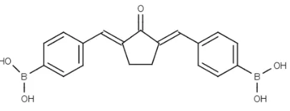
Figure 1. Chemical Structure of 2,5-bis(4-boronic acid)benzylidine cyclopentanone or Pentagamaboronon-0 (PGB-0).
Curcumin is a potential candidate of a chemotherapeutic agent to overcome the resistance phenomena on cancer due to its potency as tyrosine kinase inhibitor, including
HER2 (Hong et al., 1999). However, curcumin
is unstable at pH above 6.5 caused by its
active methylene group (Sardjiman et al., 1997).
Pentagamavunon-0 (PGV-0) and
Pentagamavunon-1 (PGV-1) are developed by modifying the active methylene groups into
cyclopentanone to overcome curcumin
instability. The previous study showed that curcumin, PGV-0, and PGV-1 possess
cytotoxic effect and inhibit NFκB activation in
vitro in MCF-7 breast cancer cells, as well as
interact with the HER2 receptor in silico
(Meiyanto et al., 2014). The interaction between
HER2 receptor with PGV-0 and PGV-1 is mediated through the H-bond between O atom on carbonyl group of PGV-0, and PGV-1
carrying pharmaceuticals for boron neutron captured therapy (BNCT) application, 2,5-bis(4-boronic acid)benzylidine cyclopentanone
or Pentagamaboronon-0 (PGB-0) (Figure 1) is designed by modifying the hydroxyl group
on benzene system into boronic acid and the
using of cyclopentanone structure to
maintain the potential binding of PGB-0 on HER2 receptor. In this study, we synthesize
a new curcumin analog, PGB-0, and explored the cytotoxic activity on HER2 overexpressed-cancer cells as a candidate of chemotherapeutic agent and boron carrying pharmaceuticals targeted on the HER2 receptor.
MATERIAL AND METHODS
Molecular docking, PLANTS
Molecular docking was performed by Protein-Ligand Ant System (PLANTS) 1.1 software. The structure of EGFR (PDB ID: 1XKK) and HER2 (PDB ID: 3PP0) proteins were downloaded from PDB. YASARA (http://www.yasara.org/viewdl.htm) was used for protein preparation and MarvinSketch (http://www.chemaxon.com/marvin/downloa
d-user.html) for ligand preparation.
Visualization 2D and 3D performed by MOE and 2mL of NaOH (Merck) 10% were mixed in 5mL of ethanol (Merck). Three hundred mg of 4-formylphenyl boronic acid (Sigma) was added then the mixture was stirred at 0°C for 30min. After settling for 1h, the mixture was neutralized by adding HCl 10% dropwise. The solid material was washed with ethanol:water 1:1 and filtered. The melting point of the
compound was determined by Buchi
instrument. Thin layer chromatography experiment was performed by using Silica
Gel 60 GF254 (Merck) and mobile phase
methanol:chloroform 3:1 with detection by UV light. HPLC analysis was conducted by C18 column (chromosorb) and mobile phase acetonitrile:water (40:60) with UV detector at wavelength 254 nm. The structure elucidation
was carried out by using 1H NMR and 13C
NMR (JEOL DELTA 500 MHz), IR spectrophotometry (Perkin-Elmer), and MS (Shimadzu).
Cell culture
MCF-7/HER2 was kindly gifted from Prof. Yoshio Inouye (Departement of Surgery, Toho University School of Medicine, Japan) through Prof. Masashi Kawaichi (Laboratory of Gene Function in Animals, NAIST, Japan). The cells NIH3T3 was kindly given by Prof. Masashi Kawaichi (Laboratory of Gene Function in Animals, NAIST, Japan). Cells were grown in
Dulbecco’s Modified Eagles Medium (DMEM)
(Sigma), HEPES, sodium bicarbonate, 1.5% Penicillin-Streptomycin and 0.5% Fungizone (Gibco). Cells were incubated at 37°C with 5%
CO2.
Cytotoxicity assay
Cells were seeded in 96-well plates with
104 cells/well. After 24h incubation with
PGB-0 with various concentrations, the culture medium was removed and cells were washed in PBS (Sigma). Then, cells were incubated with 0.5mg/mL MTT (Biovision) in 100µL culture medium each well for 4h. MTT reaction was stopped by SDS reagent (10% Sodium dodecyl sulfate (Merck) in HCl 0.01N (Merck) and was incubated overnight. The absorbance was measured by ELISA reader (Bio-Rad) at a wavelength of 595nm (Mosmann, 1983).
Cell Cycle Distribution Analysis
An amount of 5x10³ cells/well cells
were distributed to 6-well plate in DMEM medium. After 24h incubation, cells were
treated with PGB-0 ½ IC50 (135µM) and
PGB-0 IC50 (270µM). Following 24h treatment, cells
were trypsinized and centrifuged with speed 365g for 3min. Cell pellets were collected then were washed with a buffer solution as described
by the manufacturer (BD Cycletest™ Plus
incubated in tissue culture dish and was treated with PGB-0 (50µM and 150µM) for 24h. Total protein cell lysates were prepared by using radioimmunoprecipitation assay (RIPA) buffer (Santa Cruz #sc-364162) on ice. The cells suspension was sonicated and was centrifuged at 16000g for 20min at 4°C. The protein samples were denatured in 5x sample buffer and were subjected to 10% SDS-polyacrylamide gel electrophoresis. The separated proteins were transferred into PVDF membrane followed by blocking with 5% skim milk powder (w/v) in TBS (20mM Tris and 133 mM NaCl, 1M HCl) with 20% Tween 20 for 1h at room temperature. The membrane was probed using primary antibodies as follows: HER2
antibody (Santa Cruz #sc-52349) and α-tubulin
antibody (Santa Cruz #sc-32293) overnight at 4°C followed by HRP-secondary antibodies mouse (Cell signaling #7076) for 1h at room temperature and was visualized by Amersham ECL Prime (#RPN2232).
Analysis
Cytotoxicity study was analyzed by determining
IC50 value. Cell cycle distribution was acquired
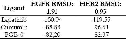
by using Modfit LT 4.1.7. Method validation of molecular docking was conducted by measuring Root Mean Square Distances (RMSD) heavy atom ligand with copy ligands. If RMSD value higher than 2.0 Å means that the molecular docking method is valid. The affinity of the compound was
evaluated by identifying fPLP score. The lower
fPLP score showed higher affinity of the binding
and vice versa.
Lapatinib -150.04 -119.55
Curcumin -88.83 -96.51
PGB-0 -82,20 -82.37
Our main purpose of this research is to develop a new compound having cytotoxic activites to specific cancer cells especially HER2 overexpressed cells. Firstly, we used
molecular docking to construct our
hypotesis regarding the spesific activities of our candidates to the target protein. We obtained that PGB-0 possessed potential candidate beside the other compounds (data not sown). Validation result showed molecular docking protocol was valid with
RMSD value <2 Å (Table I). fPLP score of
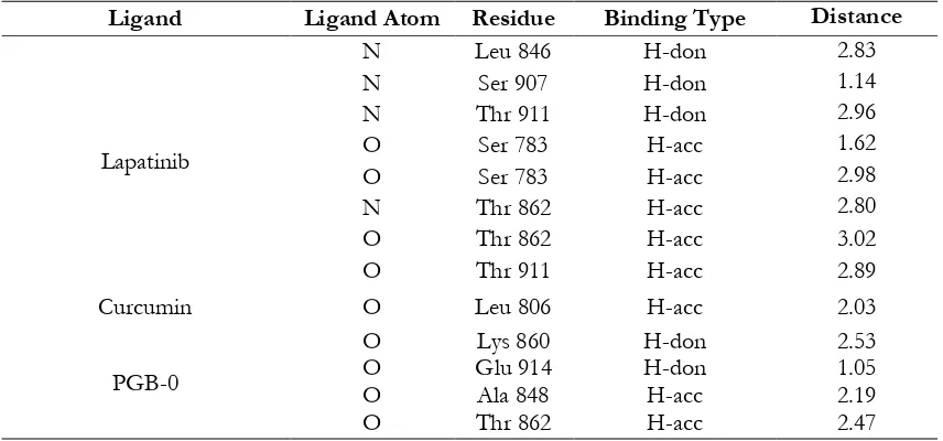
PGB-0 was higher than lapatinib and curcumin on (Table I). However, PGB-0 possessed shorter binding distance than lapatinib at Thr 862 on the HER2 receptor (Table II). Compared to curcumin, PGB-0 also possessed
PGB-0 interacts on the same amino acid residue with curcumin (Glu 906) and possessed a shorter bonding distance on EGFR receptors. However, when compared with lapatinib, PGB-0 bond on different amino acids and a longer bonding distance. (Table 3). Based on the result, we predicted that PGB-0 possibly bound to ATP-binding site of HER2 and EGFR with different binding properties
compared to lapatinib and curcumin.
Therefore, PGB-0 is a potential candidate of chemotherapeutic agent targeted on HER2 and EGFR.
Synthesis of PGB-0
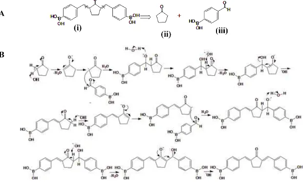
Based on the molecular docking results, PGB-0 possesed comparable affinity on EGFR and HER2 with lapatinib and curcumin, therefore in the next step we synthesize PGB-0. In order to obtain the PGB-0 compound in high purity, we performed several synthesis optimizations (data not shown). Disconnection analysis of PGB-0 showed that PGB-0
predicted to be synthesized by reacting 4-formylphenylboronic acid and cyclopentanone
based on aldol condensation reaction
(Scheme 1). PGB-0 compound was attained 33% yield. HPLC analysis showed that the purity of product was observed on 4 min with a high percentage (95%) while the starting material 4-formylphenyl boronic acid was observed in low percentage at Rt of 2min (5%) (Figure 3). The hRf of the product based on the experiment was 75. The product profile was
described as follows: mp > 200oC, 1H-NMR
(CD3OD): δ 3.17 (s, J=3.27, 4H, C-CH2-CH2
-C), 4.7 (s, J=0.68, 1H, -OH), 7.5 (s, J=1.75, 2H, ), 7.6 (s, J=3.14, 4H, -CH-CH-CH-CH-), 7.7 (s, J=1.23, 2H, -CH), and 7.8 (s, J=1, 1H,
-C-CH-C).13C-NMR (CD3OD): δ 27.7 (s, -CH2),
131 (s, -C-B-), 135 (s, -CH-), 135.1 (s, -CH-C-), 135.4 (-C-CH-), and 139 (s, -C-C=O-); IR (KBr
film) cm-1: 1620 (C=O), 3363 (OH), 1689
(C=C alkene), 1550 (C=C aromatic), 624 (C-H alkene). MS of the product was found 347.14, calculated 347.97.
Table II. Binding Interaction of Ligand on HER2
Ligand Ligand Atom Residue Binding Type Distance
Lapatinib
N Leu 846 H-don 2.83
N Ser 907 H-don 1.14
N Thr 911 H-don 2.96
O Ser 783 H-acc 1.62
O Ser 783 H-acc 2.98
N Thr 862 H-acc 2.80
O Thr 862 H-acc 3.02
O Thr 911 H-acc 2.89
Curcumin O Leu 806 H-acc 2.03
PGB-0
O Lys 860 H-don 2.53
O Glu 914 H-don 1.05
O Ala 848 H-acc 2.19
O Thr 862 H-acc 2.47
Table III. Binding Interaction of Ligand on EGFR
Ligand Ligand Atom Residue Binding Type Distance
Lapatinib O Arg 841 H-acc 2.20
Curcumin O Glu 906 H-don 2.61
O Cys 775 H-acc 2.81
Lapatinib
Curcumin
PGB-0
EGFR HER2
Figure 2. Visualization of ligand interaction on EGFR and HER2. The representative pose of lapatinib, Curcumin, and PGB-0. All ligand is depicted as balls and sticks. Green and purple cavity represent polar and hydrophobic interaction. Molecular Interaction was performed using MOE.
Figure 3. HPLC profile of PGB-0. The sample was running on C18 with mobile phase acetonitrile:water (40:60) with UV detector at wavelength 254nm. 4-formylphenyl boronic acid (i) and PGB-0 (ii)
Cytotoxic effect of PGB-0 on MCF-7/HER2 and NIH3T3 Cells.
The cytotoxic assay was carried out to evaluate the cytotoxicity of PGB-0 on HER2 overexpressed-breast cancer cells and normal cells. The cytotoxic effect of PGB-0 was
conducted by MTT assay. PGB-0inhibited cells
growth of MCF-7/HER2 with an IC50 value of
270µM and performed no effect on NIH3T3 normal cells (Figure 4). The cytotoxicity of PGB-0 was possibly through cell cycle arrest or apoptosis induction.
Figure 4. Cytotoxic effect of PGB-0 on NIH3T3 and MCF-7/HER2 cells. Cells viability was determined by using MTT assay as stated in the method. The graph represented the correlation between concentration and cells viability of PGB-0. After regression analysis
(P<0.05), we obtained IC50 of PGB-0 on
MCF-7/HER2 was 270 µM. Data expressed as the mean ± standard error of the mean (SEM) from three independent experiments.
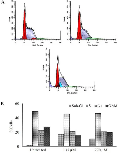
Effect of PGB-0 on Cell Cycle Modulation
To evaluate the cytotoxicity of PGB-0 whether caused by cell cycle arrest in MCF-7/HER2 cells, we performed flow cytometry analysis using PI staining. Cell cycle analysis revealed that PGB-0 increased cells accumulation in Sub-G1 phase (Figure 5).
This phenomenon needed to be confirmed by apoptosis assay to determine sub-G1 cells accumulation due to apoptosis cells or necrosis.
Figure 5. The Effect of PGB-0 on Cell Cycle Progression of MCF-7/HER2. Cells were seeded in 6-well, harvested after 24h of treatment, stained with propidium iodide and DNA content were analyzed by using flow cytometry as described in method (A) The profiles of cells in sub-G1, G1, S and G2-M phase of (i) control, (ii) PGB-0 135µM (iii) PGB-0 270µM. X-axis showed the DNA content and the Y axis showed the relative cell numbers. (B) Quantification of cell distribution in each phase.
PGB-0 Decrease HER2 Expression
on results, PGB-0 50μM did not affect HER2 expression. However, after treatment of PGB-0
150μM, HER2 expression was decreasing expression on MCF-7/HER2 cells using western blot. Cells were treated with a
concentration of PGB-0 50μM and 150 for μM
24h, then was analyzed using Western Blot as described in methods.
The purposes of this study are to synthesize and explore the anticancer activity of a new curcumin analogue, PGB-0 on HER2 overexpressed-cancer cells. Our screening using molecular docking PLANTS revealed that PGB-0 possibly bound to ATP binding site of EGFR and HER2. Lapatinib and Curcumin
were used as a native ligand. Although fPLP
score of PGB-0 is higher than lapatinib and curcumin, the binding distance of PGB-0 on both HER2 and EGFR is shorter than lapatinib and curcumin. Hence, we found that PGB-0 is comparable with lapatinib and curcumin to bind on ATP binding site of HER2 and EGFR based on molecular docking study. We succeed to synthesize PGB-0 with a simple method and reproducible. Synthesis strategy of PGB-0 is similar with common curcumin analogues synthesis which is based on aldol reaction. However, several factors such as mixing speed, temperature, pH, and the presence of boric acid on PGB-0 structure need to be controlled to minimize the by-product. Boric acid easily reacts with the other boric acid to produce
boroxine (Nishiyabu et al., 2011). Our result
demonstrated that PGB-0 inhibited cells growth of HER2 overexpressed-cancer cells and performed no effect on normal cells.
According to the IC50 values, the cytotoxicity
of PGB-0 is less potent than lapatinib
(IC50 = 8µM) and curcumin (IC50= 25µM) (the
data was not shown). However, commonly, the requirement of chemopreventive and
co-chemotherapeutic agents only need a
compound possessing specific target in cancer
cells (Meiyanto et al., 2014). On the other hand,
boron carrying pharmaceutical also requires cytotoxic compound to minimize the non-selective toxicity to the patient (Barth, 2005). Thus, the less cytotoxicity of PGB-0 tends to be an advantage compared to lapatinib and curcumin. Curcumin and its analogues are well
known inducing G2/M arrest (Meiyanto et al.,
2014) though, PGB-0 induced sub-G1 cells accumulation. Cells in sub-G1 phase were described as a hypodiploid cell with low DNA content due to the DNA fragmentation and possibly apoptotic cells. Therefore, sub-G1 accumulation induction by PGB-0 need to be confirmed by apoptosis assay.
HER2 is a transmembrane receptor that also disrupts both the intrinsic and extrinsic apoptotic pathways (Carpenter and Lo, 2013). In extrinsic pathway, HER2 suppresses the
apoptosis cell in the response to TNF-α. HER
-2 activates PI3K-Akt signaling then
Akt-mediated that leads to activation of NFκB to
transcript anti-apoptosis protein. In the western blot, the result revealed that PGB-0 decreased the expression of HER2. This result is not the first report that curcumin and its analogues or related groups possessed activity as HER2 and
tyrosine kinase inhibitor. Meiyanto et al., (2014)
reported that curcumin and PGV-1 inhibit HER2 proteins, resulting in inhibition of
CONCLUSION
PGB-0 inhibits cells growth on HER2 overexpressed-cancer cells through induction of cells death and decreasing HER2 expression. Further study must be established to evaluate the molecular mechanism of PGB-0 on HER2 downstream mechanism.
ACKNOWLEDGEMENT
We would express our gratitude to Insinas 2014-2016 Grant Ministry of Research Technology and Higher Education, No. RT-2014-810 and Nara Insitute of Science and Technology Japan who funded and supported this research.
Carpenter, R.L., Lo, H.-W., 2013. Regulation of
Apoptosis by HER2 in Breast Cancer. J.
Carcinog. Mutagen. 2013(Suppl 7), 003
D’Amato, V., Raimondo, L., Formisano, L.,
Giuliano, M., De Placido, S., Rosa, R., Bianco, R., 2015. Mechanisms of lapatinib resistance in HER2-driven
breast cancer. Cancer Treat. Rev. 41, 877–
883.
English, D.P., Roque, D.M., Santin, A.D., 2013.
HER2 Expression Beyond Breast
Cancer: Therapeutic Implications for
Gynecologic Malignancies. Mol. Diagn.
Ther. 17, 85–99.
Gibbs, J.B., 2000. Anticancer drug targets: growth factors and growth factor
signaling. J. Clin. Invest. 105, 9–13.
Hong, R.L., Spohn, W.H., Hung, M.C., 1999. Curcumin inhibits tyrosine kinase activity of p185neu and also depletes p185neu.
Clin. Cancer Res. Off. J. Am. Assoc. Cancer
Res. 5, 1884–1891.
Meiyanto, E., Hermawan, A., Anindyajati, 2011. Translational Research in Cancer Drug
Development. Ind J Canc Chemoprev. 2,
198–210.
Meiyanto, E., Putri, D.D.P., Susidarti, R.A., Murwanti, R., Sardjiman, null, Fitriasari, A., Husnaa, U., Purnomo, H., Kawaichi, M., 2014. Curcumin and its analogues (PGV-0 and PGV-1) enhance sensitivity of resistant MCF-7 cells to doxorubicin through inhibition of HER2 and NF-kB
activation. Asian Pac. J. Cancer Prev. 15,
179–184.
Mosmann, T., 1983. Rapid colorimetric assay for cellular growth and survival:
Application to proliferation and
cytotoxicity assays. J. Immunol. Methods
65, 55–63.
Moy, B., Goss, P.E., 2006. Lapatinib: Current Status and Future Directions in Breast
Cancer. The Oncologist 11, 1047–1057.
Nishiyabu, R., Kubo, Y., James, T.D., and Fossey, J.S., 2011, Boronic acid building
blocks: tools for self assembly, Chem.
Commun. 47, 1124-1150
Plavetić, N.D., Kulić, A., Vrbanec, D., 2012.
Role of HER2 signaling pathway in breast cancer: biology, detection and
therapeutical implications. Period. Biol.
114, 505–510.
Sardjiman, S.S., Reksohadiprodjo, M.S., Hakim, L., van der Goot, H., Timmerman, H.,
1997.
1,5-Diphenyl-1,4-pentadiene-3-ones and cyclic analogues as
antioxidative agents. Synthesis and
structure-activity relationship. Eur. J.
Med. Chem. 32, 625–630.
Vu, T., Claret, F.X., 2012. Trastuzumab: Updated Mechanisms of Action and
Resistance in Breast Cancer. Front. Oncol.
2, 62
Yakes, F.M., Chinratanalab, W., Ritter, C.A., King, W., Seelig, S., Arteaga, C.L., 2002.
Herceptin-induced inhibition of
phosphatidylinositol-3 kinase and Akt Is required for antibody-mediated effects on p27, cyclin D1, and antitumor action.