This book contains information obtained from authentic and highly regarded sources. Reprinted material is quoted with permission, and sources are indicated. A wide variety of references are listed. Reasonable efforts have been made to publish reliable data and information, but the authors and the publisher cannot assume responsibility for the validity of all materials or for the consequences of their use.
Neither this book nor any part may be reproduced or transmitted in any form or by any means, electronic or mechanical, including photocopying, microfilming, and recording, or by any information storage or retrieval system, without prior permission in writing from the publisher.
All rights reserved. Authorization to photocopy items for internal or personal use, or the personal or internal use of specific clients, may be granted by CRC Press LLC, provided that $1.50 per page photocopied is paid directly to Copyright Clearance Center, 222 Rosewood Drive, Danvers, MA 01923 USA. The fee code for users of the Transactional Reporting Service is ISBN 0-8493-1482-8/05/$0.00+$1.50. The fee is subject to change without notice. For organizations that have been granted a photocopy license by the CCC, a separate system of payment has been arranged.
The consent of CRC Press LLC does not extend to copying for general distribution, for promotion, for creating new works, or for resale. Specific permission must be obtained in writing from CRC Press LLC for such copying.
Direct all inquiries to CRC Press LLC, 2000 N.W. Corporate Blvd., Boca Raton, Florida 33431. Trademark Notice: Product or corporate names may be trademarks or registered trademarks, and are used only for identification and explanation, without intent to infringe.
Visit the CRC Press Web site at www.crcpress.com
© 2005 by CRC Press LLC No claim to original U.S. Government works International Standard Book Number 0-8493-1482-8
Library of Congress Card Number 2004045731 Printed in the United States of America 1 2 3 4 5 6 7 8 9 0
Printed on acid-free paper
Library of Congress Cataloging-in-Publication Data
Modern neurosurgery : clinical translation of neuroscience advances / edited by Dennis A. Turner.
p. cm. -- (Frontiers in neuroscience) Includes bibliographical references and index. ISBN 0-8493-1482-8 (alk. paper)
1. Nervous system--Surgery.
[DNLM: 1. Neurosurgery--trends. 2. Neurosurgical Procedures--trends. WL 368 M6885 2004] I. Turner, Dennis A. II. Title. III. Series: Methods & new frontiers in
neuroscience series. RD593.M627 2004
Foreword and Scope
Advances in clinical neuroscience often arise from a better understanding of brain function and hypotheses based at the cellular, system, or organ level. Recent empha-sis is on translating functions or structure-based hypotheses into clinical treatment schemes. This process of translational research depends on a number of critical steps, and in most cases, a clinical market that would make commercialization worthwhile financially. Rather than focus on current treatment schemes, this volume will critically discuss treatments in the process of development, particularly those that have arisen or will arise from advances in neuroscience knowledge. The three categories of such treatments are: (1) treatments, aids, and techniques currently in clinical trials or pending U.S. Food and Drug Administration (FDA) approval and new indications for older approved drugs and devices; (2) advances in the promising preclinical stages that may lead to a rapid progression to initial human trials over the next 5 to 10 years; and (3) approaches that failed at the clinical application level, but still offer insights into whether the initial hypothesis was invalid or significantly flawed in some respect.
Many of these advances are hypothesis-based, particularly the pharmacological approaches. However, as a surgical specialty, neurosurgery also has experienced many technical advances, both in terms of treatment and also for both diagnostic approaches and aids that enhance the technical performance of surgical procedures. Such technical advances have led the FDA to devise new methods of approval for approaches that do not directly entail treatment, for example, aids to performance of the surgery. Such aids include stereotactic frames, frameless computer-guided approaches, diagnostic ultrasound, operating microscopes, and many other devices that highlight the dominant role that technological advances continue to exert in translating neuroscience into clinical practice. However, even the application of a new technology requires the identification of a hypothesis. Clear specification of the underlying hypothesis and associated supportive data may lead logically to identifying required testing and enhancement of data both for and against a concept. This book intends to examine the interface between neuroscience progress and clinical neuroscience advances by assessing the hypotheses that drive this evolution. With this hypothesis-based approach, this book will review the relevant neuroscience underpinnings of new neurosurgical techniques, treatments, and conceptual approaches that are likely to shape clinical neuroscience over the next decade. This dynamic approach is a radical departure from more descriptive books on the topic of 21st century neurological sciences that focus on reviews of current techniques or treatment schemes with timelines to clinical application greater than 10 years.
Acknowledgments
Editor
Contributors
Table of Contents
Chapter 1
Neuroscience Hypotheses and Translation into Neurosurgery Practice Dennis A. Turner and Simon J. Archibald
Chapter 2
Clinical Prospects for Neural Grafting Therapy for Cortical Lesions Ashutosh A. Pradhan, Ashok K. Shetty, and Dennis A. Turner
Chapter 3
Advances in Treatment of Spinal Cord and Peripheral Nerve Injury Ali Zomorodi and Roger D. Madison
Chapter 4
Cellular Brain Ischemia and Stroke: Neuroprotection, Metabolism, and New Strategies for Brain Recovery
Kelley A. Foster, Christopher J. Beaver, Larry B. Goldstein, and Dennis A. Turner
Chapter 5
Imaging and Functional Mapping of Local Circuits and Epilepsy Kenneth M. Little and Michael M. Haglund
Chapter 6
Pre-ictal Seizure Detection and Demand Treatment Strategies for Epilepsy Dennis A. Turner, Miguel A.L. Nicolelis, and Kevan Van Landingham
Chapter 7
Neuroprosthetics and Clinical Realization of Brain–Machine Interfaces Dennis A. Turner, Dragan F. Dimitrov, and Miguel A.L. Nicolelis
Chapter 8
Surgical Treatment of Movement Disorders: DBS, Gene Therapy, and Beyond
Parag G. Patil and Dennis A. Turner
Chapter 9
Novel Therapeutic Approaches for High-Grade Gliomas
Chapter 10
Spinal Dysraphism: The Search For Magic Timothy M. George and David Corey Adamson
Chapter 11
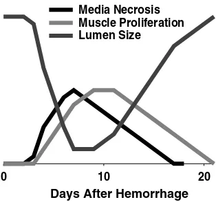
Delayed Cerebral Vasospasm: Current Hypotheses and Future Treatments Kent C. New, Cheryl Smith, Laura Niklason, and Dennis A. Turner
Chapter 12
Future Directions of Endovascular Neurosurgery Osama O. Zaidat and Michael J. Alexander
Chapter 13
Neuroscience ICU Therapeutics
Ashutosh A. Pradhan and Dennis A. Turner
Chapter 14
New Directions and Therapeutics in Surgical Spine Treatment Dennis A. Turner and William J. Richardson
Chapter 15
Clinical Research in Surgery
Ricardo Pietrobon and Dennis A. Turner
Chapter 16
1
Neuroscience Hypotheses
and Translation into
Neurosurgery Practice
Dennis A. Turner and Simon J. Archibald
CONTENTS
1.1 Introduction
1.1.1 Concept of Translational Neuroscience
1.1.2 Translational Neurosurgery versus Translational Neuroscience 1.1.3 Examples of Translational Products
1.2 Categories of Neurosurgery Advances
1.3 Critical Questions in Translational Neurosurgery
1.3.1 When Is Preclinical Data Sufficient to Proceed to Human Experimentation?
1.3.2 Who Is Involved with Translational Neurosurgery? 1.3.3 Mechanisms of Translational Neurosurgery: University
and Corporate Involvement 1.3.4 Device Development Process
1.3.5 Guidelines for Efficacious Treatment Schemes 1.4 Outline of Topics of Neurosurgery Advances
1.5 Levels of Nervous System Functioning: Cellular to Systems 1.6 Conclusions
References
1.1 INTRODUCTION
1.1.1 C
ONCEPT OFT
RANSLATIONALN
EUROSCIENCEthe preclinical scientists developing the idea for the treatment strategy, the clinicians involved with providing care to patients and using the treatment, and the formal clinical trial necessities such as trial statisticians and research nurses. However, a broader definition is provided by The American Physiological Society, which defines translational research as “the transfer of knowledge gained from basic research to new and improved methods of preventing, diagnosing or treating disease, as well as the transfer of clinical insights into hypotheses that can be tested and validated in the basic research laboratory.”3
The team concept has evolved through necessity because basic neuroscience tends to be very focused on cellular and genetic mechanisms, whereas clinical trials and subsequent applications to humans are now unique specialties in their own rights, with clinicians and neurosurgeons providing patient care in the middle course between the new specialties. In a way, almost all biological research is translational, but the timelines for clinical applications may differ considerably, i.e., 1 year, 10 years, 100 years, etc.
Neurosurgeons tend to prefer more rapid relevance (within 1 or 2 years) because of inherent impatience. However, many of the great clinical advances stem from older basic science research findings applied outside their original fields. For exam-ple, microbiologists were cataloguing bacteria for years before the process had any clinical relevance. The advent of antibiotics created an immediate need to classify and understand organisms due to their differing sensitivities to antibiotics.
The three aspects of translational research include first a great preclinical idea developed often from laboratory findings that appear to have significant potential (Figure 1.1). This seminal idea may have been tested in rodents or in nonhuman primates and hopefully a side effect profile and likely therapeutic range are in hand. For many neurosurgery devices and products, no dose-response curve is available or applicable, and often Phase I trials for side effects in normal patient populations are obviated and the device or product instead moves rapidly into Phase I/II trials in the target patient population. Thus, information obtained about side effects and dosage (if applicable) from preclinical studies can be very helpful.
The preclinical idea must then have an enthusiast or sponsor willing to do the work to begin human testing. The sponsor must obtain a relevant investigational device exemption (IDE) for a device or an investigational new drug (IND) application for a drug from the U.S. Food and Drug Administration (FDA), and usually requires an industry cosponsor to handle manufacturing compliance, assist in defraying costs, and set up the initial testing format. Individuals and companies may have varying motivations to proceed to initial clinical testing, but usually the motivation is a mix of altruism (to improve some medical condition) and a profit incentive based upon the possibility of a marketable product at some point in the future.4
sometimes be difficult because such scientists often do not have a full picture of clinical relevance and marketing aspects and, in many cases, may not understand why a product is not developed or is suppressed by a company for business reasons or because of side effects.
The second aspect then is to look for (usually) academic neurosurgery collabo-rators willing to apply the device or product to patients in the proscribed clinical trial format.5 This initiation of clinical testing in humans also requires significant paperwork and oversight, including obtaining institutional review board (IRB) approval for human research and enrolling patients — tasks for which the clinical investigator is often paid. The type of clinical trial format and patient enrollment are then closely monitored by both the institution and the FDA.
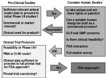
FIGURE 1.1 The transition path of a great basic science idea from preclinical studies to product. First, clinical need, commercial interest, and animal data must be sufficient before one can consider proceeding to initial human protocols. Initial consideration of human studies requires further evaluation of other factors including intellectual property rights, ability to devise a suitable human manufacturing process, market surveys to indicate clinical marketing likelihood, and initial FDA interactions. If initial feasibilities of Phase I/II clinical trials are approved, an investigation device exemption (IDE) must be obtained and the manufacturer must choose a premarket approval (PMA) or 510 K approval pathway. A PMA requires considerable additional data and clinical studies for an entirely new treatment approach; a 510 K links the product to existing FDA-approved products to show equivalence. If a pivotal trial is convincing, the FDA may finally approve marketing; then the product can be sold freely for FDA-approved indications.
The third aspect is to then assemble the data, often from multiple sources, decide on a Phase III format for a randomized trial, obtain FDA approval to proceed based on the initial side effect and dose-response profiles, and then perform the definitive trial. Even after FDA approval, a product must still meet the FDA burden of registry of late side effects and long-term issues, particularly for implanted surgical products that may involve unknown consequences years after implantation. At some point after FDA approval, more open clinical trials are often initiated by other groups, usually those with considerable skepticism about the clinical worth of the product. After these external trials are performed, many devices and products are never commonly used due to lack of efficacy, difficulty in use by those other than the core enthusiasts, or because of unanticipated side effects despite FDA approval and availability.
Since the process of translation into even simple clinical practice requires a significant burden in complying with regulations at both the institutional (IRB, ethical board, etc.) and FDA levels, the translational process also requires a clinician familiar with the treatment scheme and capable of delving adequately into regula-tions for approval and often a corporate entity to advance the significant FDA-required costs. Thus, a heavy burden is borne by the medical care system and clinicians who participate in the process of translational medicine.6-8
Translational research also implies a mechanistic understanding at the molecular, cellular, and systemic levels of the function or action of the therapy in relation to the disease mechanism or target. Many clinical advances are not translational or hypothesis-based, but rather are evolutionary or simply empiric. Thus, the concept of translational research is usually applied primarily to a situation where a hypothesis is generated, tested at the preclinical level, and then applied in sequence to initial and then final stages of clinical testing for human use. In many instances, drug development has followed this approach whenever possible, although many notable failures occurred as well.4
Translational research depends critically upon an animal model of the disease for preclinical testing of the proposed therapy, and the translational process can fail, for example, by applying results from an inappropriate animal model to a human disease. Thus, translational research continues to evolve and, in many cases, further understanding as to why a treatment does not work in an intact individual may lead to the opening of additional preclinical research avenues.
All applications into the clinical arena depend upon ample (and willing) supplies of patients for testing and clinicians willing and sufficiently enthusiastic to spend their time (beyond ordinary clinical care) for such testing. Some clinicians may also have sufficient understanding of the therapy at both the basic science and clinical levels to serve as a bridge to facilitate the transfer of the treatment to clinical care.1
1.1.2 T
RANSLATIONALN
EUROSURGERY VERSUST
RANSLATIONALN
EUROSCIENCEIn many ways, neurosurgery is very different from neuroscience in general. First, it involves far less emphasis on systemic drug treatment, although, of course, consid-erable crossover and use occur, as in the cases of anticonvulsants, antibiotics, che-motherapeutic agents, and drugs in general medical care. Second, in addition to therapeutics, a whole field of devices, most of which require FDA approval, serve as aids to surgeons performing procedures and are not directed at patient therapy. Third, many therapeutics in development and use are devices and permanent implants that may require an invasive form of delivery. The safety and efficacy requirements that must be met for FDA approval may be quite different for such therapeutics from requirements for oral or systemic drug delivery.
Because the FDA treats devices very differently from drugs, the requirements for specific types of clinical trials also differ and the entire process of translation from a preclinical state to clinical use requires different forms of expertise and knowledge of clinical trials. The focus of most of this volume is on these various categories of therapeutics, devices, and approaches to translation of preclinical advances into clinical usefulness — in other words issues more relevant to ordinary neurosurgical practice and research. These issues are rarely covered in print because the number of devices and their applicability are far fewer than medical applications for new drugs.
Another category of clinical development and advances includes the rational-ization of existing therapy. For example, most neurosurgery procedures such as craniotomy and laminectomy involve a few standard approaches that have been in development for more than a century. Since the FDA regulates surgeons in contrast to drugs and devices, little data exists on many neurosurgery procedures, their relative efficacy and safety, and their indications.
This lack of data perplexes both rational care providers and medical consumers because many different approaches to the same clinical problem may be suggested by various surgical specialists. Because the neurosurgical literature mostly involves anecdotal case series and little data generated by randomized controlled trials, few guidelines based on such data apply to management of typical problems, particularly complex issues such as brain tumors and spine therapy.
formats of clinical trials continue to percolate and develop, particularly those that go beyond traditional randomized clinical trials.
New clinical advances depend on the ability to rationally and efficiently translate new understanding of brain function into clinical neuroscience practice. Currently, most clinical neuroscience advances are purely empiric, and often are subject to clinical testing without full identification of the cellular mechanisms involved. Thus much time, energy, and money have been allocated to new treatments with minimal examination of their scientific bases and applicability. However, for many reasons, it is critical to define the hypotheses underlying the application of neuroscience to clinical use.
This definition may lead to reexamination of the data underlying advances in terms of the adequacy of support of the hypotheses and may lead to a fresh approach. However, in spite of a rational approach, the transition from preclinical studies to clinical medicine may still be difficult because of unanticipated potential side effects, clinical trial flaws or inadequacies, inappropriate disease translation, and lack of sufficient market potential.
Most current neurosurgery procedures developed from both clinical hypotheses and practice-related outcome measures to assess the worth of the hypotheses. Many stable and confirmed clinical hypotheses are common in the practice of neurosurgery, particularly the concept that “mass effect” or pressure, if relieved, may improve brain, spinal cord, or peripheral nerve functioning. However, such simple hypotheses do not work for more complex abnormalities, such as intrinsic brain tumors that involve both infiltration and mass effect. As a result, more complex hypotheses often encompassing cellular, systemic, and organ level concepts have been developed.
In many situations, neurosurgery is moving away from the simplistic mass effect hypothesis that has dominated clinical thinking for many years and into specific mechanistic approaches requiring further insight into anatomical, physiological, and pathological factors unique to the brain. This book focuses on such fresh approaches in a variety of neurosurgical fields.
Compared to pharmaceutical mechanisms of translational research, neurosurgery presents many challenges. The first is a small market throughout the world for most conditions under the neurosurgery umbrella, particularly compared to neuroscience diagnoses not involving surgical treatment, for example, Alzheimer’s disease and cardiovascular disease. This is particularly true for clinical products intended for neurosurgery centers rather than for patients (surgical instruments, diagnostics, and other intraoperative aids). This small market may preclude effective development and commercialization because its potential is often insufficient.
Because of these burdens, rarely has an experimental surgical procedure been developed commercially, except as a direct derivative of an existing procedure for which clinical payment coverage may be obtained. Examples are pain or deep brain stimulators. Rarely has a sponsoring company paid clinical study expenses except for the costs of the devices because in most cases the patients may have obtained some benefits. Obviously, this clinical coverage scheme would not be workable for a randomized, placebo-controlled clinical trial.
1.1.3 E
XAMPLESOFT
RANSLATIONALP
RODUCTSCollagen nerve guide tubes — For years, neuroscientists have tried to improve the recovery capability and ease of repair of peripheral nerves. A number of different nerve guide tubes were developed. The first used autologous materials (such as arteries and veins) because many injuries allowed insufficient autologous peripheral nerve for cable grafting of a long lesion. A simple collagen nerve guide tube was developed by Archibald et al.10 to aid in regrowth of peripheral nerves, with the advantage of absorption over time (see Chapter 3). This absorption obviated some of the problems of permanent materials such as silicone that eventually became restrictive to the nerves. After extensive testing in nonhuman primate median nerves across large gaps, the nerve guide tubes were also compared with conventional cable grafts. However, the initiation of human feasibility trials was difficult in the U.S. and European trials that were conducted first. After several years, a corporate sponsor became interested, pursued additional clinical trials, and eventually the product became FDA-approved for nerve injury repair. This time span from bench to bedside application exceeded 15 years, and the device clearly was a hypothesis-based trans-lational product.
Frameless stereotactic devices — While stereotactic frames have been in com-mon human use since the early 1950s, the difficulties in using a frame and the discomfort to the patient led to consideration of other techniques for surgical naviga-tion. As digital scans such as magnetic resonance imaging (MRI) and computerized tomography (CT) and algorithms to reconstruct the scans and provide three-dimen-sional representations became more readily available, it became possible to align a patient’s brain in the three-dimensional space of an operating room with the patient’s own computed images. The critical pieces needed to accomplish this alignment are rapid three-dimensional representations of computerized brain images and an accurate and robust three-dimensional digitizer.11,12 A variety of three-dimensional digitizing systems were developed and continue to evolve to accomplish intraoperative naviga-tion. FDA approval of frameless stereotactic devices has been expanded to require evidence of clinical usefulness because the devices are used solely by surgeons to aid intraoperative navigation. The devices have evolved into clinical products in wide use and include the Stealth (Medtronics) and BrainLAB systems. Both systems were built upon rapid advances in three-dimensional localizer technology, computer systems, and graphics. The entire laboratory-to-operating-room translational process took less than 10 years.
recombinant human protein. After several years of experimentation, nonhuman pri-mate experiments showed considerable promise for GDNF in initiating regrowth of dopaminergic collaterals within the striatum. Initial human clinical trials were begun in 1996,13 but ended prematurely due to unexpected severe side effects. After further work in nonhuman primates with both direct GDNF infusion into the putamen and gene therapy for GDNF transfection, initial human clinical trials with both methods of administration are in progress.14 GDNF continues to show significant promise and further pivotal trials will likely be conducted for at least one of these two novel methods of administration (see Chapter 8). Although FDA approval has not yet been granted, GDNF is another example of a hypothesis-driven bench-to-bedside product.
1.2 CATEGORIES OF NEUROSURGERY ADVANCES
Neurosurgery advances can be divided into three basic categories. The first category involves drugs and devices that are therapeutic and typically involve obtaining an IND or IDE for initial human use and some form of clinical trial sequence prior to full FDA approval. Examples of drugs include the Gliadel wafer (BCNU; 1,3-bis(2-chloroethyl)-1-nitroso urea), which is directly deposited into a brain tumor cavity at the time of craniotomy, the intracerebral infusion of GDNF for the treatment of Parkinson’s disease, and adenovirus vector delivery of gene therapy for GDNF enhancement in the striatum.15
Many of these new approaches are based on neural regeneration, biological plasticity, tissue grafts, and new engineering approaches.16 Examples of devices include implants such as cerebrospinal fluid (CSF) shunts, hardware for spine fixation, and deep brain stimulating (DBS) electrodes for movement disorders. Because this category generally involves permanent implants for therapy and the devices are highly invasive, extra consideration is usually involved to ensure long-term safety.
The third category is rationalization of established products the FDA has already approved and older procedures already in common clinical practice for which effi-cacy was not established.
Proceeding with a clinical trial for established surgery requires significant contention about the worth of the procedure, as when carotid endarterectomy began to be carefully scrutinized. For example, the reasons for the carotid endar-terectomy trials in the 1980s included the high cost to society for the multiple procedures performed, the lack of any valid data regarding what would happen if the procedure was not performed (contemporary natural history studies), and the prophylactic nature of the procedure, i.e., to prevent a bad event despite the risks of the procedure. Many common products have been applied to additional disease mechanisms without adequate studies of the appropriateness or risk-to-benefit ratio, and thus the great need to establish the proper patient population and determine whether existing procedures are indeed efficacious, safe, and appropriate continues to exist.
1.3 CRITICAL QUESTIONS IN TRANSLATIONAL
NEUROSURGERY
1.3.1 W
HENI
SP
RECLINICALD
ATAS
UFFICIENT TOP
ROCEEDTOH
UMANE
XPERIMENTATION?
Because devices and products involving surgical application or implantation usually involve more risk than drugs, manufacturers have somewhat higher burdens to demonstrate product worth in preclinical research before they move on to clinical trials. The pace at which a preclinical treatment or device is applied to initial patient experimentation is often dictated more by the entity developing the treatment rather than by any rational approach to quality of the preclinical data. For drug ment, the FDA has established a rigorous process of IND application and develop-ment. For devices, it uses a parallel structure of IDE approval prior to clinical trial. However, many aspects of neurosurgery and translational research are not covered by these regulatory pathways if they do not involve drugs or devices. The definition of experimental surgery has always been a complex issue for surgeons. Standard clinical practice varies considerably among surgeons. Thus, is a slight difference in surgical technique an “experiment”?
If a procedure is clearly labeled experimental, insurance carriers usually do not pay for it. This places a great burden on the patient to provide payment for an experimental procedure and assume significant risk without a known benefit. There are many examples of such deviations from surgical practice that to many observers clearly represent experimental surgery and to others constitute only small departures from current practice. The rapidity of clinical application of a new advance continues to be a highly contentious issue, clearly requiring institutional backup from the IRB and ethical support whenever a neurosurgeon engages in some form of human research.
If a corporate entity is involved with a therapeutic, then the rapidity of entry into clinical trials may be more dictated by the need for a marketable product than necessarily the quality of the preclinical evidence. Examples include many of the proprietary neural tissue graft trials.9,16 Companies have developed both porcine embryonic cell transplants (as replacements for human embryonic cells) and tumor-derived cell lines for human neural grafting protocols. These cases usually proceed from initial rodent preclinical data directly to clinical trial because of the cost and time required to adequately assess the therapy in nonhu-man primates.
One example is a trial of cultured human neuronal neurons for deep hemorrhagic stroke; they were applied to humans in early clinical trials after only a few rodent studies showed cell survival and presence of grafted cells.9,17 Because the therapy may be headed for FDA approval, the promoters usually have a burden to demon-strate safety and efficacy before the therapy reaches final clinical trials. However, if a corporate entity is less involved or is not as enthusiastic as the investigators, as was the case with GDNF gene therapy trials for Parkinson’s disease, much more careful nonhuman primate work may be performed before initial human experimen-tation is considered.13,14 Thus, the source of the therapeutic, the need for clinical product development, and marketing for later commercialization may all dictate both the manner and pace of the translational process.
Do neurosurgeons jump too quickly to human experimentation without proper consideration for appropriate human risk and benefit? Clearly, the example discussed earlier of autologous adrenal medulla transplants for Parkinson’s disease involved premature human application, and fairly rapid abandonment of the procedure because of significant risk and lack of efficacy. Thus, convincing preclinical data in a vali-dated animal model of the disease (whenever possible) is required to support the transition to initial clinical trials.
1.3.2 W
HOI
SI
NVOLVED WITHT
RANSLATIONALN
EUROSURGERY?
Ideally, the application of devices, drugs, and surgeons’ aids should be performed by clinicians with sufficient background to understand and critically assess the value of a new approach and suggest optimal application.5,19 Because a preclinical team may have minimal clinical experience or knowledge, obvious clinical problems in the translational process may easily be avoided by an astute clinician who has the necessary experience. Also, clinicians involved in the translational process should understand FDA procedures and regulations, IRBs, and ethics and should have the expertise to fully validate initial clinical trials. Particularly for new approaches fresh from a preclinical scheme, knowledge of the goal of the underlying application may considerably aid the translational process because appropriate translation usually requires changes and scaling from preclinical applications.
Thus translational effort of taking a therapy or device from discovery to clinical application is generally provided by a team, and rarely by a lone neurosurgeon. The FDA requires rigorous consideration of the manufacture and construction of devices by a company with knowledge of human applications and materials to preclude “garage” level implementations. Thus, a team may include preclinical scientists who foster an idea and clinicians who are knowledgeable about the basic science side and the initial clinical application and who have access to appropriate patients, statisticians, trial designers, research nurses, and database and analysis personnel. This team usually requires outside funding to fully implement the translational advance, either through a grant or from a corporate entity with visions of a marketable and profitable product within a specific timeline.
Considering that training may be needed in a variety of disciplines beyond neurosurgery, the typical academic neurosurgeon often may be overwhelmed by the needs of even a simple clinical trial. The degree of paperwork, oversight, and IRB approval is astounding without significant administrative help, and often the design of a clinical trial from an industry-funded approach is insufficient to answer a scientific question even though it may be sufficient for FDA approval.
Academic neurosurgeons interested in translational approaches must be aware of and understand many of the basic neuroscience implications of the research, have captive patient groups who can be recruited, have the necessary clinical skills to adequately institute the methodology, and be aware of the relevant clinical trial needs for the study. This is a wide range of skills and the training to obtain most of them (beyond ordinary clinical skills) is not readily available through medical school or neurosurgery training — it requires additional time.19,20 As is the case for most academic medicine efforts, developing a team is critical, and a shortage of neuro-surgeons interested in and sufficiently enthusiastic about such research to become translational “bridges” continues to exist.1
1.3.3 M
ECHANISMSOFT
RANSLATIONALN
EUROSURGERY:
U
NIVERSITY ANDC
ORPORATEI
NVOLVEMENTtrans-lation through clinical trial into a treatment. Usually this approach is sponsored by a drug company, and therapies are developed because of market forces. An initial market survey is necessary to determine how much a drug would cost to develop, patent, and manufacture, how much profit is required to recapture development costs, and the size of the potential market. However, a large number of drug and device companies often take ideas from academics and then perform the translational work to prepare for clinical trials without actually proceeding on to clinical trials due to the cost. Instead, the marketable preclinical products may then be sold to more traditional drug firms or the translational companies themselves converted or sold into a different entity for further product development.
The drug field provides many examples of successes and failures. Many start-up drug companies went bankrstart-upt in the search for new drugs, often at an initial level because the research was too far from a direct path to clinical development. In some cases, such as drug trials for stroke, a drug appeared promising in animal trials, but failed at the initial clinical trial level. This may have been caused by incorrect application of the animal model to the human situation (exploiting drugs that work in focal stroke to treat global ischemia), failure to understand how a drug may work in an intact system, side effects (a psychotropic profile for N-methyl-D-aspartate [NMDA] antagonist), or failure of clinical trial design. However, for devices and particularly for experimental surgeries, translational research often means something completely different. Obviously, a device may be patented, but it may be difficult ethically and legally to patent a surgical procedure.
1.3.4 D
EVICED
EVELOPMENTP
ROCESSSpecific device or therapeutic development begins with a great idea, but a complex process usually involving commercialization must be followed before considering human application of the concept (Figure 1.1). Commercialization of an idea for clinical use involves consideration of many critical issues before development pro-ceeds further. The critical issues include exclusiveness and the availability of patent rights, market size and access to markets, and the feasibility of commercial produc-tion. Once a process is deemed feasible for production using accepted standards for devices and drugs (good laboratory and manufacturing practices), a number of parties may decide whether to proceed with initial human feasibility trials. For most devices, no equivalent of Phase I volunteer testing in healthy subjects exists, so most initial trials for feasibility follow Phase I/II in patient populations relevant to the product. Considerable interaction with the FDA is required during design and perfor-mance of feasibility and then pivotal clinical trials. Finally, the path to FDA approval can include a full premarket application (PMA) or a comparison of equivalence to an existing device or therapeutic (510K application). Defining the selected indica-tions for use and patient populaindica-tions for potential use are critical in order to obtain the widest FDA approval possible.
products are essentially dormant despite FDA approval. It may take several years for a product to find a market niche, even though the FDA approved it. Further rigorous clinical studies for postmarket approval may be required to fully define indications, risks, and efficacy.
1.3.5 G
UIDELINESFORE
FFICACIOUST
REATMENTS
CHEMESIn many instances, a product becomes a standard aspect of clinical treatment schemes, for example, immunization vaccines for many childhood diseases. Multiple studies confirmed high degrees of efficacy and the clinical evidence was consider-able. Clinical guidelines developed for many medical care situations and diseases usually incorporate treatments that are generally agreed to be efficacious as parts of the standard clinical treatment scheme. However, a second type of translation8 involves convincing practitioners to routinely provide care based on guidelines. This requires considerable education of practitioners. However, family practitioners now face so many guidelines, particularly for long-term health care maintenance, that the time required merely to follow the guidelines is considerably more than that required to provide ordinary health care. In a way, postmarket guideline development is critical for the transference of information from tight clinical trials with rigorous patient populations to the general patient population at large.
1.4 OUTLINE OF TOPICS OF NEUROSURGERY
ADVANCES
This volume is not meant to be all-inclusive. It is intended to provide an overview of several exciting areas of neurosurgery. We selected fields that lend themselves to translational work and discuss examples of translational products. For example, little translational science is now ongoing in the field of skull base surgery, but functional neurosurgery to treat epilepsy and movement disorders, insert neuroprosthetics, and perform neural grafting is well represented because of many hypotheses at the preclinical level. Neurosurgery involves a broad range of subdisciplines, but is generally divided into a few basic categories or mechanisms of disease:
Brain tumors and meoplasias — Examples of translational research include new brain tumor therapeutics such as antibodies, radiation, infusions, drugs, Gliadel wafers, and many promising new approaches (see Chapter 9).
Pediatrics and congenital — Examples include folic acid for prevention of meningomyelocele, clinical trials underway on fetal surgery, and development of new CSF shunt techniques (see Chapter 10).
Head injury, peripheral nerve regeneration, and trauma — A significant amount of work is focused on central and peripheral regeneration, improved intensive care unit (ICU) therapy, and enhanced recovery after injury (see Chapter 2, Chapter 3 and Chapter 13).
new neuroprosthetics devices including new stimulators and pumps for medicine delivery are also in development (see Chapter 2 and Chapter 5 through 8).
Cerebrovascular, stroke, and endovascular — Challenging topics include treatment of stroke, delayed cerebral vasospasm, new approaches for endovascular treatments such as catheters, balloons, and coils, and improved postoperative care in ICU settings (see Chapter 4 and Chapter 11 through 13).
Spine and peripheral nerves — A large number of spine implants have been approved and are now in common use; their usefulness is poorly characterized. Spine surgery needs considerable rationalization as to when it is appropriate, and what exactly should be done under various circumstances (see Chapter 3 and Chapter 14). Although the subject is not disease based, the history of neurosurgery has also become a very popular topic at neurosurgery meetings over the past 20 years. While the history of neurosurgery is clearly beyond the scope of this volume, interesting failures of the past may provide excellent guidance as to how not to proceed in the future. Other topics are aspects of clinical trials as applied to developing translational approaches (see Chapter 15) and new approaches to the teaching of neurosurgery skills (see Chapter 16).
The focus is the discussion of advances beyond the current clinical domain for two main reasons. The first is that current and conventional treatments are well treated in many aspects of neurosurgery literature — both books and journal articles. The second is that much can be learned about flawed hypotheses, pitfalls of the translational approach in general, and untested hypotheses and clinical advances from past (and now unused) treatments. This volume focuses on promising new concepts still in preclinical development, those already applied to some clinical trial phase, and those that failed during application to patients. It should provide a worthwhile opportunity to review whether the basic mechanisms, animal models, and translational processes were flawed. Clearly, many of these topics fall outside the scope of a traditional, detailed, and comprehensive neurosurgery textbook.
1.5 LEVELS OF NERVOUS SYSTEM FUNCTIONING:
CELLULAR TO SYSTEMS
Diseases are caused by mechanisms and affect levels ranging from subcellular (genetic, mitochondrial, etc.) to organ. In general, most surgical disciplines primarily consider therapeutics aimed at the organ level (e.g., resecting brain tumors), whereas medical disciplines often test cellular or subcellular approaches involving medicines and drugs. Because knowledge about brain function is accumulating at every level, the approaches at different levels such as subcellular (genetic, molecular, organelle), cellular (electrical integration, channels, and regeneration), local circuits, systems, and organs should be considered. Eventually all translation from preclinical findings to clinical testing depends in one form or another on clinical hypotheses and appro-priate clinical trial design for adequate testing of hypotheses and devices where a hypothesis rests at one particular level.
or functions. In many cases translational research in neurosurgery can take advantage of all three levels of brain functioning loosely defined as follows:
1. Brain function at the cellular level — The primary cell type and basic functional building block of the nervous system is the neuron. Neurons are assembled together in local circuits. Important additional types include glial cells and Schwann cells. Several disorders are based on disturbed aspects of cellular and local circuit function (epilepsy, movement disorders, demyelinating diseases, and aberrant regen-eration). In many situations, treatment schemes for these disorders may include pharmacotherapy directed at single neurons or circuits, gene therapy to alter indi-vidual cell functioning, cellular replacement and transplantation, and other forms of restorative treatment.
2. Brain function at the system level — Systems within the nervous system include various local circuits and regions working together for a common modality. Examples of modalities include a variety of motor planning, modulation, and execution systems, sensory systems dedicated to particular types of inputs, cog-nitive and memory systems, and basic systems that control alertness, respiration, and cardiac status. Each system forms a unit of functioning based on a certain modality and assembled for cooperative nervous system functioning. When a system dysfunctions, for example with movement disorders, treatment such as deep brain stimulation may be directed at the systems level to alter the function of the system.
3. Brain structure and function at the organ level — Regardless of the function of the nervous system, the brain remains an organ that requires adequate nutrition, blood flow, oxygenation, removal of waste products, and mechanical support from the skull and spinal column. It can also develop mass lesions such as tumors. The treatment of such disorders, although specific to the brain, is similar to other clinical treatments at the organ level, i.e., resecting of mass lesions, enhance-ment of blood flow, CSF diversion, and mechanical restoration of the spinal column. Many of these treatments are empirical and may require assessment of their clinical efficacy separate from any cellular basis for the treatment scheme.
1.6 CONCLUSIONS
REFERENCES
1. Bast, R.C., Mills, G.B., and Young, R.C., Translational research: traffic on the bridge, Biomed. Pharmacother., 55, 565–571, 2001.
2. Birmingham, K., What is translational research? Nature Med., 8, 647, 2002. 3. Hall, J.E., The promise of translational physiology, Am. J. Physiol. Heart Circ.
Physiol., 51, 803–804, 2002.
4. Choi, D.W., Exploratory clinical testing of neuroscience drugs, Nature Neurosci., 5S, 1023–1025, 2002.
5. Carrel, T., The relationship between surgeon and basic scientist, Transplant Immunol., 9, 331–337, 2002.
6. Finkelstein, R., Miller, T., and Baughman, R., The challenge of translational research: a perspective from the NINDS, Nature Neurosci., 5S, 1029–1030, 2002.
7. Pober, J.S., Neuhauser, C.S., and Pober, J.M., Obstacles facing translational research in academic medical centers, FASEB J., 15, 2303–2313, 2001.
8. Sung, N.S. et al., Central challenges facing the national clinical research enterprise, JAMA, 289, 1278–1287, 2003.
9. Kondziolka, D. et al., Transplantation of cultured human neuronal cells for patients with stroke, Neurology, 55, 565–569, 2000.
10. Archibald, S.J. et al., Monkey median nerve repaired by nerve graft or collagen nerve guide tube, J. Neurosci., 15, 4109–4123, 1995.
11. Barnett, G.H. et al., Intraoperative localization using an armless, frameless stereotactic wand: technical note, J. Neurosurg., 78, 510-514, 1993.
12. De la Porte, C., Technical possibilities and limitations of stereotaxy, Acta Neurochir., 124, 3-6, 1993.
13. Nutt, J.G. et al., Randomized, double-blind trial of glial cell line-derived neurotrophic factor (GDNF) in PD, Neurology, 60, 69–73, 2003.
14. Gill, S.S. et al., Direct brain infusion of glial cell line-derived neurotrophic factor in Parkinson’s disease, Nature Med., 9, 589–595, 2003.
15. Janson, C.G. et al., Viral-based gene transfer to the mammalian CNS for functional genomic studies, Trends Neurosci., 24, 706–712, 2001.
16. Hodges, C.J. and Boakye, M., Biological plasticity: the future of science in neuro-surgery, Neurosurgery, 48, 2–16, 2001.
17. Zivin, J.A., Cell transplant therapy for stroke: hope or hype? Neurology, 55, 467, 2000. 18. Sugarman, J. and McKenna, W.G., Ethical hurdles for translational research,
Radia-tion Res., 160, 1–4, 2003.
19. Nathan, D.G., Careers in translational clinical research: historical perspectives, future challenges, JAMA, 287, 2424–2427, 2002.
2
Clinical Prospects for
Neural Grafting Therapy
for Cortical Lesions
Ashutosh A. Pradhan, Ashok K. Shetty, and
Dennis A. Turner
CONTENTS
2.1 Introduction
2.2 Graft Cell Integration: Preclinical Studies 2.2.1 Graft Cell Labeling
2.2.2 Graft Cell Survival
2.2.3 Graft Cell Migration and Dispersion 2.2.4 Synoptic Graft–Host Interactions 2.2.5 Preclinical Graft Cell Sources 2.3 Clinical Contexts
2.3.1 Cortical Lesions: Early Postlesion Grafting 2.3.2 Cortical Lesions: Late Postlesion Grafting 2.3.3 Neural Grafts for Treatment of Epilepsy 2.3.4 Neural Grafts for Treatment of Stroke
2.3.5 Neural Grafts for Treatment of Severe Head Injury 2.4 Clinically Appropriate Graft Cell Sources
2.4.1 Embryonic Neural Cells 2.4.2 Cultured Cell Lines 2.4.3 Non-Neural Cells
2.4.4 Pluripotent Progenitor and Stem Cells 2.5 Side Effects of Neural Grafts
2.6 Clinical Applicability and Challenges in Translation References
2.1 INTRODUCTION
grafting for lesions of the cerebral cortex arising from epilepsy, stroke, and head injury. Additional chapters will include further information on neural grafting for spinal cord injury (Chapter 3) and Parkinson’s disease (Chapter 8). Although clinical neural grafting has been performed primarily in the context of Parkinson’s disease,6 many issues relevant to this disease do not necessarily transfer to lesions of the cerebral cortex and hippocampus.
For example, Parkinson’s disease involves grafting of a dopaminergic phenotypic cell that possesses a large, diffuse axonal elaboration with a modulatory function, rather than specific synaptic relay systems as noted in glutaminergic synapses within the cortex and hippocampus. Thus, the goal of grafting in cortical lesions is usually to functionally replace part of a highly organized relay system, whereas in Parkin-son’s disease the goal is to replace dopaminergic innervation nonspecifically to the striatum.
Clinical treatments for acute lesions of the cerebral cortex and hippocampus such as head injury and stroke have focused almost exclusively on cytoprotection and prevention of secondary damage within the early period after the lesion.4 While a moderate reduction in the number of neurons damaged may facilitate recovery, in many instances this format of treatment was clearly insufficient clinically because recovery was less than optimal. Moreover, the spontaneous neuronal replacement that transpires via proliferation of endogenous stem/progenitor cells after injury appears to be very restricted, ephemeral, and nonfunctional.7
Few available treatments are aimed at enhancing recovery of function of surviv-ing neuronal elements.2 Additionally, recovery can be accompanied by aberrant axonal plasticity of surviving neurons, characterized by inappropriate innervation of denervated synaptic regions.8,9 One consequence of such inappropriate recovery is the late occurrence of epilepsy due in many instances to isolation of hyperexcitable regions, but which may still exert an untoward effect on the intact brain.8,10 Resto-ration of normal afferent brain control over autonomous, hyperexcitable regions may be critical to both restore function and alleviate epilepsy. Exogenous transplants of multipotent progenitor or stem cells may play a role not only in epilepsy, but also in head injury, stroke, and degenerative disease.11–17
At both early and late time points after hippocampal or cortical lesions, one method to enhance recovery and restore function may be grafting of committed embryonic neural cells even though the goals may differ.1,4 Neural grafting acutely after a lesion may provide additional unformed neuronal elements that may then insinuate and become integrated into the host circuitry, potentially enhancing overall recovery.1,18,19 Early grafting may also change the acute milieu, decreasing death among host cells. Late after a lesion, when the damage is stable, neural grafts may be competent to enhance actual appropriate circuitry reconstruction.3 This is likely accomplished by:
1. Providing correct target neurons for host axons 2. Furnishing proper afferent axons to host neurons
These events together may suppress hyperexcitability and restore afferent control in autonomous regions. Embryonic neural grafts have the dual advantage of surviving the transplantation trauma and anoxia and possessing competence for considerable axonal growth into the adult host CNS.1,3,20 In Parkinson’s disease, for example, neural grafts have been used to treat a stable, long-term disorder by adding ectopic but critical dopaminergic re-innervation.6
The goal of circuitry reconstruction with neural grafts requires appropriate neuronal elements for the host region that are capable of becoming functionally integrated within the host. Many other possible goals and mechanisms can be achieved by neural transplantation including release of neurotrophic factors or neu-rotransmitters and replacement of glial cells.16,21–23 However, the specific requirement for circuitry reconstruction leads to a hypothesis as to what an ideal graft may be.3 An ideal graft would have certain characteristics:
1. Adequate survival of the transplanted neurons within the host environment (at least 20% of grafted neurons)
2. Appropriate dispersion or migration of the transplanted cells to restore host neuronal cell layers (leaving few cells at the transplant site) 3. Normal cellular development including acquisition of region-specific
den-dritic complexity, synapses and intrinsic characteristics
4. Appropriate elaboration of both local circuit and long-distance axons for synaptic connectivity into the host
5. Attraction of a significant number of specific afferent axons from the host
While these requirements are rigorous, the quantitative measurement of these characteristics may lend credence to exertion by the graft of a specific, defined role in the host, as opposed to a nonspecific or non-neuronal effect.1,3
Grafting into cerebral cortex or hippocampus to facilitate circuitry reconstruction may be radically different from the grafting treatment of Parkinson’s disease. For example, grafts into the striatum of dopamine-enriched tissue are intentionally ectopic, and do not appear to develop long-distance axonal growth despite the fact that embryonic dopaminergic axons are inherently capable of such growth.6 The other major difference is the type of neuron that is grafted and its neurotransmitter type because dopamine neurons possess much more diffuse and larger axonal ter-minal synaptic fields than the more typical glutamatergic neurons and GABAergic neurons considered in hippocampal or cortical grafting. Thus, only some parallels may be noted between the two different regions, but issues of graft tissue survival and integration remain paramount for both.3,24,25
limitations. Finally, the bridge between preclinical research and clinical usefulness and applicability will be discussed.
2.2 GRAFT CELL INTEGRATION: PRECLINICAL STUDIES
We defined graft integration into the host on a quantitative, cellular basis specifically to assess circuitry reconstruction.26–28 Neural grafting has many other goals, for example, provision of an enzyme or neurotransmitter, furnishing cells to form myelin sheaths for host axons, and production of growth factors or metabolic products. Our hypothesis of cellular integration applies primarily to the goal of making a graft an integral part of synaptic circuitry within the brain. The specific measurable aspects of integration include:
1. Cell survival, directly comparing the number of cells transplanted and those recovered in vivo at different postgrafting time points
2. Cell dispersion and migration away from the graft site
3. Graft cell differentiation into region-specific neuronal phenotypes 4. Graft cell local and long-distance efferent synaptic interactions with the
host neurons
5. Graft cell afferent connectivity with appropriate host axons
Graft integration may be differentially analyzed for various cell types, including embryonic neurons and immature stem cells.28,29 Figure 2.1 is a schematic of the results of these preclinical studies.
2.2.1 G
RAFTC
ELLL
ABELINGAssessment of graft integration requires a unique label for the grafted neurons so that their survival, migration, and differentiation fate after transplantation may be followed.30 Genetically engineered cells may be labeled with a permanent, gene-based label (such as green fluorescent protein or beta galactosidase).31 Prior to harvesting embryonic postmitotic cells, embryonic neurons may be efficiently labeled with a DNA label such as the thymidine analog 5-bromodeoxyuridine (BrdU)26 by injecting the maternal host during times of neurogenesis for those cells. Because the cells are postmitotic and committed after embryonic harvesting, the neurons retain the BrdU label permanently.
unbiased cell counting methods. Unique labels form the critical basis for evaluation of graft integration within the host to unambiguously identify the grafted cells within the host.26
2.2.2 G
RAFTC
ELLS
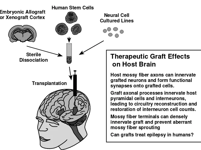
URVIVALCell survival can be assessed in terms of the number of cells grafted compared to those recovered later in vivo. In normal or intact hosts and grafts performed late after lesions, only 18 to 30% of grafted hippocampal cells survived.26 In contrast, dopaminergic grafts in models of Parkinson’s disease showed far poorer survival ranging from 3 to 20% in various studies. However, at early time points following lesions, the degree of survival was much greater (60 to 80%), particularly in young adults.26 This enhanced survival is due primarily to the enhanced neurotrophic factor environment present up to 10–14 days following a lesion and the potential effects of denervation.32 Grafts soon after lesions were well tolerated and considerably FIGURE 2.1 (See color insert following page 146.) Cortical grafting studies. First, cell sources include human or porcine embryonic cortex or hippocampus, various types of pro-genitor or stem cells, or cultured cell lines, most derived from neuronal tumors. After disso-ciation and transplantation, the fate of the transplanted cells can be assessed for synaptic integration within the host. In rodent models, therapeutic graft effects on the host include the formation of mossy fiber synapses onto grafted neurons, amelioration of postlesion interneu-ron loss, and prevention of aberrant mossy fiber sprouting. Whether grafts can ameliorate epilepsy remains to be analyzed in rodents and humans although the framework has been established.
Human Cell Sources
Therapeutic Graft Effects
on Host Brain
Host mossy fiber axons can innervate grafted neurons and form functional synapses onto grafted cells.
Graft axonal processes innervate host pyramidal cells and interneurons, leading to circuitry reconstruction and restoration of interneuron cell counts. Mossy fiber terminals can densely innervate graft and prevent aberrant mossy fiber sprouting
Can grafts treat epilepsy in humans? Embryonic Allograft
or Xenograft Cortex
Human Stem Cells
Sterile Dissociation
Transplantation
enhanced, compared to the intact cortex and the situation after resolution of the lesion.26 At time points equivalent to a fully healed human lesion or with aging, the hippocampus and cortex become much less receptive to grafts. Graft augmentation techniques are required to enhance graft survival and integration.33,34
2.2.3 G
RAFTC
ELLM
IGRATION ANDD
ISPERSIONBreaking down the separate aspects of integration led to some surprising results for embryonic hippocampal grafts. First, during development time when embryonic neurons are removed (at embryonic day 19), these cells have completed programmed migration along the radial glia into their respective layers. Second, the total distance of migration for embryonic hippocampus is short — less than 0.1 mm. Therefore, after grafting, these cells show minimal specific migration to appropriate cell body layers, and most remain clumped within 0.5 mm of the grafting site.26 In an attempt to enhance dispersion, grafts were transplanted as a suspension rather than as tissue, but migration still remained minimal. This lack of capability for migration within the host requires more accurate placement of multiple grafts directly within the degenerated cell layer because only appropriately placed grafts of certain cell types show capacity for specific connectivity.27,35,36 One of the promises of stem cells may be that enhanced migration capability could lead to recapitulation of hippocampal and cortical architecture.15
Grafts that are nonspecific to the lesioned region, such as striatal tissue placed within the lesioned hippocampus, demonstrate poor survival and little capacity for integration.9 Thus, it is critical that appropriate cellular elements are placed into locations most suitable for their development. They differ from dopaminergic grafts for Parkin-son’s disease that appear to function better when placed ectopically within the striatum, and grafted as strands rather than as a dissociated cell suspension.6 In the hippocampus, the various types of functions that improve with appropriate placement include afferent circuitry (mossy fiber terminals on grafted CA3 neurons) and efferent circuitry to both the deafferented CA1 region and commissural projection areas.9,27,35
2.2.4 S
YNAPTICG
RAFT–H
OSTI
NTERACTIONSConnectivity between graft and host requires that host fiber tracts have access to the graft and that graft axons are able to recognize and follow host axon guidance pathways.3,27,36 A graft placed as a chunk of tissue, rather than as dissociated cells, encourages local connections to form within the graft, discouraging connections between the surrounding brain and the graft.37 Axon guidance pathways differ, depending on the inherent wiring and pattern of connections.20,27 For example, hippocampal CA3 cell grafts can demonstrate contralateral commissural efferent projections if located near the degenerated CA3 cell layer, which apparently provides the appropriate molecular signals for such long-distance connectivity. We have termed this capability the axon guidance pathway, which for commissural connec-tions appears to be specific.
fibers into the septum. These embryonic graft neuron axons demonstrate compe-tence to follow innate host axon guidance pathways and are not susceptible to inhibitory molecules such as myelin-associated glycoproteins along the host path-ways, unlike adult host axons. The locations of these axon guidance pathways in the host may be highly specific, requiring accurate placement of the grafts to achieve access. Afferents into the graft develop readily, particularly mossy fiber ingrowths, if the grafted cells are their natural target cells (i.e., CA3 cells), demonstrated both physiologically by direct slice neuronal recordings and ana-tomically by Timm’s histochemical staining.9,20,30 Likewise, short-distance out-growth from embryonic grafts was demonstrated to be both dense and appropriate (from CA3 grafts to denervated regions of the CA1 subfield and the dentate gyrus), indicating that embryonic grafts can develop both appropriate afferent and efferent connections in the hippocampus.28
2.2.5 P
RECLINICALG
RAFTC
ELLS
OURCESAspects of integration have been well defined for embryonic hippocampal cells, as discussed above.26 However, these cells are not optimal in the sense that they are not “ideal” grafts, particularly because of limited supply, ethical issues, and lack of ability to migrate within the host after transplantation. Therefore, hippocampal stem cells with pyramidal neuronal phenotypes have also been analyzed as alternatives to embryonic cells.12,28,29 These cells likely arise from the posterior subventricular zone and form neurospheres in vitro in the presence of mitogenic factors such as epidermal growth factor (EGF) or fibroblast growth factor (FGF).29,38–40
Neurospheres are large collections of undifferentiated cells that develop in spe-cific culture conditions from clonal stem cells removed from in vivo subventricular zones and contain both stem cells and their progeny. However, these cells show limited differentiation into neurons in vitro and in vivo, and may require conditioning with appropriate neurotrophic factors to enhance neuronal differentiation both prior to and after transplantation.29 For example, physiological development and fiber outgrowth may be limited, even for cells resembling pyramidal neurons, due to their limited differentiation and axon growth. Further, the milieu of the injured brain could adversely affect differentiation of stem cells into neurons as a result of inadequate positional cues. Thus, hippocampal stem cells (and neural stem cells in general) are very promising, but will clearly require priming into partially differentiated region-specific neurons prior to grafting to fully achieve their differentiation and connec-tivity specific to the site of grafting. Whether this differentiated phenotype will be maintained after grafting, particularly for prolonged periods, will require further research.11,13,15,38–44
2.3 CLINICAL CONTEXTS
This chapter focuses on clinical entities — primarily lesions of the cerebral cortex including the hippocampus. Grafting proposed for spinal cord treatment will be discussed in Chapter 3; subcortical grafting for Parkinson’s disease is discussed in Chapter 8.
2.3.1 C
ORTICALL
ESIONS: E
ARLYP
OSTLESIONG
RAFTINGCommon lesions of the cerebral cortex (including the hippocampus) include head injuries, particularly cerebral contusions, and cerebral infarcts. Both head injury and stroke may be accompanied by extensive tissue damage and early neuronal replace-ment via grafts may facilitate structural and functional recovery by adding unformed neural elements to assist in circuitry reconstitution.18,19,47 Such facilitation could consist of enhancing regional recovery and also preventing aberrant regeneration that may accompany cortical recovery in the form of compensatory sprouting of neighboring axons and inappropriate innervation of denervated synaptic sites.9 The relatively unformed neuronal characteristics of neural grafts and their enhanced short- and long-distance axonal collateral growth in the adult, host CNS, may facilitate host recovery far beyond what would be obtained with either innate axonal regrowth alone or endogenous stem/progenitor cell proliferation and differentiation.7 The environment, within a few days after the lesion, appears particularly con-ducive to graft survival and integration.26 This favorable host environment may be due to an enhancement in the level of neurotrophic factors in the vicinity of the lesion32 as a result of astrocytic hypertrophy, microglial activation, and enhanced neurotrophic gene expression in surviving neurons.
For clinical grafting purposes, hippocampal grafts could be placed directly within the appropriate cell layers by stereotactic injection. However, neocortical suspension grafts may require multiple small injections into the neocortex on the border of the damaged region because direct injection into a severely damaged (or ischemic) area may provide minimal tissue nutrition and support for initial growth of axons. Preclinical studies of grafts into ischemic regions suggest excellent integration of embryonic cells into the appropriate tissue.18,47 Histolog-ical demonstration of surviving cells and behavioral changes are considered to define “graft” effect in many studies. Depending on the goal of the graft, if circuitry reconstitution is desired, clear evidence of actual restoration of the damaged circuitry at the cellular level of analysis should be present. In other words, graft cell presence in the host does not necessarily imply circuitry recon-struction or appropriate synaptic interactions with host neurons. Physiological study of the grafted neurons and their synaptic interactions with the host is critical to fully define mechanisms of graft action on circuitry.30