O R I G I N A L A R T I C L E
Motor dysfunction of the ‘‘non-affected’’ lower limb: a kinematic
comparative study between hemiparetic stroke and total knee
prosthesized patients
Sergio Bagnato
Æ
Cristina Boccagni
Æ
Filippo Boniforti
Æ
Antonia Trinchera
Æ
Giovanni Guercio
Æ
Giulia Letizia
Æ
Giuseppe Galardi
Received: 11 February 2008 / Accepted: 26 January 2009 / Published online: 13 February 2009 ÓSpringer-Verlag 2009
Abstract
In patients with hemispheric stroke, abnormal
motor performances are described also in the ipsilateral
limbs. They may be due to a cortical reorganization in the
unaffected hemisphere; moreover, also peripheral
mecha-nisms may play a role. To explore this hypothesis, we
studied motor performances in 15 patients with hemispheric
stroke and in 14 patients with total knee arthroplasty, which
have a reduced motility in the prosthesized leg. Using the
unaffected leg, they performed five superimposed circular
trajectories in a prefixed pathway on a computerized
foot-board, while looking at a marker on the computer screen.
The average trace error was significantly different between
the groups of patients and healthy subjects [
F
(2,25)=
7.9;
p
=
0.003]; on the contrary, the test time execution did not
vary significantly. In conclusion, both groups of patients
showed abnormal motor performances of the unaffected
leg; this result suggests a likely contribution of peripheral
mechanisms.
Keywords
Stroke
Knee arthroplasty
Ipsilateral motor impairment
Hemiparesis
Introduction
Several reports demonstrated an abnormal function of
ipsilateral limbs in hemispheric stroke patients [1–3]. It has
been documented in both upper [1,
2,
4,
5] and lower limbs
[6] and it was related to strength [7], speed and dexterity
[8], temporal coordination and complex manual task
exe-cution [9].
The reasons of this dysfunction are still debated and may
be due to multiple factors. It may be caused by cognitive
deficits affecting perception and control of action. In
patients with left hemisphere damage, ipsilateral motor
dysfunction may be due to ideomotor apraxia, while in
patients with right hemisphere injury, it may be caused by
visuospatial impairments [8,
10]. However, impaired
ipsi-lateral function has also been reported in patients without
cognitive impairment [2,
3].
The abnormal ipsilateral descending output, which is
associated with an unilateral cortical lesion, may
con-tribute to ipsilateral limbs abnormal function too [10–12].
The presence of a bilateral component in motor control is
supported by the anatomical evidences of a small
ipsi-lateral component into the corticospinal tract [13] and by
observations that a functional activation might have a
bilateral expression in cortical areas [14]. Also, spinal
reflexes abnormalities might contribute to spread
abnor-mal motor command to the ipsilateral side. Actually,
spinal reflexes can show bilateral long-lasting changes in
hemiparetic subjects [15]. In stroke patients descending
commands, by interacting with a network of spinal
interneurons and motoneurons, might cause an abnormal
level of excitability in both sides. Thus, the combination
of abnormal descending commands and altered spinal
excitability might be responsible for the ipsilateral motor
impairment.
S. BagnatoC. BoccagniA. TrincheraG. Guercio G. Galardi (&)
Rehabilitation Department,
Fondazione Istituto San Raffaele-G.Giglio, 90015 Cefalu` (PA), Italy
e-mail: [email protected]
F. Boniforti
Orthopaedic Department,
Fondazione Istituto San Raffaele-G. Giglio, Cefalu` (PA), Italy
G. Letizia
Physical Medicine and Rehabilitation Department, University of Palermo, Palermo, Italy
Moreover, in patients with stroke other impairments of the
affected side, such as weakness, loss of weight sense, pain,
immobilization, etc., might be accounted for ‘‘other side leg’’
motor impairment. About this topic, there is a reduced
number of studies underlining the role of peripheral
mech-anisms [
2
]. For instance, an abnormal postural stabilization
too may play a role in ipsilateral motor impairment [
3
,
16
].
To explore the role of peripheral mechanisms in the
genesis of ipsilateral motor limb abnormalities, we
mat-ched two different groups of patients with unilateral leg
impairment, respectively, caused by a cerebral lesion and
by a joint lesion. Thus the two groups of patients were
chosen because they had an impaired motility of the
affected leg as a result of stroke and knee prosthesis. In
both groups we may hypothesize, as common peripheral
factor, the presence of a reduced flow of sensory afferents
due to an impaired motility of the affected leg. We
mea-sured the motor function of the ‘‘other side leg’’ in stroke
patients and in patients with recent total knee arthroplasty
by using a computerized proprioceptive kinematic
foot-board (Pro-Kin system, Tecnobody, Italy). Results were
compared with those obtained in healthy subjects.
Methods
Participants
Three groups of subjects have been included: patients with
hemiparesis caused by stroke (HS), patients with total knee
arthroplasty (TKA) and control subjects (CS). None of the
participants used neuroactive drugs; patients affected by
diabetes, polineuropathy and mielopathy were excluded.
HS group
Out of the 15 patients initially included in the study, 5 were
excluded later due to mistakes in the execution of the test
or to its early interruption. Out of the 10 patients, who
performed a correct test, 7 were men and 3 women (mean
age 64.4
±
6.4 years) (see Table
1
). All patients were at
their first stroke event and they presented a severe spastic
hemiparesis, with absence or just minimal motor activity in
the arm and a residual strength not more than 3 at the
Medical Research Council (MRC) scale in the leg. A
thorough neurological examination was performed in all
patients, without evidence of deficit of strength, sensation
and apraxia in the ipsilateral limbs. Moreover, no patient
had spatial impairments. All of them had an adequate trunk
control, so that they were able to maintain a comfortable
seated position during the text execution and were able to
walk for some meters with assistive devices. No synkinetic
movements were present during the test execution. Patients
participated to the study as long as no relevant cognitive
(mini
mental
state
examination
[
25/30)
and
visual
impairments were present. Neuroimaging studies did not
show any lesion in the non-affected hemisphere. The
interval time from the acute event to the experiments was
6.8
±
6.2 months.
TKA group
Out of the 14 patients initially included in the study, 6 were
excluded later due to mistakes in the execution of the test
or to its early interruption. Out of the 8 patients, who
performed a correct test, 4 were men and 4 women (mean
age 65
±
3.9 years). Four had a right TKA and 4 had a left
TKA. All patients were right handed. None had
experi-enced other joint or bone diseases and all of them were able
to walk before surgery. At the time of the examination, no
sensory disturbances or pain in the affected limb were
referred; a standard nerve conduction and
electromyo-graphic study was performed in all patients to exclude post
surgery nervous lesions. The functional normality of the
unaffected side was evaluated by an orthopaedic history
and clinical examination and by X-rays, without evidence
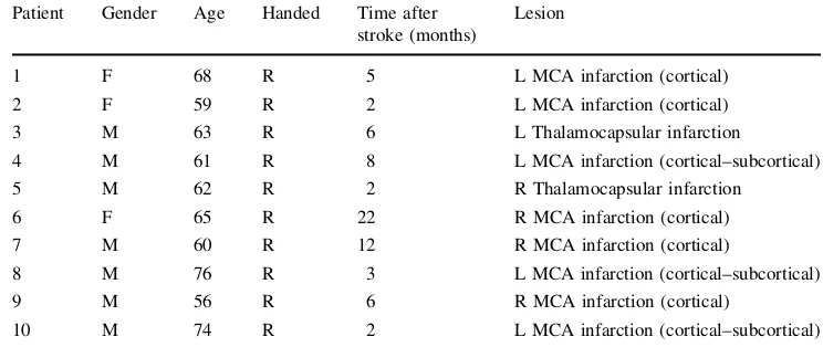
Table 1 Demographic and clinical characteristics of the 10 patients with stroke who performed the test correctly
Ffemale,Mmale,Rright, Lleft,MCAmiddle cerebral artery
Patient Gender Age Handed Time after stroke (months)
Lesion
1 F 68 R 5 L MCA infarction (cortical)
2 F 59 R 2 L MCA infarction (cortical)
3 M 63 R 6 L Thalamocapsular infarction
4 M 61 R 8 L MCA infarction (cortical–subcortical)
5 M 62 R 2 R Thalamocapsular infarction
6 F 65 R 22 R MCA infarction (cortical)
7 M 60 R 12 R MCA infarction (cortical)
8 M 76 R 3 L MCA infarction (cortical–subcortical)
9 M 56 R 6 R MCA infarction (cortical)
of sensory-motor abnormalities. The hip, knee and ankle
arthritic changes were graded less than II [17]. All patients
were passively mobilized since the day after surgery; they
were able to flex the knee of 110°
in order to put their foot
on the plank behind the footboard and to stabilize the trunk
during the test execution. Time from surgery to the
eval-uation was 6.1
±
3.7 days.
CS group
Ten
subjects,
6
men
and
4
women
(mean
age
61.4
±
7.5 years). They were patients close relatives. All
subjects were right handed and in good health. Tests were
performed using the right or the left foot in a randomized
way (in 6 and in 4 subjects, respectively); none of the CS
was excluded because of mistakes or early interruption.
CS and patients gave their informed consent according
to the Declaration of Helsinki.
ProKin system
The system is an electronic transducer board swinging on a
pivot that allows angular movements in sagittal and frontal
planes. When the foot of the patients moves the footboard,
data are transmitted from the transducers to an analogic/
digital converter and sent to the PC. The software processes
data and shows in real time traces of the mobile footboard
movement on the computer screen.
All subjects were seated on a comfortable chair,
adjustable in height, to allow horizontal position of the
thigh, knee bent at 110°, ankle plantar flexed at 10°. The
foot was kept on the centre of the footboard and the
affected leg on the plank apart the footboard in a rest
position (Fig.
1). The head was kept in primary position, to
allow the subjects to comfortably watch the monitor.
The experimental protocol consisted of two consecutive
phases: the training and the evaluation.
The training phase
was applied to gain confidence with
the system; it consisted of three series of exercises. The
subject was asked to use his/her lower limb to follow
consecutive vertical (Fig.
2a), horizontal (Fig.
2b) and
oblique (Fig.
2c) sequences, each one lasting 120 s. They
moved the cursor on the screen tilting the board and tracing
the route with an active movement of the foot and inverting
direction when the target was signalled. The exercise
stopped automatically at the end of the 120 s. There was an
interval time of 5 min between each series.
The evaluation phase. The subject was asked to use his/her
lower limb to follow a circular route in a predefined corridor
(Fig.
2d) five consecutive times. A bell rang anytime the
cursor crossed the border. The direction of the exercise was
clockwise if the right limb was used and anti-clockwise if the
left one was used. To follow the requested route the board had
to be tilted at 7.5°
in each plane. The test stopped
automati-cally at the end of the five turns. Patients had to perform the
tests as quickly as possible and to the best of their ability.
Mistakes or interruptions before the end caused the exclusion
of the subject from the study to avoid the possibility of a
training effect if the exercises were repeated. The
experi-mental session (training and evaluation phases) lasted for
about 25 min and was well tolerated by all patients.
The average trace error (ATE) and time to carry out the
test time execution (TTE) were evaluated.
The ATE was calculated for each of the five turns
required in absolute value, without positive or negative
signs, as ATE
¼
ðOPRP
RPÞ100
;
where OP was the length of
the observed path (executed by subjects), RP was the
length of the requested (ideal) path. Large ATE values
mean large errors in path control, 0 means a perfect task
execution (the trace of the patient’s path could perfectly
overlap the requested path). TTE was the time (in seconds)
from the start to the end of the trial.
Statistical analysis
For the studied variables all data are expressed as
mean
±
standard deviation (SD). A one-way analysis of
var-iance (ANOVA) was used to compare each variable between
CS, HS and TKA patients. When appropriate, Tukey’s honest
significant difference test was used for post hoc analysis.
Results
The ATE was 55.9
±
18.7 for CS, 95.6
±
34.1 for HS and
104.2
±
33.0 for TKA (Fig.
3). ANOVA showed that the
ATE was significantly different between CS, HS and TKA
(F(2,25)
=
7.9;
p
=
0.003). In particular, post hoc
t
tests
revealed that ATE was significantly greater in HS
(
p
=
0.01) and TKA patients (
p
=
0.005) compared to CS
(Fig.
3
). Figure
4
reports examples of the traces in one CS
and in one HS and TKA patient.
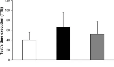
The TTE was 39.7
±
15.9 s in CS, 65.9
±
29.3 s in HS
and 51.6
±
25.6 s in TKA (Fig.
5
). No significant
differ-ence was found between the three groups (
F
(2,25)=
2.9;
p
=
0.07).
Six HS patients had parameters exceeding the cut-off of
two standard deviations from mean normal value: three
exceeded in both ATE and TTE, two in ATE alone and one
in TTE alone. Of these patients, two had right hemiparesis
and four left hemiparesis.
Six TKA patients had parameters exceeding the cut-off
of two standard deviations from mean normal value: one
exceeded in both ATE and TTE, four in ATE alone and one
in TTE alone. Of these patients, three have had right knee
replacement surgery and three left.
Discussion
This study has been designed in order to understand the
mechanisms responsible of ipsilateral leg motor
impair-ment in patients with stroke and, particularly, to evaluate
the possible contribute of peripheral mechanisms. We
adopted the term ‘‘other side leg’’ to indicate the ‘‘normal
limbs’’ of both groups of patients in order to avoid
mis-understanding between ‘‘ipsilateral limb’’ used in HS
patients and ‘‘contralateral limb’’ in TKA. TKA group was
chosen since the motor disability of the ‘‘other side leg’’
Fig. 2 The trajectories that patients and control subjects had to follow on the screen moving their foot.a,bandcdescribe the exercises of the training phase.dis the evaluation test in which subjects had to trace on the screen 5 circular trajectories superimposed on themidline (the ideal line); thearrowis the cursor that subjects had to move
might be caused by an abnormal sensory afferents flow due
to reduced motility in the prosthesized leg. In HS group,
the expected motor disability of the ‘‘other side leg’’ might
instead be caused both by the lesion in the contralateral
hemisphere and by the abnormal sensory afferents due to
the reduced motility resulting from the paresis.
Both HS and TKA patients exhibited abnormal motor
performances of the ‘‘other side leg’’.
In this study, we found the following: (a) a significant
increment of the ATE, (b) an increment of the TTE,
although without statistical significance, (c) a number of
patients in which the ATE and the TTE exceeded the
cut-off of two standard deviation from normal values and (d)
an high percentage of patients who were unable to
con-clude the test. Since clinical evaluation did not show
neuromuscular deficits, functional errors, such as mistakes
in direction and amplitude, differences in velocity and
acceleration, failure to control the inertial properties of the
limb in the initial trajectory may be accounted for these
results. These results suggest that the impaired sensory
afferents flow play a determinant role in TKA and a likely
concurrent role in HS in the abnormal test execution of the
‘‘other side leg’’. Abnormalities in the peripheral afferent
input or in the brain response to sensory input may interfere
with motor programs processing in the cortical motor areas.
We cannot state if this result correlates, in HS patients,
with specific sites of the cerebral lesion or with the phase of
disease (acute, sub-acute or chronic stroke) and if, in all
patients, it is due to a specific cinematic alteration (peak of
velocity, acceleration, etc.). It would be interesting to be
able to evidence a typical pattern of movement abnormality
in HS and TKA patients. However, the test used in our
study could highlight only an overall error rate; so, further
studies are necessary in order to answer to this question.
Due to the nature of the TKA patients disability,
mechanisms such as cognitive deficits [
8
,
10
], abnormal
ipsilateral descending output and spinal reflexes [
10
–
12
,
15
,
18
] have not been discussed. On the contrary, reduced
motility and proprioceptive impairments are themes of
discussion which involve both groups of patients. Motor
impairment of the ‘‘other side leg’’ may be a consequence
of abnormal postural stabilization [
3
,
16
]. In HS patients,
the force produced by the ‘‘ipsilateral limb’’ is reduced
because of the difficulty in supplying contralateral
stabil-ization using the paretic side owing to weakness, spasticity
and abnormal postures [
19
]. This difficulty might explain
the deficits observed in functional tasks such as gross and
fine dexterity, global performance and motor coordination
[
20
]. However, this abnormal postural stabilization is
unlikely in TKA patients, in whom the trunk muscle
con-trol operates in order to compensate the actual loading
deficit of the immobilized leg. Actually, TKA patients were
able to put their affect ed foot on the plank behind the
footboard, in order to stabilize the ‘‘other side leg’’ during
the text execution. It is possible to suppose that the
abnormal performances of the ‘‘other side leg’’ in TKA
patients are a consequence of the reduced motility, with a
loss of sensory afferents from the affected leg. This
reduced flow of sensory afferents, due to an impaired
Fig. 4 Evaluation test. Example the traces in one control subject (a), in one hemiparetic patient (b) and in one patient with knee replacement surgery (c)
motility, is a ‘‘peripheral’’ factor present also in HS
patients. More likely, abnormal motor performances in
both group of patients were due to the involvement of
complex mechanisms, requiring the integrity of both
hemispheres and suitable information from the periphery.
Gordon et al. [
21
] reported impairments of reaching
movements in patients without proprioception. Ting et al.
[
22
] outlined how the sensory state of the contralateral leg
may affect ipsilateral muscle coordination of pedaling
movements. In both groups of patients, afferent inputs from
muscles, tendons and capsular joints were altered due to
their reduced motility. Moreover, patients with stroke may
have motor and sensitive impairments in ‘‘contralateral
limb’’ which may interfere with motor control of the
ipsi-lateral one; likewise, patients with TKA may have
abnormal proprioception as a consequence of the surgical
introduction of a prosthetic joint [
23
,
24
], which may
interfere with motor control of the contralateral one. The
disruption of the sensory afferents to central nervous
sys-tem may be the trough for the ‘‘other side’’ abnormal motor
performances by inducing changes into cerebral
connec-tivity [
25
]. It is well known that sensory information from
the peripheral receptors, the continuous feedback process at
the spinal level and their central processing, may play an
important regulatory role on motor performances [
26
–
28
].
In this way, the abnormal performances of the ‘‘other
side leg’’, plausibly acting on central nervous system level,
might be seen as the core of a neuroplastic process,
pro-duced by both central and peripheral causes. In HS
patients, the involvement of the intact hemisphere may be
due to the contralateral hemispheric lesion as well as to the
sensory loss of peripheral afferents, caused by the reduced
motility; in TKA patients, the involvement of the ipsilateral
hemisphere, by means of the contralateral one, may be due
only to a loss of sensory afferents [
29
]. Therefore, in both
groups of patients, it is possible to hypothesize that a
reorganization in the ipsilateral hemisphere may be crucial
to explain the altered performances in our test. A surprising
finding is that TKA patients, in whom only peripheral
mechanisms should operate, have similar or slightly
increased values of ATE compared with HS patients. We
may formulate some hypothesis to explain this result: (a)
HS patients may have various neuroplastic compensatory
strategies during the test execution, because they are
studied months after the stroke, unlike TKA patients, who
are studied some days after the surgery; (b) there is a
predominant role for the peripheral mechanisms also in HS
patients during the test execution.
Animal studies demonstrated that a neocortical
infarc-tion leads to hyper-excitability in contralateral hemisphere,
contributing to a functional reorganization after stroke [
30
,
31
]. In humans, neurophysiological studies showed an
intact hemisphere increased excitability [
32
], likely due to a
transhemispheric diaschisis [
33
]. Moreover, functional MRI
studies confirmed an increased activation of ipsilateral
motor areas in patients after stroke, while moving the
affected limb [
34
–
36
]. Another possible mechanism is that
damage in one hemisphere may alter trans-callosal signals
and disrupt neural processing in the opposite one. In
humans, the transient deafferentation by means of regional
anaesthesia or ischemic nerve block induces cortical
chan-ges such as modifications in the amplitude of motor evoked
potentials and in regional cerebral blood flow [
37
,
38
]. This
cortical reorganization may imply an adaptive change to
compensate for motor deficits or it may be the expression of
the unmasking of normally inhibited pathways [
39
], even if
its meaning in stroke patients is still unclear. These
exper-imental observations lead us to suggest that a loss of
the normal pattern of interactions between the affected
hemisphere and the non-affected one could underlie the
abnormal motor performance in the ‘‘other side leg’’.
In conclusion, this study might introduce new insights
into: (1) the role of peripheral sensory input in the
inter-hemispheric connectivity, (2) the pathophysiology of the
‘‘other side leg’’ altered performances and (3) the
rehabil-itative programs to achieve the best recovery. In fact, it is
well known that early rehabilitation prevents complications
such as skin sores, joint rigidity and muscular stiffness;
moreover, it might contribute actively to the recovery and
to avoid the ‘‘other side leg’’ abnormalities by conveying
sensory information to cortical motor areas, by facilitating
the sensory-motor integration and the processes of neuronal
plasticity and by sustaining the execution of motor
pro-grams. Furthermore, our results suggest to extend the
attention to the ‘‘other side’’ in stroke patients as well as in
other kind of unilateral motor impairment in order to
achieve better rehabilitative results [
40
].
Acknowledgment The authors thank Davide Cattaneo, Pht, from Fondazione Don Gnocchi of Milan, for his contribution on technical and methodological aspects of this study.
References
1. Jebsen RH, Griffith ER, Long EW, Fowler R (1971) Function of the ‘‘normal’’ hand in stroke patients. Arch Phys Med Rehabil 52:170–174
2. Jones RD, Donaldson IM, Parkin PJ (1989) Impairment and recovery of ipsilateral sensory-motor function following unilat-eral cerebral infarction. Brain 112:113–132
3. Desrosiers J, Bourbonnais D, Bravo G, Roy PM, Guay M (1996) Performances of the ‘‘unaffected’’ upper extremity of elderly stroke patients. Stroke 27:1564–1570
4. Colebatch JG, Gandevia SC (1989) The distribution of muscular weakness in upper motor neuron lesions affecting the arm. Brain 112:749–763
contralateral and ipsilateral upper extremity function in hemi-plegia. Phys Ther 69:195–203
6. Kim SH, Pohl PS, Luchies CW, Stylianou AP, Won Y (2003) Ipsilateral deficits of targeted movements after stroke. Arch Phys Med Rehabil 84:719–724
7. Andrews AW, Bohannon RW (2000) Distribution of muscle strength impairments following stroke. Clin Rehabil 14:79–87 8. Sunderland A, Bowers MP, Sluman S-M, Wilcock DJ, Ardron
ME (1999) Impaired dexterity of the ipsilateral hand after stroke and the relationship to cognitive deficit. Stroke 30:949–955 9. Yelnik A, Bonan I, Debray M, Lo E, Gelbert F, Bussel B (1996)
Changes in the execution of a complex manual task after ipsi-lateral ischemic cerebral hemispheric stroke. Arch Phys Med Rehabil 77:806–810
10. Winstein C, Pohl P (1995) Effects of unilateral brain damage on the control of goal-directed hand movements. Exp Brain Res 105:163–174
11. Carey JR, Baxter TL, Di Fabio RP (1998) Tracking control in the nonparetic hand of subjects with stroke. Arch Phys Med Rehabil 79:435–441
12. Hermsdorfer J, Laimgruber K, Kerkhoff G, Mai N, Goldenberg G (1999) Effects of unilateral brain damage on grip selection, coordination, and kinematics of ipsilesional prehension. Exp Brain Res 128:41–51
13. Nathan PW, Smith MC, Deacon P (1990) The corticospinal tracts in man. Course and location of fibres at different segmental levels. Brain 113:303–324
14. Cramer SC (1999) Stroke recovery. Lessons from functional MR imaging and other methods of human brain mapping. Phys Med Rehabil Clin N Am 10:875–886
15. Dewald JP, Beer RF, Given JD, McGuire JR, Rymer WZ (1999) Reorganization of flexion reflexes in the upper extremity of hemiparetic subjects. Muscle Nerve 22:1209–1221
16. Bertrand AM, Mercier C, Shun PL, Bourbonnais D, Desrosiers J (2004) Effects of weakness on symmetrical bilateral grip force exertion in subjects with hemiparesis. J Neurophysiol 91:1579– 1585
17. Petersson IF, Boegard T, Saxne T, Silman AJ, Svensson B (1997) Radiographic osteoarthritis of the knee classified by the Ahlback and Kellgren & Lawrence systems for the tibiofemoral joint in people aged 35–54 years with chronic knee pain. Ann Rheum Dis 56:493–496
18. Thilmann AF, Fellows SJ, Garms E (1991) The mechanism of spastic muscle hypertonus. Variation in reflex gain over the time course of spasticity. Brain 114:233–244
19. Gauthier J, Bourbonnais D, Filiatrault J, Gravel D, Arsenault AB (1992) Characterization of contralateral torques during static hip efforts in healthy subjects and subjects with hemiparesis. Brain 115:1193–1207
20. Massion J (1992) Movement, posture and equilibrium: interaction and coordination. Prog Neurobiol 38:35–56
21. Gordon J, Ghilardi MF, Ghez C (1995) Impairments of reaching movements in patients without proprioception I. Spatial errors. J Neurophysiol 73:347–360
22. Ting LH, Raasch CC, Brown DA, Kautz SA, Zajac FE (1998) Sensorimotor state of the contralateral leg affects ipsilateral muscle coordination of pedaling. J Neurophysiol 80:1341–1351
23. Pap G, Meyer M, Weiler HT, Machner A, Awiszus F (2000) Proprioception after total knee arthroplasty: a comparison with clinical outcome. Acta Orthop Scand 71:153–159
24. Wada M, Kawahara H, Shimada S, Miyazaki T, Baba H (2002) Joint proprioception before and after total knee arthroplasty. Clin Orthop Relat Res 403:161–167
25. Hanlon CA, Buffington AL, McKeown MJ (2005) New brain networks are active after right MCA stroke when moving the ipsilesional arm. Neurology 64:114–120
26. Hasan Z (1992) Role of proprioceptors in neural control. Curr Opin Neurobiol 2:824–829
27. Pearson KG (1995) Proprioceptive regulation of locomotion. Curr Opin Neurobiol 5:786–791
28. Devanne H, Maton B (1998) Role of proprioceptive information in the temporal coordination between joints. Exp Brain Res 119:58–64
29. Zanette G, Manganotti P, Fiaschi A, Tamburin S (2004) Modula-tion of motor cortex excitability after upper limb immobilizaModula-tion. Clin Neurophysiol 115:1264–1275
30. Buchkremer-Ratzmann I, August M, Hagemann G, Witte OW (1996) Electrophysiological transcortical diaschisis after cortical photothrombosis in rat brain. Stroke 27:1105–1111
31. Buchkremer-Ratzmann I, Witte OW (1997) Extended brain dis-inhibition following small photothrombotic lesions in rat frontal cortex. NeuroReport 8:519–522
32. Butefisch CM, Netz J, Wessling M, Seitz RJ, Homberg V (2003) Remote changes in cortical excitability after stroke. Brain 126:470–481
33. Andrews RJ (1991) Transhemispheric diaschisis. A review and comment. Stroke 22:943–949
34. Cramer SC, Nelles G, Benson RR et al (1997) A functional MRI study of subjects recovered from hemiparetic stroke. Stroke 28:2518–2527
35. Cao Y, D’Olhaberriague L, Vikingstad EM, Levine SR, Welch KM (1998) Pilot study of functional MRI to assess cerebral activation of motor function after post-stroke hemiparesis. Stroke 29:112–122
36. Jaillard A, Martin CD, Garambois K, Lebas JF, Hommel M (2005) Vicarious function within the human primary motor cor-tex? A longitudinal fMRI stroke study. Brain 128:1122–1138 37. Brasil-Neto JP, Cohen LG, Pascual-Leone A, Jabir FK, Wall RT,
Hallett M (1992) Rapid reversible modulation of human motor outputs after transient deafferentation of the forearm: a study with transcranial magnetic stimulation. Neurology 42:1302–1306 38. Sadato N, Zeffiro TA, Campbell G, Konishi J, Shibasaki H,
Hallett M (1995) Regional cerebral blood flow changes in motor cortical areas after transient anesthesia of the forearm. Ann Neurol 37:74–81
39. Manganotti P, Patuzzo S, Cortese F, Palermo A, Smania N, Fiaschi A (2002) Motor disinhibition in affected and unaffected hemisphere in the early period of recovery after stroke. Clin Neurophysiol 113:936–943