Plasma HIV-1 copy number and in vitro infectivity of
plasma prior to and during combination
antiretroviral treatment
Stefano Vella, M.C. Galluzzo, G. Giannini, M.F. Pirillo, M. Andreotti,
C. Tomino, V. Fragola, R. Bucciardini, D. Ricciardulli, A. Binelli,
L.E. Weimer, M. Floridia*
Laboratory of Virology,Istituto Superiore di Sanita`,Viale Regina Elena,299-00161 Rome, Italy
Received 20 December 1999; accepted 24 May 2000
Abstract
Some studies on untreated patients have shown a general correlation between plasma HIV copy number and plasma infectivity in in vitro models. Recent observations also indicate that HIV-RNA level is an important predictor of perinatal transmission and may also have a role in heterosexual transmission. To further analyse the correlation between HIV viral load and plasma infectivity, we studied the relationship between HIV-1 plasma copy number and plasma infectivity prior to and during treatment with antiretroviral combination regimens in HIV-1 infected adults. Plasma infectivity was assessed in vitro by coculture of plasma from HIV-positive patients with PHA-stimulated fresh PBMC from uninfected donors. A positive plasma isolation, in almost all cases (43/45) and irrespective of treatment status, was associated with an HIV viral load higher than 100 000 copies per ml, with higher plasma HIV-1 RNA values in isolation-positive samples compared with isolation-negative samples (median values, 710 000 vs. 37 500 copies per ml, respectively). SI and NSI strains had similarly high viral load values (470 000 vs. 790 000 copies per ml), but CD4 counts were lower in the SI phenotype group. Our data indicate that low levels of viral load are only exceptionally associated with isolation from plasma in the in vitro model we used. This observation confirms indirectly the presence of an association between viral load and infectivity. The requisite of a high plasma viral load in order to obtain infectivity (i.e. positivity of HIV isolation from plasma) also seems maintained under antiretroviral treatment, adding confidence in the conclusion that reductions in viral load translate into reduction of plasma infectivity. Due to the extreme complexity of factors determining transmission, a very prudent interpretation of the results is essential when information from experimental studies has to be transferred to clinical situations requiring assessment of risks or clinical decisions. © 2000 Elsevier Science B.V. All rights reserved.
Keywords:HIV; Plasma infectivity; Plasma viral load; Antiretroviral treatment
www.elsevier.com/locate/antiviral
All patients signed an informed consent which had been approved by the Ethical Committee at each institution. Presented in part: ECEAR Conference, Tampere, Finland, June 18 – 22, 1999.
* Corresponding author. Tel.: +39-6-49903228; fax: +39-6-49387199.
E-mail address:[email protected] (M. Floridia).
1. Introduction
Plasma HIV copy number is an important factor in predicting progression of HIV disease and survival. HIV viral load is also generally accepted as an indirect marker of ongoing or residual HIV replication and represents the main parameter used to manage antiretroviral ther-apy. HIV copy number is also considered a marker of infectivity for plasma and other bio-logical samples, with higher values linked to higher probability of HIV transmission, as sug-gested by studies on HIV perinatal and sexual transmission (Garcia et al., 1999; Mofenson et al., 1999; Pedraza et al., 1999). In discordant couples at high-risk of transmission, both viral load and viral isolation are risk factors for het-erosexual transmission of HIV-1 (Pedraza et al., 1999).
An in vivo assessment of the risk of HIV transmission is difficult and assessment and quantitation of HIV infectivity is usually per-formed through in vitro experiments based on coculture models, which use biological samples from HIV-infected patients and cells from unin-fected donors. In this setting, we had already shown a good correlation between high RNA copy number and HIV plasma isolation in asymptomatic and early symptomatic HIV-1 in-fected patients with low CD4 (Ercoli et al., 1995). This correlation indicates that plasma HIV-1 copy number reflects viral infectivity of plasma in patients not pretreated with antiretro-viral therapy. Little is known about this relation in patients undergoing antiretroviral treatment. In general, the reduction in plasma viral load obtained through antiretroviral combina-tion regimens is expected to translate into an overall reduction of plasma infectivity. At the start of the antiretroviral therapy, however, a strong evolutionary selection is introduced into a steady state in which total HIV-1 copy number in plasma is probably composed of a mixture of variants with variable charac-teristics. In a group of predominantly un-treated patients (80% unun-treated, 20% on zi-dovudine monotherapy) Piatak et al. have
shown an average ratio of 1/60 000 between titers of infectious HIV-1 and circulating levels of plasma virus determined by QC-PCR, with several factors possibly responsible for this phe-nomenon, including production of genetically defective virus (Piatak et al., 1993). The ratio between infectious particles and total viral load can be considered a measure of the degree of infectivity of plasma. During antiretroviral treat-ment, this ratio could be higher than in pre-treatment status because of the additional selection pressure introduced by antiretroviral treatment. We decided to investigate this issue using samples collected prior to and during an-tiretroviral combination treatment.
defining the above correlations, HIV isolation from plasma was considered an overall measure of plasma infectivity.
2. Patients and methods
2.1. Patient population
Samples were obtained from patients enrolled in a double blind, randomised multicentre trial with inclusion criteria of AIDS (defined as a previous history of at least one clinical AIDS-defining event) or CD4+ below 200/mm3
and no previous antiretroviral treatment (ISS 047) (Floridia et al., 1999). Treatments during the study were zidovudine (AZT, ZDV, 200 mg tid) plus didanosine (ddI, 200 mg bid), plus nevirap-ine, 200 mg bid or zidovudine plus didanosine plus nevirapine-matching placebo.
2.2. CD4 count determination
The lymphocyte phenotyping of PBMCs for CD4 count was performed in the same laboratory at each centre, using licensed test kits according to existing quality assurance programs.
2.3. Preparation of plasma samples
All plasma samples were processed within 4 h of sampling. Heparinized (for plasma isolation) and EDTA-treated (for RNA quantitation) blood samples obtained from each patient were cen-trifuged at 200×gfor 15 min to separate plasma and cellular fractions. Samples were processed at each centre site, preparing aliquots of 1.5 ml which were locally stored at −70°C and subse-quently analysed at a single laboratory (Labora-tory of Virology, Istituto Superiore di Sanita`, Rome, Italy) at the end of the study.
2.4. HIV-1 RNA quantitation
HIV-1 RNA plasma copy quantification was performed centrally using the NASBA technique (cut-off, 4000 copies per ml) (Van Gemen et al., 1993), with samples below the cut-off reanalysed
with a more sensitive assay (detection threshold 400 copies per ml). All samples below the detec-tion threshold with the more sensitive assay were assigned the value of 399 copies per ml in quanti-tative analyses.
2.5. Plasma isolation
Heparinized plasma (1 ml) from the patient was added to 2 ml of culture medium containing 10×106
PHA stimulated HIV-seronegative donor PBMCs and 20 U/ml IL-2. This suspension was incubated in a T25 flask for 2 h at 37°C in 5% CO2 atmosphere. After incubation, 8 ml of
medium containing 20 U/ml of IL-2 were added; cultures were incubated at 37°C in a 5% CO2
atmosphere for 28 days. Feeding was performed twice a week: half of the supernatant was re-moved and replaced once with fresh culture medium and once with fresh culture medium in-cluding 10×106 PHA-stimulated donor PBMCs.
The supernatant was tested weekly for HIV-1 p24 antigen using a commercial ELISA kit (Abbott HIVAG-1 monoclonal). Positivity was defined by two consecutive HIV p24 antigen ‘out of range’ (O.D.\2) values.
2.6. SI/NSI phenotype
Supernatants (50 ml) from HIV-1 positive
cul-tures were added to duplicate wells containing 50 000 MT-2 cells in 150 ml medium in 96 well
2.7. Statistical analysis
Plasma isolation data (positive/negative, SI/NSI phenotype) were tabled by plasma HIV-1 RNA copy number (median and geometric mean), CD4+ count and treatment status (on treatment vs. no treatment) at the time of sam-pling. Comparison of continuous variables was performed using Wilcoxon’s two-sample test. Probabilities to be isolation-negative according to treatment status were expressed by relative risk plus 95% confidence interval. In order to evaluate the effect of treatment on plasma isola-tion controlling for its effect on RNA, we used a logistic regression model which considered as out-come variable a positive plasma isolation and as explicative variables RNA (dichotomised, B 100 000 or ]100 000 copies per ml), CD4 count and treatment status. Statistical analyses were made using the statistical software EPI-INFO, version 6.04 (Centers For Disease Control & Pre-vention, USA; World Health Organization, Geneva, Switzerland, 1997) and the statistical package SAS (SAS Inc., Cary, NC, USA), version 6.12.
3. Results
3.1. Plasma isolation and HIV-1 RNA copy number
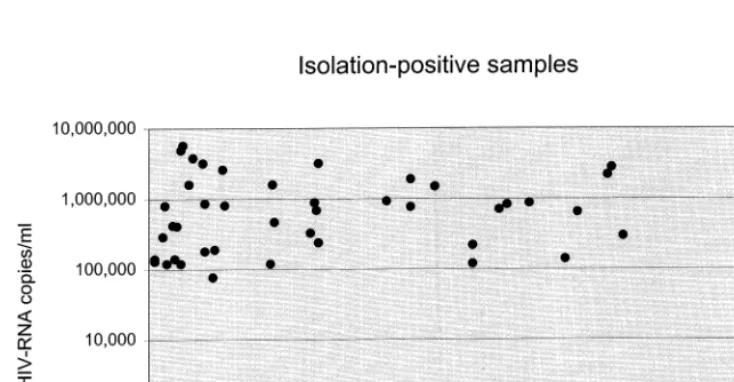
Plasma isolation (qualitative coculture of plasma from HIV-positive patients with unin-fected PHA-stimulated PBMC from uninunin-fected donor) was performed on 124 samples from 57 patients. Forty-eight samples (39%) tested positive and 76 negative (61%). Thirty-six patients (63%) had at least one isolation-positive sample.
Data on both HIV-1 RNA copy number and isolation results were available for 119 samples: a positive plasma isolation, in almost all cases (43/ 45) and irrespective of treatment status, was asso-ciated with an HIV viral load higher than 100 000 copies per ml (Fig. 1). A negative isolation result, however, did not exclude high RNA levels (Fig. 2). Overall, isolation-positive samples had higher plasma HIV-1 RNA values compared with isola-tion-negative samples (copies per ml: mean, 546 175; median 710 000 versus: mean, 13 643; median 37 500; PB0.000001, Wilcoxon’s two
Fig. 2. HIV plasma copy number and CD4 cell count associated to isolation-negative samples.
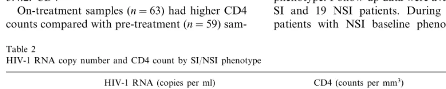
Table 1
HIV-1 RNA copy number and CD4 count by isolation results (positive/negative)
CD4 (counts per mm3) HIV-1 RNA (copies per ml)
N Mean Median Range N Mean S.D. Median Range
55 068 120 000 399–5 700 000 119
All samples 119 142.1 107.4 131 3–472
45 (37.8%) 546 175
Isolation-positive 710 000 5300–5 700 000 46 (38.7%) 92.3 88.3 62.5 3–330
74 (62.2%) 13 643 37 500 399–1 370 000 73 (61.3%) 173.5 107.0 171
Isolation-negative 9–472
3.3. Isolation-positi6e samples: HIV-1 RNA copy number and CD4 cell count according to SI/NSI phenotype
Data on SI/NSI status were available for 45 of the 48 isolation-positive samples. SI and NSI strains were not different in terms of concomitant HIV-1 RNA copy number (mean, 475 480; me-dian 470 000 for SI vs. mean 585 372; meme-dian, 790 000 for NSI; P=0.33). SI strains, compared with NSI strains, tended to be associated with lower CD4 counts at sampling (mean, 69/mm3,
median 27/mm3for SI vs. mean 104/mm3, median
84/mm3 for NSI; P=0.11, Table 2).
sample test). HIV-1 RNA data in relation to isolation results are shown in Table 1.
3.2. Plasma isolation and CD4 cell count
Data on both CD4 counts and isolation results were available for 119 samples. Overall, isolation-positive samples had lower CD4 counts compared with isolation-negative samples (mean, 92.3/mm3, median 62.5
/mm3 vs. mean
173.5/mm3, median 171/mm3; P=0.000034,
3.4. Effect of treatment
3.4.1. RNA
On-treatment samples were collected between week 24 and 48 of treatment. Pre-treatment sam-ples had a lower probability to be isolation-negative with respect to on-treatment samples (23/57 vs. 53/67, relative risk=0.51, 95% CI 0.36 – 0.72). As expected, on-treatment samples had overall lower RNA values (mean, 8954; median, 9400) compared with pre-treatment samples (mean, 424 912; me-dian, 575 000; PB0.000001). Isolation-positive samples were characterised by high RNA values in both on-treatment and pre-treatment samples, al-though values were lower for the samples collected on-treatment (mean and median, 238 166 and 180 000, respectively, compared with pretreatment samples, 738 593 and 810 000, respectively; P= 0.026). The results of the logistic regression analy-sis, performed to estimate the strength of the association between a positive plasma isolation and high RNA values and to assess the role of treatment controlling for its effect on RNA, showed that only HIV-1 RNA was associated significantly with an increased risk of a positive isolation result (OR for HIV copy number\100 000 compared with a value below 100 000: 41.3, CI 95% 9.1 – 186.8, PB0.0001), with treatment status and CD4 count not associated with an increased risk of a positive plasma isolation (odds ratios 1.32 and 1.00; P= 0.63 and 0.65, respectively). RNA and CD4 data by treatment status and isolation outcome are summarised in Table 3.
3.4.2. CD4
On-treatment samples (n=63) had higher CD4 counts compared with pre-treatment (n=59)
sam-ples (mean, 189.7/mm3, median 194
/mm3 vs. 88.6
and 62.5/mm3, respectively). In contrast with the
RNA results, however, the difference between iso-lation-positive samples and isolation-negative re-sults observed in the whole group (i.e. not considering treatment status) was evident only among pre-treatment samples (mean, 63.9/mm3,
median 33/mm3vs. 124 and 139/mm3, respectively),
with on treatment samples showing only a minor difference between positive and isolation-negative groups (mean, 164.2/mm3
, median 162/ mm3
vs. 196.4 and 196/mm3
, respectively) (Table 3).
3.4.3. HIV-isolation results
At baseline, 34 patients were isolation-positive and 23 isolation-negative. Follow-up isolation re-sults were available for 28 out of 34 isolation-pos-itive patients and for 19 of 23 isolation-negative patients. During treatment, most of isolation-posi-tive patients became isolation-negaisolation-posi-tive, with a trend for a higher negativisation rate among pa-tients receiving the triple combination (total, 20; triple combination 13 vs. double combination 7). Most of the patients who were isolation-negative at baseline remained isolation-negative during follow-up (17 out of 19 with available data). Only two patients who were isolation-negative at baseline became isolation-positive during treatment. Both had very high baseline viral load (640 000 and 1 370 000 copies per ml, respectively).
3.4.4. SI/NSI phenotytpe
At baseline, among the 34 isolation-positive patients, 11 had an SI phenotype and 23 an NSI phenotype. Follow-up data were available for nine SI and 19 NSI patients. During follow-up, 15 patients with NSI baseline phenotype and five
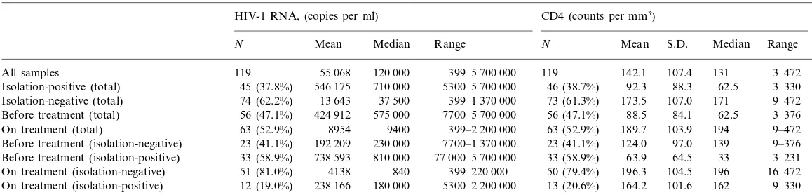
Table 2
HIV-1 RNA copy number and CD4 count by SI/NSI phenotype
CD4 (counts per mm3) HIV-1 RNA (copies per ml)
Mean N Mean S.D. Median
N Median Range Range
3–330 45 546 175 710 000
Isolation-positive 5300–5 700 000 46 92.3 88.3 62.5
(SI+NSI)
30 (66.7%) 585 372 790 000 5300–5 700 000 30 (65.2%) 104.2 92.0
Vella
et
al
.
/
Anti
6
iral
Research
47
(2000)
189
–
198
195
Table 3
HIV-1 RNA copy number and CD4 count by isolation results and treatment status
HIV-1 RNA, (copies per ml) CD4 (counts per mm3)
Mean S.D. Median Range
N
N Mean Median Range
399–5 700 000 119 142.1 107.4 131 3–472 55 068
All samples 119 120 000
92.3 88.3 62.5 3–330 46 (38.7%)
45 (37.8%)
Isolation-positive (total) 546 175 710 000 5300–5 700 000
73 (61.3%)
74 (62.2%) 13 643 37 500 399–1 370 000 173.5 107.0 171 9–472 Isolation-negative (total)
56 (47.1%) 424 912 575 000 56 (47.1%) 88.5 84.1 62.5 3–376
Before treatment (total) 7700–5 700 000
189.7 103.9 194 9–472 63 (52.9%)
On treatment (total) 63 (52.9%) 8954 9400 399–2 200 000
124.0 97.0 139 9–376 Before treatment (isolation-negative) 23 (41.1%) 192 209 230 000 7700–1 370 000 23 (41.1%)
63.9 64.5 33 3–231 33 (58.9%)
Before treatment (isolation-positive) 33 (58.9%) 738 593 810 000 77 000–5 700 000
399–220 000 50 (79.4%) 196.3 104.5 196 16–472 On treatment (isolation-negative) 51 (81.0%) 4138 840
164.2 101.6 162 9–330 13 (20.6%)
238 166
patients with SI baseline phenotype became isola-tion-negative. Five patients remained isolation-positive maintaining their baseline phenotype (NSI, 3; SI, 2) and three patients remained isola-tion-positive switching to the opposite phenotype (1 switch NSI-SI, 2 SI-NSI).
4. Discussion
Infectivity of plasma and other biological sam-ples from HIV-positive patients is a relevant issue in several situations in which an assessment of the risk of HIV transmission may be necessary. Meth-ods to assess infectivity must rely on in vitro models and are not easily applicable to most of the situations occurring in clinical practice. There-fore, the risk of HIV transmission is commonly assessed estimating the amplitude of the exposure using data relating to quantity and type of sam-ple, and amount of virus contained in the sample. Plasma samples of equal volume may differ sig-nificantly in terms of viral load. Based on previ-ous data from others and us a general direct relationship between plasma viral load and plasma infectivity can be assumed in untreated patients. The main goals of our study were to define this relationship during antiretroviral treat-ment and to evaluate whether the same level of viral load was associated with characteristics of infectivity (i.e. positivity of HIV isolation from plasma) irrespective of status of antiretroviral treatment.
The main finding of our study was that isola-tion positivity was almost invariably associated with a viral load of at least 100 000 copies per ml, in both pre-treatment and on-treatment samples. These results indicate that in the in vitro model we used, low levels of viral load are only excep-tionally associated with isolation from plasma, confirming the association already found by oth-ers between HIV-1 viral load and plasma infectivity.
The maintained presence of this association also in samples collected during treatment and the existence among isolation-positive samples of only a limited viral load difference (about 0.5 log) be-tween before-treatment and on-treatment samples
suggests that therapy does not induce major changes in the proportion of infectious/ non-infec-tious HIV particles which compose global HIV viral load and that on-treatment viral load levels can be considered roughly equivalent to pre-treat-ment viral load levels in terms of infectivity. This conclusion also is supported by the results of a logistic regression analysis, which showed no sig-nificant effect of treatment after controlling for RNA values.
Our data confirm clearly the positive effect of combination regimens on plasma viral load and CD4 cell count. The concomitant negativisation of plasma isolation observed during treatment in most of the patients who were isolation-positive at baseline indicates that combination treatment re-duces infectivity through a reduction in viral load. This assumption is also supported by the trend for a higher negativisation rate that we observed among patients receiving the more potent an-tiretroviral regimen.
Two studies on perinatal transmission (Garcia et al., 1999; Mofenson et al., 1999) showed no HIV transmission from mother to child with viral load levels below 500 and 1000 copies per ml, respectively. Similarly, our in vitro results showed no positive isolation among samples with a viral load below 1000 copies, supporting the concept of a low or absent risk of infectivity in the presence of a low or undetectable viral load.
We therefore support the hypothesis that high viral load, although representing a major factor influencing transmission and infectivity, is not per se sufficient to determine infectivity and that other factors, particularly in vivo, may be involved and need to be studied, with particular reference to host response factors, HIV biological characteris-tics and replication dynamics. Among HIV bio-logical characteristics, we could not estimate a possible influence of HIV drug resistance on plasma infectivity because samples were not tested for resistance status.
CD4 counts in on-treatment samples were simi-lar in isolation-positive and isolation-negative samples. This is not unexpected, being a conse-quence of the CD4 increase induced by treatment. It also confirms that, as expected, plasma infectiv-ity is mainly related to HIV viral load and not to CD4 levels.
SI/NSI phenotype was also studied. Given the fast-growth kinetics reported for SI isolates, higher viral load values could have been expected for SI compared with NSI isolates. We did not find major viral load differences between SI and NSI isolates, but SI phenotype resulted associated to lower CD4 counts, as already reported by others (Fenyo, 1995). This observation is compat-ible with the assumption that SI strains can deter-mine CD4 decline not only by direct destruction but also by formation of syncytia in which both infected and uninfected cells are involved.
Assuming that an SI phenotype is associated with an increased risk of progression, it may also be important to define to what extent treatment may induce reversal of SI phenotype. In our sample, almost half of the isolation-positive, SI-positive patients became isolation-negative during treatment, and two patients switched from an SI to an NSI phenotype. These changes may be considered as positive, and indicate that treatment can revert, at least transiently, negative prognostic markers. Although favourable changes (such as negativisation of plasma isolation results or switch from SI to NSI phenotype) involved the majority of patients, some patients remained iso-lation-positive during follow-up and a limited number switched from negative to positive or from an NSI to an SI phenotype. These
observa-tions confirm that combination regimens can in-duce positive changes in most but not all patients treated, and that a proportion of non-responders can be expected for CD4, viral load and other virological markers. The identification of baseline characteristics predicting treatment failure would represent an important tool to individualise treat-ment and improve its effectiveness. In the whole trial population studied, we found that high base-line viral load was a factor linked to lower re-sponse to treatment (Floridia et al., 1999). Although this might lead to the use of more potent regimens in patients with high baseline viral load, this strategy has not yet been confi-rmed by specifically designed controlled trials.
From a methodological point of view, plasma isolation has clearly some limits as a measure of infectivity. Use of plasma may determine a carry-over of some amounts of drugs present in the plasma, with a possible interference in cell infec-tion. This effect, which is difficult to remove in all plasma isolation procedures, is, however, tem-pered by dilution procedures occurring prior to challenging cells. We did not perform experiments to assess the magnitude of this effect in our study. However, similar experiments to exclude this ef-fect were conducted by Zhang et al. (1996) in a study assessing HIV virion kinetics at different time points following initiation of nevirapine. The results showed that, on treatment, the virus-inacti-vated plasma had only very modest effects on slowing viral load in culture, with the infectivity titers of pretreatment plasma samples not altered by adding in a 1:1 ratio HIV-inactivated plasma from patients receiving nevirapine (Zhang et al., 1996).
factors determining transmission, a very prudent interpretation is essential when in vitro data have to be transferred to clinical situations requiring assessment of risk or clinical decisions.
Acknowledgements
We thank Tony Sofia for his help in the revi-sion of the manuscript. This study was supported by grants from Progetto Nazionale Terapia An-tivirale AIDS 1996 (980/A/1). Boehringer Ingel-heim provided nevirapine and matching placebo, supported monitoring and sample shipment.
References
Alkhatib, G., Combadiere, C., Broder, C.C., Feng, Y., Kennedy, P.E., Murphy, P.M., Berger, E.A., 1996. CC CKR5: A RANTES, MIP1-alpha, MIP1-beta receptor as a fusion cofactor for macrophage-tropic HIV1. Science 272, 1955 – 1958.
Ercoli, L., Sarmati, L., El-Sawaf, G., Cochi, S., Lanti, T., Ludicone, P., Guglielmetti, M., Giannini, G., Galluzzo, C., Tomino, C., 1995. Plasma viremia titration and RNA quantitation in ICD-p24 negative HIV type-1 infected pa-tients. AIDS Res. Hum. Retroviruses 11, 1203 – 1207. Fenyo, E.M., 1995. HIV biological phenotype: prognosis
marker for transmission, disease progression and therapy. J. Biol. Regul. Homeostatic Agents 9, 88 – 90.
Floridia, M., Bucciardini, R., Ricciardulli, D., Fragola, V., Pirillo, M.F., Weimer, L.E., Tomino, C., Giannini, G., Galluzzo, C.M., Andreotti, M., Cargnel, A., Alberici, F., De Rienzo, B., Leoncini, F., Fiaccadori, F., Francisci, D., Grillone, W., Ortona, L., Piazza, M., Scalzini, A., Nigra, E., Tumietto, F., Vella, S., 1999. A randomized, double-blind trial on the use of a triple combination including nevirapine, a non-nucleoside reverse transcriptase HIV in-hibitor, in antiretroviral-naive patients with advanced dis-ease. J. AIDS Hum. Retrovirol. 20, 11 – 19.
Garcia, P.M., Kalish, L.A., Pitt, J., Minkoff, H., Quinn, T.C., Burchett, S.K., Kornegay, J., Jackson, B., Moye, J., Han-son, C., Zorrilla, C., Lew, J.F., 1999. Maternal levels of
plasma human immunodeficiency virus type 1 RNA and the risk of perinatal transmission. New Engl. J. Med. 341, 394 – 402.
Katzenstein, T.L., Nielsen, C., Bruun, L., et al., 1996. Quan-tification of HIV-1 RNA during antiretroviral therapy: association with viral phenotype and development of resis-tance. Antiviral Ther. 1, 246 – 254.
Koot, M., Keet, I.P.M., Vos, A.H.V., de Goede, R.E., Roos, M.T., Coutinho, R.A., Miedema, F., Schellekens, P.T., Tersmette, M., 1993. Prognostic value of HIV-1 syncytium-inducing phenotype for rate of CD4+ cell depletion and progression to AIDS. Ann. Intern. Med. 118, 681 – 688. Mofenson, L.M., Lambert, J.S., Stiehm, E.R., Bethel, J.,
Meyer, W.A. III, Whitehouse, J., Moye, J. Jr, Reichelder-fer, P., Harris, D.R., Fowler, M.G., Mathieson, B.J., Neom, G.J., 1999. Risk factors for perinatal transmission of human immunodeficiency virus type 1 in women treated with zidovudine. New Engl. J. Med. 341, 385 – 393. Pedraza, M.-A., del Romero, J., Roldan, F., Garcia, S.,
Ayerbe, M.C., Noriega, A.R., Alcami, J., 1999. Heterosex-ual transmission of HIV-1 is associated with high plasma viral load levels and a positive viral isolation in the infected partner. J. AIDS 21, 120 – 125.
Piatak, M., Saag, M.S., Yang, L.C., Clark, S.J., Kappes, J.C., Luk, K.C., Hahn, B.H., Shaw, G.M., Lifson, J.D., 1993. High levels of HIV-1 in plasma during all stages of infec-tion determined by competitive PCR. Science 259, 1749 – 1754.
Richman, D.D., Bozzette, S.A., 1994. The impact of the syncytium-inducing phenotype of human immunodefi-ciency virus on disease progression. J. Infect. Dis. 169, 968 – 974.
Rusconi, S., De Pasquale, M.P., Mainini, F., et al., 1996. Viral load, viral phenotype modification, zidovudine susceptibil-ity and reverse transcriptase mutations during the first 6 months of zidovudine monotherapy in HIV-1-infected peo-ple. Antiviral Ther. 1, 211 – 219.
Van Gemen, B., Kievits, T., Nara, P., Huisman, H.G., Jurri-aans, S., Goudsmith, J., Lens, P., 1993. Qualitative and quantitative detection of HIV-1 RNA by nucleic acid sequence-based amplification. AIDS 7 (Suppl. 2), S107 – S110.
Zhang, H., Dornadula, G., Wu, Y., Havlir, D., Richman, D.D., Pomerantz, R.J., 1996. Kinetic analysis of in-travirion reverse trancription in the blood plasma of hu-man immunodeficiency virus type 1-infected individuals: direct assessment of resistance to reverse transcriptase in-hibitors in vivo. J. Virol. 70, 628 – 634.