Prefrontal Cortex Distinguishes Younger from Older
Adults in Major Depressive Disorder
Jose´ Javier Miguel-Hidalgo, Christie Baucom, Ginny Dilley, James C. Overholser,
Herbert Y. Meltzer, Craig A. Stockmeier, and Grazyna Rajkowska
Background: Recent postmortem studies in major depres-sive disorder (MDD) provide evidence for a reduction in the packing density and number of glial cells in different regions of the prefrontal cortex; however, the specific types of glia involved in those morphologic changes are unknown.
Methods: The territory occupied by the astroglial marker glial fibrillary acidic protein (GFAP) was measured as an areal fraction in cortical layers III, IV, and V in sections from the dorsolateral prefrontal cortex (dlPFC) of MDD and control subjects. In addition, the packing density of GFAP-immunoreactive somata was measured by a direct three-dimensional cell counting method.
Results: The mean areal fraction and packing density of GFAP-immunoreactive astrocytes in the dlPFC of MDD subjects were not significantly different from those in control subjects; however, in MDD there was a significant strong positive correlation between age and GFAP immu-noreactivity. When the MDD group was divided into younger (30 – 45 years old) and older (46 – 86) adults, in the five younger MDD adults, areal fraction and packing density were smaller than the smallest values of the control subjects. In contrast, among older MDD subjects these parameters tended to be greater than in the older control subjects.
Conclusions: The present results suggest that the GFAP-immunoreactive astroglia is differentially involved in the pathology of MDD in younger compared with older adults.
Biol Psychiatry 2000;48:861– 873 © 2000 Society of
Bi-ological Psychiatry
Key Words: Astroglia, dorsolateral prefrontal cortex,
human postmortem, neuropathology, aging, quantitative immunohistochemistry.
Introduction
A
lterations in the physiology and morphology of neu-rons or glial cells in the cerebral cortex are hallmarks of neurodegenerative disorders such as Alzheimer’s dis-ease and Huntington disdis-ease. These alterations are mani-fested by loss of neurons and glial proliferation and hypertrophy. In psychiatric disorders, however, changes at the cellular level are not as prominent; consequently, strong evidence that those changes may be a part of the neuropathology of these disorders is fairly recent (Benes 1993; Bogerts 1993; Falkai et al 1988; Heckers 1997; O¨ ngu¨r et al 1998; Rajkowska et al 1998, 1999; Selemon et al 1995, 1998). Postmortem histopathologic studies in mood disorders reveal significant morphologic alterations in glial cells in several regions of the prefrontal cortex (O¨ ngu¨r et al 1998; Rajkowska et al 1999). In major depressive disorder (MDD), a significant reduction in the packing density of glial cells is observed in layers III and V of the dorsolateral prefrontal cortex among other pre-frontal regions (Rajkowska et al 1999). This reduction is accompanied by an increase in the average size of glial cell nuclei in layer III. In addition, cell packing density and size of neurons are decreased in layer III of the dlPFC in MDD. Thus, structural alterations of glial cells may be related to the abnormal morphology of cortical neurons and to the gross functional (metabolic) and structural alterations observed with neuroimaging techniques in the dlPFC in MDD (for reviews, see Ketter et al 1996; Winsberg et al 2000). Nonetheless, in MDD, it is not yet known whether a specific type of neuroglia is mainly affected by the observed morphologic and functional changes.The three major types of neuroglia found in the central nervous system, microglia, oligodendroglia, and astroglia, are known to respond to variations in the neural environ-ment (Coyle and Schwarcz 2000; de Vellis 1993; Mu¨ller 1995). Microglia are mainly involved in immune re-sponses within the nervous system and in the elimination of cellular debris. Oligodendroglia are crucial for the From University of Mississippi Medical Center, Jackson (JJM-H, CB, CAS, GR),
Case Western Reserve University, Cleveland, Ohio (GD, JCO), and Vanderbilt University, Nashville, Tennessee (HYM).
Address reprint requests to Jose´ J. Miguel-Hidalgo, University of Mississippi Medical Center, 2500 North State Street, Box 127, Jackson MS 39216. Received April 13, 2000; revised July 7, 2000; accepted July 25, 2000.
© 2000 Society of Biological Psychiatry 0006-3223/00/$20.00
conduction of action potentials along axons. Among glial cells, astrocytes deserve special attention because they are involved in numerous and complex functions, such as response to injury, participation in immune defense (to-gether with microglia), promoting the transfer of nutrients to neurons, maintenance of synaptic transmission, ionic homeostasis of the neuropil, and the support of cerebral glucose metabolism (Coyle and Schwarcz 2000; Magis-tretti and Pellerin 1999; MagisMagis-tretti et al 1999). Moreover, astrocytes are known to express transporters and receptors for the neurotransmitters glutamate,g-aminobutyric acid, serotonin, and norepinephrine (Blankenfeld and Ketten-mann 1992; Cohen et al 1999; Flott and Seifert 1991; Hirst et al 1998; Kimelberg and Katz 1985; Russ et al 1996; Schousboe and Westergaard 1995; Sutin and Shao 1992). Finally, astroglia generate specific changes in plasma membrane ionic conductance and second messenger re-sponses when exposed to these neurotransmitters (Mc-Carthy et al 1995; Walz 1995). Thus, astrocytes are able to respond with functional and morphologic changes to alterations in the neurotransmitter systems implicated in both the pathophysiology of mood disorders and the therapeutic response to psychotropic medications.
In MDD, the packing density and size of different types of glial cells as determined by Nissl-staining is altered in several regions of the prefrontal cortex, including the dorsolateral prefrontal cortex (dlPFC; Rajkowska et al 1999). Interestingly, in cortical layer III of the dlPFC, there is a reduction in the packing density of medium-sized glial cells associated with an increase in the density of glial cells with extra large nuclei; in layer V, there is a reduction in the density of glial cells with large nuclei. Glial cells with medium to large nuclei are known to include mainly astrocytes, whereas cells with medium to small nuclear sizes are most likely to be oligodendrocytes or microglia (Blinkov and Glezer 1968, 237–253). En-largements in the size of glial cell nuclei occur in some neurodegenerative brain pathologies, and, in the case of astrocytes, those changes parallel the changes in GFAP expression (Berciano et al 1995; Levison et al 1998; Weis et al 1993). Thus, the changes in the density of glial cell nuclei with the largest sizes observed in Nissl material in MDD suggest that there may be a reduction in the packing density of astrocytes in this disease, or that astrocyte morphology and the expression of the astrocytic cytoskel-etal marker glial fibrillary acidic protein (GFAP) are different in MDD compared with normal control subjects. Astrocytes react to neuronal injury or degeneration by increasing the synthesis of the cytoskeletal component GFAP and by expanding and elongating their processes (Bignami and Dahl 1995; Norenberg 1996). In astrocytes, GFAP immunoreactivity also is regulated through activa-tion of serotonergic and adrenergic receptors (Griffith and
Sutin 1996; Le Prince et al 1990), which are considered to play a crucial role in the pathophysiology of mood disorders (Potter 1996). Accordingly, the aim of our study was to examine in postmortem tissue the relative extent of GFAP immunoreactivity and the packing density of GFAP-immunoreactive astrocytes in the dlPFC from sub-jects with MDD compared with psychiatrically normal control subjects.
Methods and Materials
Human Subjects
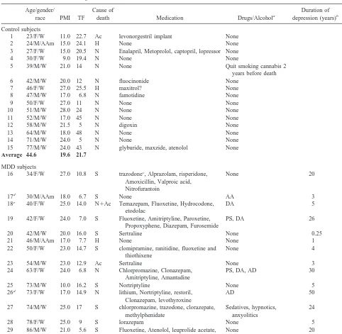
All human postmortem brain tissue was collected from autopsies that took place at the Cuyahoga County Coroner’s Office in Cleveland, Ohio. In all cases, retrospective psychiatric assess-ments were conducted in accordance with the Institutional Review Board policies, and written consent was obtained from the next of kin. Consensus diagnoses of MDD were made in accordance with the Diagnostic and Statistic Manual of Mental Disorders-Revised (DSM-III-R; American Psychiatric Associa-tion 1987). AddiAssocia-tional informaAssocia-tion about previous hospital med-ical records, prior medmed-ical or substance abuse problems, medi-cation history, and postmortem toxicology also was gathered. Evidence of head trauma, neurologic disease, or psychoactive substance use disorder within the last year of life was sufficient basis to exclude a case from the study. Only brains with a postmortem delay of less than 30 hours and a fixation time no longer than 4 years were included (Table 1). Further details about the diagnostic procedures and methods for collecting information on the subjects are provided elsewhere (Rajkowska et al 1999).
Tissue Sampling
The tissue samples were obtained from the left prefrontal cortex of 14 subjects retrospectively diagnosed with MDD (nonpsy-chotic) and 15 psychiatrically normal control subjects. Morpho-metric parameters were measured in the left dlPFC (Brodmann’s area 9). Previously established cytoarchitectonic criteria for area 9 (Rajkowska and Goldman-Rakic 1995a, 1995b) were used to select the cortical regions of interest for the present study.
Blocks of tissue (23 2 cm) sampled from the dlPFC were embedded in celloidin and cut into 40mm thick sections. Three sections evenly spaced 400 mm apart were chosen from each subject to be immunostained for GFAP. These sections were adjacent to or within 100mm of the Nissl-stained sections that were used in our previous morphometric study demonstrating reduced glial cell packing density in MDD (Rajkowska et al 1999). The Nissl-stained sections were then used to draw the boundaries between individual cortical layers. These laminar boundaries were imposed as guides on the GFAP immunostained sections to determine the laminar distribution of GFAP-immu-noreactive cells.
Immunohistochemistry
developed in our laboratory as a modification of existing proto-cols (Miguel-Hidalgo and Rajkowska 1999). Briefly, celloidin was removed from the sections with a saturated 50% solution of NaOH in methanol, and sections were rinsed three times in 100% methanol. The sections were then washed in phosphate buffer saline (pH 7.4) followed by Tris-HCl buffer saline (pH 7.6).
Subsequently, the floating sections were incubated with a mouse monoclonal GFAP antibody at a 1:500 dilution, and the binding of this antibody was detected with a biotinylated secondary antibody according to the ABC method (ABC kit, Vector Laboratories, Burlingame, CA). The monoclonal antibody against GFAP used as first antibody in our study was obtained Table 1. Characteristics of Subjects
Age/gender/
race PMI TF
Cause of
death Medication Drugs/Alcohola
Duration of depression (years)b
Age of onset
Control subjects
1 23/F/W 11.0 22.7 Ac levonorgestril implant None
2 24/M/AAm 15.0 24.1 H None None
3 27/F/W 15.0 20.5 N Enalapril, Metoprolol, captopril, lopressor None
4 30/F/W 9.0 19.4 N None None
5 39/M/W 21.0 14 N None Quit smoking cannabis 2
years before death
6 42/M/W 20.0 12 N fluocinonide None
7 46/F/W 27.0 25.5 H maxitrol? None
8 47/M/W 17.0 6.8 N famotidine None
9 50/F/W 27.0 11 N None None
10 51/M/W 28.0 24 N None None
11 52/M/W 17.0 45 N None None
12 58/M/W 21.5 5 N digoxin None
13 64/M/W 18.0 48 N None None
14 71/M/W 24.0 5 N None None
15 77/M/W 24.0 43 N glyburide, maxzide, atenolol None
Average 44.6 19.6 21.7
MDD subjects
16 34/F/W 27.0 10.8 S trazodonec, Alprazolam, risperidone,
Amoxicillin, Valproic acid, Nitrofurantoin
None 20 14
17d 30/M/AAm 18.0 6.7 S None AA 3 27
18e 40/F/W 25.0 14.0 N1Ac Temazepam, Fluoxetine, Hydrocodone,
etodolac
DA 5 35
19 42/F/W 24.0 7.0 S Fluoxetine, Amitriptyline, Paroxetine, Propoxyphene, Diazepam, Furosemide
PS, DA 26 15
20 42/M/W 20.0 16.0 S Sertraline None 0.25 41
21 46/M/AAm 17.0 7.7 H None None 1 45
22 50/F/W 23.0 14.7 S clomipramine, ranitidine, fluoxetine and thiothixene
None 4 45
23 54/M/W 23.0 12.9 Ac Sertraline None 3 51
24 63/F/W 24.0 6.8 N Chlorpromazine, Clonazepam, Amitriptyline, Amantadine
PS, DA, AD 30 33
25 73/M/W 10.0 16.2 S Nortriptyline None 5 68
26d 73/F/W 17.0 14.9 N lithium, Nortriptyline, restoril,
Clonazepam, levothyroxine
AD 50 23
27 74/M/W 25.0 17 S chlorpromazine, trazodone, clorazepate, methylphenidate
Sedatives, hypnotics, anxyolitics
24 50
28 78/F/W 25.0 9 S lorazepam None 5 73
29 86/M/W 21.0 5.6 S Fluoxetine, Atenolol, leuprolide acetate, lupron
None 20 56
Average 52.8 21.4 11.4
PMI, postmortem interval (hour; defined as the time between death and the beginning of the formalin fixation); TF, time in formalin (months); F, female; W, white; Ac, accident; M, male; AAm, African American; H, homicide; N, natural, MDD, major depressive disorder; S, suicide; AA, alcohol abuse; DA, drug addiction; PS, polysubstance abuse; AD, alcohol dependence.
aDefined as psychoactive substance use disorder.
bThe duration of illness covers the time between the first display (and not necessarily diagnosis) of symptoms of a depressive illness and the date of death. cCapitalized drugs were prescribed in last month of life.
from Sigma (catalogue no. G 3893, clone G-A-5). This antibody was originally developed in mouse using GFAP from pig spinal cord as immunogen. It specifically stains GFAP in immunoblots and in brain sections (Debus et al 1983; Franke et al 1991). In our experiments, omission of the first antibody or the biotinylated secondary antibody resulted in the absence of any immunostain-ing. It is important to stress that the Nissl-stained sections used in our previous study for estimates of neuronal and glial densities were closely adjacent to the sections processed for immunohis-tochemistry. To reduce to a minimum the variability in the intensity of staining due to uncontrollable changes from experi-ment to experiexperi-ment, each immunostaining experiexperi-ment involved sections from both the control group and the MDD group. In each of three of the experiments, 23 sections were selected per experiment, all of them processed simultaneously. Out of these sections, 11 were from different subjects of the control group, and 12 were from the corresponding subjects of the MDD group. In each of three additional experiments, six sections were selected per experiment. Out of these sections, four were from the control group, and two were from the MDD group. In total, six experiments were conducted resulting in three sections per subject used for the quantitative immunohistochemistry studies.
Extent of GFAP Immunoreactivity
The area occupied by GFAP-immunoreactivity in astrocytes on histologic sections was examined according to a modification of the method for the measurement of GFAP-immunoreactivity described by Zilles et al (1991). The area occupied by GFAP-immunoreactivity in layers III1IV and in layer V was examined. These layers were chosen for analysis because significant changes in glial cell density and glial nuclear size in these same layers have been previously reported (Rajkowska et al 1999). Layers III and IV were analyzed together since the pattern of distribution of immunoreactivity is very similar in the lower layer III and in layer IV. Moreover, in area 9 the boundary between layer III and layer IV is considerably tortuous, which prevents establishing a reliable separation between these two layers in the GFAP-immunoreactive sections. In contrast, there is a much denser appearance to GFAP immunoreactivity in layer V, which permits a reliable distinction of layer V from the neigh-boring layers.
An image analysis system working with images from a video camera attached to a microscope was used. A box of fixed width (1530mm) was positioned spanning either layers III1IV or layer V. In each of those boxes, an image was obtained with the video camera and digitized into gray level images. Immunopositive structures were segmented by defining a background level in an area of the section with no specific immunoreactivity and thresholding with a fixed level of 20 gray values over the background; gray levels were from 0 (brightest) to 255 (darkest). By this procedure, a binary image of the area occupied by immunoreactivity was obtained. The areal fraction occupied by glial immunoreactivity was calculated by dividing the immuno-reactive area by the total area occupied by the cortical layers in the outlined box. The immunoreactivity was expressed as a percentage of the total area. The average of three measurements
obtained in the samples per subject was considered as the value of areal fraction and used for statistical analysis.
THREE-DIMENSIONAL CELL COUNTING. To estimate the number of GFAP-immunoreactive cell bodies, a direct, three-dimensional cell counting setup was used (Williams 1989; Williams and Rakic 1988). This system is based on the optical disector method, which allows the estimation of the number of cells without introducing biases due to cell size, cell shape, or section thickness. To avoid a selective bias when counting astrocytes in GFAP-immunostained sections, a probe was con-sistently located in the middle of the region used to measure the areal fraction of GFAP immunoreactivity. The probe consisted of a series of consecutive boxes spanning either layers III1IV or layer V. The dimensions of each box were 25mm thick, 60mm wide, and 90 mm high. In layers III1IV and in layer V, the packing densities of GFAP-immunoreactive cell bodies were obtained according to the procedure described elsewhere (Raj-kowska et al 1999). The packing densities of glial cells are noted as the number of cells3103
/mm3.
One probe per section was sampled in each of three sections per subject. The probe contained an average of 13 boxes for layers III1IV and three boxes for layer V. The averages of all morphometric parameters measured in the three sections per brain sampled from dlPFC (area 9) were subjected to a single-factor (disease) analysis of variance (ANOVA) between the depressed and control groups. Other pairwise comparisons were performed with the Student’s t test. Differences between the groups were defined at the level of
p, .05. Influence of age, postmortem delay, storage time in formalin, duration of illness and onset of illness on the areal fraction of GFAP immunoreactivity, and the packing density of GFAP immunoreactive cells was examined by Pearson correla-tional analysis. The same correlation analyses were used to establish the relationship between GFAP parameters and the packing densities of glial cells in the Nissl-stained sections.
Results
Distribution of GFAP Immunoreactivity: Qualitative Observations
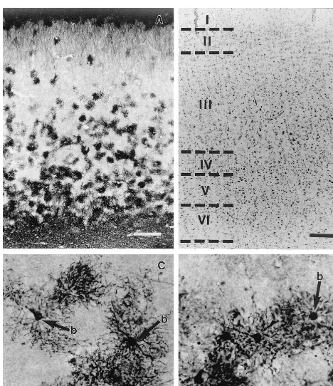
dense in layer V. In these three layers, however, the processes and cell bodies of individual astrocytes were clearly distinguishable for the most part (Figure 1, C and D). Layer VI was covered by GFAP-immunoreactive processes, and although the overall immunoreactivity appeared less intense than in layer I, in most cases it was difficult to identify individual GFAP-immunoreactive cell bodies in this layer (Figure 1A).
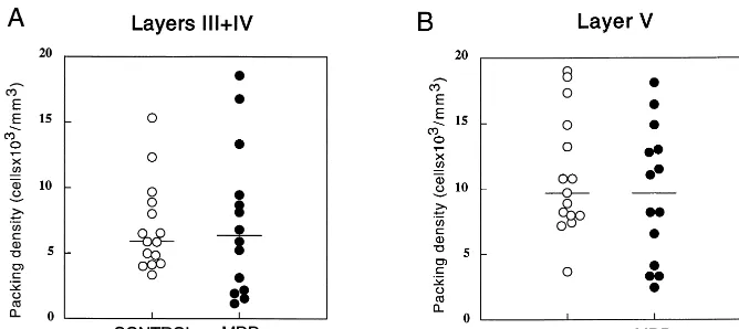
The high variability in GFAP immunostaining was reflected in dramatic interindividual differences in the amount of tissue covered by GFAP-immunoreactive struc-tures and in the packing density of GFAP-immunoreactive cell bodies (Figure 2). This difference was more prominent in the MDD group. Some subjects had a very small area occupied by GFAP immunoreactivity and almost no im-munoreactive cell bodies; in others, the areal fraction
covered was as high as 95%, and the cell packing density was maximal (Figure 2, A and B).
Comparison of the Areal Fraction Covered by GFAP Immunoreactivity in Control and MDD Subjects
LAYERS III1IV. The areal fraction covered by the
processes and cell bodies of GFAP-immunoreactive astro-cytes was measured in layers III1IV and in layer V both in subjects with MDD and in the control group. In layers III1IV, the average areal fraction (%) measured in control subjects (44.84618.46; mean6 SD) was lower although not significantly different (F50.018, p5.895) from that in MDD subjects (46.13632.30); however, the higher SD in MDD and the distribution of values (Figure 3) suggested that the subjects with MDD could be separated into two sub-groups, one of them with very low GFAP immunoreactivity, the other with high immunoreactivity.
Figure 1. Distribution of glial fibrillary acidic protein (GFAP)– immunoreactive astrocytes in area 9 of the human dorsolateral prefrontal cortex in a normal control subject. (A) Low-power picture showing the distribution of GFAP immunoreactivity across the six cortical layers. (B) Picture of a section adjacent to the one in A, but stained with cresyl violet where the borders between layers can be distinguished. (C) High-power photograph of GFAP-immunoreactive astrocytes in the lower part of cortical layer III. (D) High-power picture of GFAP immunoreactive astrocytes in cortical layer V. Note in C and D that the immunoreactive cell bodies of astrocytes are clearly visible. In both layers, GFAP immunoreactivity was mostly contained in abundant processes stemming from the cell bodies of astrocytes, producing a bushy or starry appearance. Calibration bars, 200
mm (A, B) and 50mm (C, D).
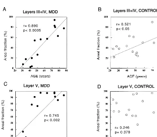
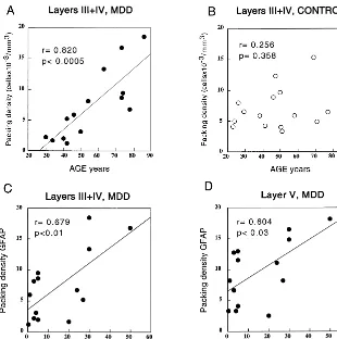
Demographic variables of the subjects were examined for possible correlations with the widely different values of the areal fraction covered by GFAP immunoreactivity. In the MDD subjects, there was a significant positive correlation (r 5 .890, p , .0005) between age and the areal fraction occupied by immunoreactivity in layers III1IV (Figure 4A). In the control group, there was a tendency toward higher GFAP immunoreactivity in older ages in layers III1IV (Figure 4B) that reached statistical significance (r 5 .521, p , .05). The slope of the regression line was smaller than that in the MDD group, however. It was also remarkable that the areal fraction values (range of 2.46 –19.73%) in the five youngest
individuals in the MDD group were lower that the lowest value (21.8%) in the control group. Based on these results, the control and MDD groups were both sorted into an older subgroup (46 – 86 years old) and a younger subgroup (23– 45 years old). Within the control group, comparison of the younger subgroup with the older subgroup showed that there was no significant difference in the areal fraction (t5 20.629, p5.540); within the MDD group, however, the analogous comparison revealed a significant difference between the younger and the older subgroup (t 5 5.786,
p,.0005).
Pairwise statistical comparisons revealed a significant 75% reduction in the GFAP areal fraction in layers III1IV Figure 3. Plots showing the distribution of individual values for the areal fraction cov-ered by glial fibrillary acidic protein–im-munoreactive astrocytes in the control and major depressive disorder (MDD) groups.
between the six subjects of the younger control subgroup (41.08613.09) and the five subjects of the younger MDD subgroup (10.2967.95; Student’s t test, t54.796, p5 .001); however, no such significant difference was appar-ent between the older control subgroup (47.34 6 21.72) and the older MDD subgroup (66.03 6 20.39). On the contrary, a statistically nonsignificant tendency (t 5 21.882, p5 .078) for higher values of the GFAP areal fraction appeared in the older MDD subgroup.
LAYER V. The results for layer V followed a pattern
similar to that observed in layers III1IV (Figure 3B). Although the average GFAP areal fraction did not differ between the studied groups, there was a highly significant positive correlation (r5.745, p5.002) between age and the areal fraction covered by GFAP immunoreactivity (Figure 4C) in the MDD group. In layer V, however, the segregation of the MDD group into two age subgroups was even more marked than in layers III1IV. In contrast, among the control group, there was no correlation between age and the areal fraction of GFAP (r5.246, p 5.378; Figure 4D). The average value of the GFAP areal fraction (38.22623.32) in the younger MDD subgroup was also significantly lower (t 5 23.423, p , .01) than in the younger control subgroup (73.35 6 9.05). In addition, there was a nonsignificant increase in GFAP areal fraction (t 5 1.972, p 5 .066) in the older MDD subgroup (92.0566.99) compared with the older control subgroup (78.606 19.24).
Packing Density of GFAP-Immunoreactive Cell Bodies
To establish whether the reduced GFAP areal fraction in the younger MDD subjects was related to a reduced packing density of GFAP-immunoreactive astrocytes, the density of GFAP-immunoreactive cell bodies was esti-mated in MDD and control subjects.
The packing density of GFAP-immunoreactive cell
bodies in the MDD group (layers III1IV, 7.35 6 5.6 cells3103/mm3; layer V, 9.5765.17 cells3103/mm3) was not significantly different (layers III1IV, F 5 0.05,
p 5 .82; layer V, F 5 0.66, p 5 .43) from that in the control group (layers III1IV, 6.96 6 3.38 cells 3 103/ mm3; layer V, 11.0364.55 cells3103/mm3; Figure 5, A and B); however, as in the case of the GFAP areal fraction, the cell packing density showed a strong significant correlation with age in the MDD group both in layers III1IV (r5 .820, p, .0005) and in layer V (r5.811,
p, .0005). In contrast, among the control group, there was no significant correlation with age (layers III1IV, r5 .256, p5 .358; layer V, r5 .079, p5 .780; Figure 6, A and B; graphs for layer V not shown).
The packing density of GFAP-immunoreactive cell bodies in layers III1IV was also positively correlated with the areal fraction of GFAP immunoreactivity in both the control group (r5.578, p,.03) and the MDD group (r5 .824, p,.0005).
The areal fraction and the packing density of GFAP-immunoreactive cells in layers III1IV and in layer V were not correlated either with the postmortem interval or the time in formalin in either the MDD group or the control group. Although the average time the brain tissue was stored in formalin was significantly higher in the control group than in the depressed group (Table 1), there was no significant correlation of areal fraction or packing density and the time in formalin (for all comparisons p. .2).
Correlation of GFAP Immunoreactivity with the Duration and Onset of Illness
layers III1IV (r5.679, p,.01) and in layer V (r5.604,
p,.03; Figure 6, C and D). By contrast, neither the areal fraction (layers III1IV r5 .487, p5 .077; layer V r5 .343, p5.231) nor the GFAP cell packing density (layers III1IV r5 .264, p5.361; layer V r5 .319, p5.267) was significantly correlated with the age of onset of the disease.
Comparison with Nissl Morphometric Studies of Glia
Among MDD subjects, 12 out of 14 individuals included in our study were the same as those used previously in the morphometric analysis of the dlPFC in MDD (Rajkowska et al 1999). In that study, the packing density of the entire population of glial cells stained nonspecifically with cresyl violet for the Nissl method was markedly reduced in the MDD group compared with control subjects. Moreover, when the Nissl-stained glia were divided into four size classes (small, medium, large, or extra large based on their nuclear size; details on size criteria are described in Rajkowska et al 1998, 1999), the reductions in densities were confined to extra large, large, and medium classes of glia. Therefore, we tested the hypothesis that the densities of specific cell classes in the Nissl-stained material corlated with the measures of GFAP immunoreactivity
re-ported in our present study. Because all the samples in both studies were taken from the same region of area 9 and they were adjacent to one another, the areal fraction and packing density of GFAP-immunoreactive astrocytes in layers III1IV and in layer V were directly compared with the packing densities of the different size classes of glial cell nuclei studied in neighboring Nissl-stained sections. The comparison revealed that the GFAP areal fraction and the GFAP packing density in layers III1IV was positively correlated with the density of Nissl-stained large glial nuclei in all cortical layers combined (GFAP area r 5 .590, p,.05; GFAP packing density r5.636, p,.03). The two GFAP parameters were also significantly corre-lated with the density of Nissl-stained large glial nuclei in layer III (GFAP area r 5 .622, p , .04: GFAP packing density r5.604, p,.04). In layer V, the packing density of GFAP-immunoreactive astrocytes was also signifi-cantly correlated with the density of large glial nuclei in all layers combined (r5.640, p,.03) and in layer Va (r5 .591, p,.05). In contrast, GFAP areal fraction and GFAP packing density were not correlated with the density of any of the other glial size classes (small, medium, extra large) measured in Nissl-stained preparations.
significant, for an age-dependent higher density of large glial nuclei in all layers combined (r5.508, p5.092) and in sublayer IIIa (r 5 .540, p 5 .07). Moreover, this correlation was significant when layers III (r5.591, p, .05) and V (r5 .586, p , .05) and sublayers IIIc (r 5 .636, p,.03) and Va (r5.589, p,.05) were analyzed separately. In contrast, the packing density of small, medium, or extra large nuclei did not show any tendency to be correlated with age (p. .3).
Discussion
Our present study tested the hypothesis that changes in GFAP-immunoreactive astroglia in the dorsolateral pre-frontal cortex are involved in the histopathology of MDD. The results revealed that the area occupied by GFAP immunoreactivity and the packing density of GFAP-immunoreactive astrocytes were strongly correlated with age in layers III1IV and in layer V in the MDD group; in the control group, however, the correlation was weaker, only reaching statistical significance for the areal fraction but not for packing density. The data further indicate that GFAP-positive structures are greatly reduced in depres-sive subjects younger than 45 years old, compared with both older depressive subjects and with the nonpsychiatric control group. In the MDD group, there was a tendency, although nonsignificant, for higher values of GFAP im-munoreactivity in older subjects compared with the older subjects of the control group. Thus, the main difference between the MDD and control groups appears to rest on a very low density of GFAP immunoreactive structures in the younger depressives (under 45 years old).
Other studies in the normal aging human brain using a larger number of subjects also indicate an age-dependent increase in the amount of GFAP immunoreactivity and GFAP mRNA in the neocortex and hippocampus (David et al 1997; Nichols et al 1993; Takashima and Becker 1983). These studies also report a high interindividual variability not explainable solely by age, however. In addition, the age-dependent increases in GFAP immuno-staining are more marked in some brain regions than in others. They are more prominent in the hippocampus than in the neocortex (David et al 1994, 1997). They tend also to be noticeable only after the sixth decade of age (David et al 1997; Hansen et al 1987; Nichols et al 1993). On the other hand, the great age-independent variability of GFAP content in the brain of normal subjects is understandable in the light of animal studies showing that the richness of the environment, and the metabolic and nutritional status of the aging animals greatly influence the age-related in-crease in the GFAP level (Morgan et al 1999; Soffie et al 1999). It is proposed that in the brain of subjects diagnosed with MDD, the environmental circumstances related to
their behavior and the brain metabolic and functional changes may influence the expression of GFAP in the frontal cortex. Conversely, a primary defect in the expres-sion of GFAP or astrocytic function could influence the normal functioning of neurons and result in metabolic and structural alterations; however, this line of argument would not apply to the older depressive subgroup in our study, which showed a nonsignificant tendency for higher GFAP immunoreactivity than did the older control subjects.
One way to make compatible the low GFAP content in younger depressives with a high content in older adults would be to consider the lack of GFAP-immunoreactive astrocytes in younger depressives as a reflection of low-ered activity or responsiveness in these cells. This dimin-ished activity would lead to a reduced support of neuronal activity during the life span of the depressive subject as MDD progresses. Eventually, the lack of support would produce degenerative features in neurons and their con-nections that would trigger an enhanced response in the GFAP-immunoreactive astrocytes. Additionally, the en-hanced GFAP response might be compounded with a normal age-related increase in the expression of GFAP. Supporting the above explanation is the fact that both in layers III1IV and in layer V, there is a significant positive correlation between the packing density of GFAP-immu-noreactive astrocytes and the estimated duration of illness. Thus, the absence of correlation with the onset of disease further implies that even depressed subjects with a rela-tively early onset of disease would increase dramatically the content of GFAP-immunoreactive elements with a long duration of depression.
relationship to age-dependent differences in the prefrontal glial pathology of MDD.
Another possibility that cannot be ruled out is that prolonged antidepressant treatment increases astrocyte activation and GFAP expression. Most MDD subjects in our study had received treatment with antidepressants for variable periods of time and different combinations of psychotropic medications. Nearly all effective antidepres-sant drugs predominantly target the adrenergic and sero-tonergic neurotransmitter systems (for review, see Potter 1996), and there is evidence that changes in those systems influence the astroglial reaction and the expression of GFAP. Formation of reactive astrocytes in vivo is influ-enced by noradrenergic axons (Griffith and Sutin 1996), and blockade of b-adrenergic receptor greatly reduces reactive astrocyte formation, as determined by GFAP labeling (Sutin and Griffith 1993). Conversely, in the absence of injury, b-adrenergic receptor activation in-creases GFAP immunoreactivity (Hodges-Savola et al 1996). By contrast, serotonin decreases GFAP immunola-beling in cultured brainstem astrocytes (Le Prince et al 1990). The direct influence of antidepressants on glial morphology has not been studied to date. Interestingly, increases in glial cell packing density have been reported recently in the prefrontal cortex of rhesus monkeys treated with neuroleptics (Selemon et al 1999); however, the augmentation was detected only in layer I (with typical antipsychotics) or layer IV (with atypical antipsychotics). An argument could be made that our data are biased because of some unknown effect of prolonged exposure to formalin. Increasing the time in formalin tends to reduce immunoreactivity for some antigens (Battifora et al 1986); however, in a previous study using the same antigen recovery technique as that in our present study, we found no evidence of reduced immunoreactivity for various antigens in brain tissues from normal control subjects with periods in formalin up to 2 years (Miguel-Hidalgo and Rajkowska 1999). Furthermore, in our present study, the lowest values of immunoreactivity parameters were found in the MDD group, in which also the lowest values for areal fraction and packing density of GFAP immunoreac-tive were observed. This fact, together with the lack of any correlation or tendency toward correlation between GFAP immunoreactivity and fixation time, indicates that the differences in GFAP immunostaining found in our mate-rial cannot be attributed to differences in the fixation times.
Size Classes of Glial Cells and GFAP Immunoreactivity
Comparison of the present findings with the data from our previous study on Nissl-stained sections sampled from the
same cortical area in MDD subjects suggests that the glial cell nuclei classified as large in that study correspond to the GFAP-immunopositive elements in our present study. This would further implicate that a reduced GFAP immu-noreactivity coincides with a reduced packing density of astrocytes with large nucleus and, consequently, that changes in GFAP expression are related to changes in the size of the astrocyte nucleus. Because GFAP immunore-activity increases with age, an age-related increase in the packing density of astrocyte nuclei should also be ex-pected in MDD. In fact, a close examination of the data from our previous Nissl study revealed a significant correlation between age and the density of large glial cell nuclei in layer III and layer V; however, no correlation between age and the density of any of the other glial size classes (small, medium, and extra large) was found.
The involvement of the cells with large nuclei (probably astrocytes) in MDD suggested by the combined analysis of data from our present study and our previous report does not rule out the participation of other glial cell types in the pathophysiology of MDD. In our previous Nissl study, the density of medium-sized and extra large glia in layer III, and medium and large glia in layer V, was significantly different in the MDD brains compared with control brains. By contrast, in our present study, there was no significant difference in the average areal fraction or packing density of GFAP positive astrocytes either in layers III1IV or in layer V. These contrasting findings suggest that other types of glia (microglia or oligodendroglia) also might be implicated in the histopathology of major depression.
Comparison with Other Psychiatric Disorders
In schizophrenia, possible changes in GFAP have been examined with different methods of assessing in situ GFAP immunoreactivity (Arnold et al 1996; Falkai et al 1999; Roberts et al 1987). These studies reveal an absence of GFAP-related gliosis in schizophrenia, which is consis-tent with the lack of significant changes in the density of Nissl-stained glial cells or sizes or glial nuclei in postmor-tem brains of schizophrenics (Rajkowska et al 1998; Selemon et al 1995). The absence of major changes in glial parameters in schizophrenia would predict a relationship between GFAP immunoreactivity and age similar to the one observed in normal control subjects. This correlation has not been tested, however. Future studies on the relationship between GFAP expression and age in schizo-phrenia are needed to ascertain that prediction.
observed in corresponding regions of MDD (O¨ ngu¨r et al 1998; Rajkowska et al 1997; G. Rajkowska et al, unpublished data). In the subgenual prefrontal cortex of subjects with familial bipolar disorder, O¨ ngu¨r et al (1998) have demonstrated a decrease in the number and density of glial cells, although in this study neither a distinction between different cortical layers nor an analysis of specific size classes in individual layers was made. In contrast, analysis of Nissl-stained cells in individual layers of dlPFC revealed a reduction in glial cells with medium-sized nuclei in all six cortical layers (G. Rajkowska et al, unpublished data). Interest-ingly, no reductions in the other size classes of glial cells were observed in this material. In contrast to BPD, reductions in packing density in MDD involve both medium-sized and large glial cells (Rajkowska et al 1999). Thus, it seems that different types of glial cells are involved in MDD and BPD or that the changes in these types are specific for each mood disorder. Further research with specific markers or methods to distin-guish types of neuroglia and with larger samples of subjects are needed to ascertain the relative involve-ment of the various types of glial cells in the neuropa-thology of different mood disorders. A link between the possible involvement of different glial cell types in MDD and BPD and the different medications used in the treatment of each of these disorders also remains to be ascertained.
This study was supported by funds from VISN 16 MIRECC (GR), NARSAD Independent Investigator Award (GR), and NIMH Grants Nos. MH55872 (GR) and MH45488 (CAS). We acknowledge the excellent assistance of the Cuyahoga County Coroner’s Office in Cleveland, Ohio. The authors thank Zoltan Makkos, M.D., (National Institute of Psychiatry and Neurology, Budapest, Hungary) for technical help with the GFAP staining, and Lisa Konick (Case Western Reserve University, Cleveland, Ohio, USA) for assistance in collecting information on many of the subjects under study.
References
Alexopoulos GS, Meyers BS, Young RC, Kakuma T, Silbers-weig D, Charlson M (1997): Clinically defined vascular depression. Am J Psychiatry 154:562–565.
American Psychiatric Association (1987): Diagnostic and
Sta-tistical Manual of Mental Disorders, 3rd ed rev. Washington,
DC: American Psychiatric Association.
Arnold S, Franz B, Trojanowski J, Moberg P, Gur R (1996): Glial fibrillary acidic protein-immunoreactive astrocytosis in elderly patients with schizophrenia and dementia. Acta
Neu-ropathol (Berl) 91:269 –277.
Battifora H, Kopinski M (1986): The influence of protease digestion and duration of fixation on the immunostaining of keratins. A comparison of formalin and ethanol fixation.
J Histochem Cytochem 34:1095–1100.
Benes FM (1993): Neurobiological investigations in cingulate cortex of schizophrenic brain. Schizophr Bull 19:537–549. Berciano MT, Andres MA, Calle E, Lafarga M (1995):
Age-induced hypertrophy of astrocytes in rat supraoptic nucleus: A cytological, morphometric, and immunocytochemical study. Anat Rec 243:129 –144.
Bignami A, Dahl D (1995): Gliosis. In: Kettenmann H, Ransom BR, editors. Neuroglia. New York: Oxford University Press, 843– 858.
Blankenfeld Gv, Kettenmann H (1992): Glutamate and GABA receptors in vertebrate glial cells. Mol Neurobiol 5:31– 41. Blinkov SM, Glezer II, editors (1968): The Human Brain in
Figures and Tables. New York: Plenum Press.
Bogerts B (1993): Recent advances in the neuropathology of schizophrenia. Schizophr Bull 19:431– 444.
Clayton PJ (1985): Suicide. Psychiatr Clin North Am 8:203–214. Cohen Z, Bouchelet I, Olivier A, Villemure JG, Ball R, Stan-imirovic DB, Hamel E (1999): Multiple microvascular and astroglial 5-hydroxytryptamine receptor subtypes in human brain: Molecular and pharmacologic characterization. J Cereb
Blood Flow Metab 19:908 –917.
Coyle JT, Schwarcz R (2000): Mind glue. Implications of glial cell biology for psychiatry. Arch Gen Psychiatry 57:90 –93. David JP, Fallet-Bianco C, Vermersch P, Frigard B, Di Menza C,
Delacourte A (1994): Normal cerebral aging: Study of glial reaction. C R Acad Sci III 317:749 –753.
David JP, Ghozali F, Fallet-Bianco C, Wattez A, Delaine S, Boniface B, et al (1997): Glial reaction in the hippocampal formation is highly correlated with aging in human brain.
Neurosci Lett 235:53–56.
Debus E, Weber K, Osborn M (1983): Monoclonal antibodies specific for glial fibrillary acidic (GFA) protein and for each of the neurofilament triplet polypeptides. Differentiation 25: 193–203.
de Vellis J (1993): Supporting cells: Central and peripheral. In: Gorio A, editor. Neuroregeneration. New York: Raven, 61–75.
Falkai P, Bogerts B, Rozumek M (1988): Limbic pathology in schizophrenia: The entorhinal region—a morphometric study.
Biol Psychiatry 24:515–521.
Falkai P, Honer WG, David S, Bogerts B, Majtenyi C, Bayer TA (1999): No evidence for astrogliosis in brains of schizo-phrenic patients. A post-mortem study. Neuropathol Appl
Neurobiol 25:48 –53.
Flott B, Seifert W (1991): Characterization of glutamate uptake systems in astrocyte primary cultures from rat brain. Glia 4:293–304.
Franke FE, Schachenmayr W, Osborn M, Altmansberger M (1991) Unexpected immunoreactivity of intermediate fila-ment antibodies in human brain and brain tumors. Am J
Pathol 139:67–79.
Griffith R, Sutin J (1996): Reactive astrocyte formation in vivo is regulated by noradrenergic axons. J Comp Neurol 371:362– 375.
Heckers S (1997): Neuropathology of schizophrenia: Cortex, thalamus, basal ganglia, and neurotransmitter-specific projec-tion systems. Schizophr Bull 23:403– 421.
Hirst WD, Cheung NY, Rattray M, Price GW, Wilkin GP (1998): Cultured astrocytes express messenger RNA for multiple serotonin receptor subtypes, without functional coupling of 5-HT1 receptor subtypes to adenylyl cyclase. Brain Res Mol
Brain Res 61:90 –99.
Hodges-Savola C, Rogers SD, Ghilardi JR, Timm DR, Mantyh PW (1996): Beta-adrenergic receptors regulate astrogliosis and cell proliferation in the central nervous system in vivo.
Glia 17:52– 62.
Ketter TA, George MS, Kimbrell TA, Benson BE, Post RM (1996): Functional brain imaging, limbic function, and affec-tive disorders. Neuroscientist 2:55– 65.
Kimelberg HK, Katz DM (1985): High-affinity uptake of sero-tonin into immunocytochemically identified astrocytes.
Sci-ence 228:889 – 891.
Krishnan KR, Hays JC, Blazer DG (1997): MRI-defined vascular depression. Am J Psychiatry 154:497–501.
Krishnan KR, Hays JC, Tupler LA, George LK, Blazer DG (1995): Clinical and phenomenological comparisons of late-onset and early-late-onset depression. Am J Psychiatry 152:785– 788.
Kumar A (1993): Functional brain imaging in late-life depression and dementia. J Clin Psychiatry 54(suppl):21–25.
Le Prince G, Copin MC, Hardin H, Belin MF, Bouilloux JP, Tardy M (1990): Neuron-glia interactions: Effect of serotonin on the astroglial expression of GFAP and of its encoding message. Brain Res Dev Brain Res 51:295–298.
Levison SW, Hudgins SN, Crawford JL (1998): Ciliary neuro-trophic factor stimulates nuclear hypertrophy and increases the GFAP content of cultured astrocytes. Brain Res 803:189 – 193.
Magistretti PJ, Pellerin L (1999): Cellular mechanisms of brain energy metabolism and their relevance to functional brain imaging. Philos Trans R Soc Lond B Biol Sci 354:1155–1163.
Magistretti PJ, Pellerin L, Rothman DL, Shulman RG (1999): Energy on demand. Science 283:496 – 497.
McCarthy KD, Enkvist K, Shao Y (1995): Astroglial adrenergic receptors: Expression and function. In: Kettemann H, Ran-som BR, editors. Neuroglia. New York: Oxford University Press, 354 –366.
Miguel-Hidalgo JJ, Rajkowska G (1999): Immunohistochemistry of neural markers for the study of the laminar cytoarchitecture in celloidin sections from the human cerebral cortex. J
Neu-rosci Methods 93:69 –79.
Morgan TE, Xie Z, Goldsmith S, Yoshida T, Lanzrein AS, Stone D, et al (1999): The mosaic of brain glial hyperactivity during normal aging and its attenuation by food restriction.
Neuro-science 89:687– 699.
Mu¨ller CM (1995): Glial cells and activity-dependent central nervous system plasticity. In: Kettenmann H, Ransom BR, editors. Neuroglia. New York: Oxford University, 805– 814.
Nichols NR, Day JR, Laping NJ, Johnson SA, Finch CE (1993): GFAP mRNA increases with age in rat and human brain.
Neurobiol Aging 14:421– 429.
Norenberg M (1996): Reactive astrocytosis. In: Aschner M, Kimelberg H, editors. The Role of Glia in Neurotoxicity. Boca Raton, FL: CRC Press, 93–107.
O¨ ngu¨r D, Drevets WC, Price JL (1998): Glial reduction in the subgenual prefrontal cortex in mood disorders. Proc Natl
Acad Sci U S A 95:13290 –13295.
Potter WZ (1996): Adrenoceptors and serotonin receptor func-tion: Relevance to antidepressant mechanisms of action.
J Clin Psychiatry 57:4 – 8.
Rajkowska G, Goldman-Rakic PS (1995a): Cytoarchitectonic definition of prefrontal areas in the normal human cortex: I. quantitative criteria for distinguishing areas 9 and 46. Cereb
Cortex 4:307–322.
Rajkowska G, Goldman-Rakic PS (1995b): Cytoarchitectonic definition of prefrontal areas in the normal human cortex: II. Variability in locations of areas 9 and 46. Cereb Cortex 4:323–337.
Rajkowska G, Miguel-Hidalgo JJ, Wei J, Dilley G, Pittman SD, Meltzer HY, et al (1999): Morphometric evidence for neuro-nal and glial prefrontal cell pathology in major depression.
Biol Psychiatry 45:1085–1098.
Rajkowska G, Selemon LD, Goldman-Rakic PS (1997): Marked neuronal neuropathology in prefrontal cortex dis-tinguishes bipolar disorder from schizophrenia. Schizophr
Res 24:41.
Rajkowska G, Selemon LD, Goldman-Rakic PS (1998): Neuronal and glial somal size in the prefrontal cortex: A postmortem morphometric study of schizophrenia and Hun-tington disease. Arch Gen Psychiatry 55:215–224.
Roberts GW, Colter N, Lofthouse R, Johnstone EC, Crow TJ (1987): Is there gliosis in schizophrenia? Investigation of the temporal lobe. Biol Psychiatry 22:1459 –1468.
Russ H, Staust K, Martel F, Gliese M, Schomig E (1996): The extraneuronal transporter for monoamine transmitters exists in cells derived from human central nervous system glia. Eur
J Neurosci 8:1256 –1264.
Schousboe A, Westergaard N (1995): Transport of neuroactive aminoacids in astrocytes. In: Kettenman H, Ransom BR, editors. Neuroglia. New York: Oxford University Press, 246 –258.
Selemon L, Rajkowska G, Goldman-Rakic P (1998): Elevated neuronal density in prefrontal area 46 in brains from schizo-phrenic patients: Application of a 3-dimensional, stereologic counting method. J Comp Neurol 392:402– 412.
Selemon LD, Lidow MS, Goldman-Rakic PS (1999): Increased volume and glial density in primate prefrontal cortex associ-ated with chronic antipsychotic drug exposure. Biol
Psychi-atry 46:161–172.
Selemon LD, Rajkowska G, Goldman-Rakic PS (1995): Abnor-mally high neuronal density in the schizophrenic cortex: A morphometric analysis of prefrontal area 9 and occipital area 17. Arch Gen Psychiatry 52:805– 818.
Soffie M, Hahn K, Terao E, Eclancher F (1999): Behavioural and glial changes in old rats following environmental enrichment.
Behav Brain Res 101:37– 49.
Sutin J, Shao Y (1992): Resting and reactive astrocytes express adrenergic receptors in the adult rat brain. Brain Res Bull 29:277–284.
Takashima S, Becker LE (1983): Developmental changes of glial fibrillary acidic protein in cerebral white matter. Arch Neurol 40:14 –18.
Walz W (1995): Acetylcholine and serotonin receptor activation. In: Kettenmann H, Ransom BR, editors. Neuroglia. New York: Oxford University Press, 346 –353.
Weis S, Haug H, Budka H (1993): Astroglial changes in the cerebral cortex of AIDS brains: A morphometric and immu-nohistochemical investigation. Neuropathol Appl Neurobiol 19:329 –335.
Williams RW (1989): Three-dimensional counting: An accurate and direct method to estimate numbers of cells in sectioned material (erratum). J Comp Neurol 281:335.
Williams RW, Rakic P (1988): Three-dimensional counting: An accurate and direct method to estimate numbers of cells in sectioned material. J Comp Neurol 278:344 –352. Winsberg ME, Sachs N, Tate DL, Adalsteinsson E, Spielman D,
Ketter TA (2000). Decreased dorsolateral prefrontal N-acetyl aspartate in bipolar disorder. Biol Psychiatry 47:475– 481. Zilles K, Hajos F, Kalman M, Schleicher A (1991): Mapping of