L
Journal of Experimental Marine Biology and Ecology, 241 (1999) 207–221
Evidence for changing symbiotic algae in juvenile tridacnids
a,b ,* a a a a
C.A. Belda-Baillie , M. Sison , V. Silvestre , K. Villamor , V. Monje ,
a a,b
E.D. Gomez , B.K. Baillie a
Marine Science Institute, University of the Philippines, P.O. Box 1, Diliman, Quezon City 1101,
Philippines b
Marine Biotechnology Institute Co., Ltd., Kamaishi Institute, Heita, Kamaishi City, Iwate 026-0001, Japan Received 21 December 1998; received in revised form 21 May 1999; accepted 2 June 1999
Abstract
This study investigated the effects of different clonal strains of Symbiodinium sp. (zoox-anthellae) on clam growth and survival, while monitoring the persistence of the induced symbioses in outdoor tanks and in the field using allozyme and random amplified polymorphic DNA (RAPD) analyses. Aposymbiotic clam larvae that were inoculated with homologous zooxanthellae (cultured or freshly isolated from the same host species) or heterologous zooxanthellae (cultured from different host species) had significantly-different survival rates at harvest (3 months post-spawning) with small growth differences. The improved survival rates in juvenile Hippopus
hippopus (heterologous infection) and Tridacna gigas (homologous infection) were maintained
through 3 months onshore and 3 months offshore. However, isozyme and RAPD analyses of re-isolated zooxanthellae after 3, 6, and 9 months revealed a high genetic diversity of symbionts (ca. 99% variation in 200 re-isolates) from individual hosts, within and between treatments. Furthermore, the genetic patterns of the re-isolated algae following clonal culture were different from those of the introduced clones, which, in contrast, retained their unique genetic patterns over many culture generations in the laboratory. These results demonstrate that the subsequently-established symbiont populations in juvenile clams were not clonal. The allozyme and RAPD techniques detected individual genetic differences in clam symbionts, but not differences between algal taxa. The presence of significant survival trends suggests possible differences between subsequently-established dominant symbiont taxa, which were uncultureable or undetectable using these genetic markers. The implications of this symbiont diversity in giant clams are discussed.
1999 Elsevier Science B.V. All rights reserved.
Keywords: Zooxanthella; Dinoflagellate; Diversity; Genetic marker; Isozyme; RAPD; Tridacnid clam; Survival
*Corresponding author. Present address: Marine Biotechnology Institute Co., Ltd., Kamaishi Institute, Heita, Kamaishi City, Iwate 026-0001, Japan.
E-mail address: [email protected] (C.A. Belda-Baillie)
1. Introduction
The majority of symbiotic dinoflagellates (zooxanthellae) occurring in marine invertebrates, specifically those harboured by many cnidarians and tridacnid clams, have been placed in the genus Symbiodinium due to their lack of distinguishing morphological characteristics and elusive sexual reproduction (see Freudenthal, 1962; Schoenberg and Trench, 1980a,b; Trench, 1987; Rowan and Powers, 1991a). Recently, however, the use of restriction fragment length polymorphism (RFLP) of the 18S rRNA gene has revealed the existence of high genetic diversity and several major clades among cnidarian zooxanthellae (Rowan and Powers, 1991a,b, 1992; Rowan and Knowlton, 1995). The increasing use of molecular genetic techniques has significantly advanced our under-standing and appreciation of the role of these algal symbionts, whose precise taxonomic status is still unresolved.
For some time now it has been realised that single hosts may contain more than one taxon in residence at any one time (Rowan and Knowlton, 1995). Recent work on corals has shown that the greater resistance to high temperatures and irradiation of certain zooxanthellae taxa is indicative that resistance to bleaching may be a property of the zooxanthellae and not the animal (Fitt and Warner, 1995). Thus, the particular strain of zooxanthellae establishing symbiosis with the animal host could have a major influence on the stability and properties of the intact association.
This raises important questions relating to clam production. If zooxanthellae strains have different efficiencies in terms of photosynthesis and ability to transfer photo-synthates to the host (see Iglesias-Prieto and Trench, 1994), then there is likely to be a relationship between the infecting strain and clam performance. Previous work on induced symbiosis of giant clams with zooxanthellae from various sources (different host species or fast-growing hosts) demonstrated differences in growth and survival rates in the laboratory (Fitt and Trench, 1981; Fitt, 1985) and outdoor culture tanks (Molea and Munro, 1994). Indeed, the need to evaluate the different ‘strains’ of zooxanthellae in tridacnids was underscored as among the research priorities at the previous international Giant Clam Genetics Workshop (Macaranas, 1993).
2. Materials and methods
2.1. Establishment and maintenance of clonal zooxanthellae cultures
Zooxanthellae were isolated from different species of giant clams held in culture or captivity at the University of the Philippines Marine Science Institute’s (UPMSI) Bolinao Marine Laboratory in northwestern Philippines. Clippings of the clam mantle
were rinsed in sterile 0.45mm Millipore-filtered sea water (MFSW). The zooxanthellae
were scraped out into a petri dish, resuspended in MFSW, and washed by centrifugation at 4000 rpm for 5 min, until easily resuspended in MFSW. These were brought into crude culture in f / 2 medium (Guillard and Ryther, 1962) at a constant temperature of
22 21
288C, irradiance level of 150 mE m s , and photoperiod of 12 h:12 h (Belda and
Yellowlees, 1995). After a few days of incubation, single motile cells from serial dilutions were isolated under an inverted microscope using freshly-pulled sterile pasteur pipettes. Single-cell isolations were incubated in test tubes for at least a month. Viable clones were aseptically transferred, then treated with a triple-antibiotic solution of
21 21 21
penicillin G (125 g l ), streptomycin (62.5 g l ), and chloramphenicol (12.5 g l ) for
24 h (Belda, 1994). Sterility tests in nutrient broth were made periodically to check for bacterial contamination. Uncontaminated cultures were subcultured into 50 ml cultures and routinely transferred every 2 weeks. Clonal cultures were purified further by re-isolating a single cell from each parent culture.
2.2. Production of clam larvae
Two separate groups of 30 Hippopus hippopus (22–30 cm shell length) and 40 T.
gigas (54–63 cm shell length) were transported on consecutive occasions from the ocean
nurseries off Silaqui Island (168259N, 1198559E) to the outdoor hatchery of the marine
laboratory. These clams were brushed free of epiphytes, left exposed to the air and sun
for 1 h, and placed in a spawning tank containing UV-treated, 0.2mm filtered sea water
(UV-FSW). Representatives of each group were injected in the gonad with 2–4 ml of 2 mM serotonin via the anterior side of the exhalent siphon. Needles were rinsed in methylated spirits and in distilled water between use to minimise infection. Six clams of each species released sperm immediately after serotonin injection, while a couple of clams of each species released eggs half an hour later. Released sperm was scooped out into separate buckets and kept under shade. Egg-releasing clams were rinsed with UV-FSW, and allowed to release more eggs in separate containers of UV-FSW. For the large T. gigas, sperm and eggs were separately caught in plastic bags upon forceful
6
expulsion from the clams’ exhalent siphon. Eggs (ca. 8310 for H. hippopus and
6
65310 for T. gigas) were immediately fertilised with a mixture of sperm (ca. 1 ml
dense sperm suspension per litre egg suspension) from conspecific clams which did not release eggs. Fertilised eggs of H. hippopus and T. gigas were transferred at stocking
21
rates of 5 and 20 eggs ml into three and six hatching tanks, respectively, containing
cultures were siphoned from 5 cm off the bottom of the tanks onto a series of 53mm filter sieves to retrieve swimming veligers and replace the sea water. Beginning on day 3 and after each water change, the larvae were fed on cultured Tetraselmis sp. (2000–7000
21
cells ml larval medium).
2.3. Design and maintenance of experiments
Two similar experiments were conducted in parallel for 9 months. Eight-day-old pediveliger larvae of H. hippopus and T. gigas were stocked at 58,400 and 55,000 larvae
21 22
bin , respectively (i.e., 26 and 25 pediveligers cm of bin bottom, respectively), into
60-l plastic bins containing gently aerated UV-FSW and a slab of roughened cement substrate for larval settlement. On day 9, cultured (CZ) or freshly-isolated zooxanthellae (FIZ) were harvested or extracted, washed, and introduced into each larval bin at a
21
density of ca. 200 cells larva . H. hippopus bins were randomly allocated into one of
four treatments in triplicate bins: CZ originally isolated from (1) T. derasa (PHMS TD1A), (2) T. squamosa (PHBO TS3A), (3) H. hippopus (PHMS HH1A), and (4) FIZ
] ] ]
extracted from a parent H. hippopus (conspecific control). For T. gigas larvae, there were three treatments in four replicate bins: (1) a mixture of CZ from T. gigas (PHSO TG1A), T. derasa (PHMS TD1A), H. hippopus (PHMS HH1A), and H. porcellanus
] ] ]
(PHSU HP1A), (2) CZ from T. gigas alone, and (3) FIZ from a parent T. gigas.]
Zooxanthellae infection was repeated on day 13 to ensure intake of the algal symbionts
by the metamorphosing juveniles. Flow-through but serially-filtered (50, 10, 1mm) sea
water was used during the third and fourth weeks to keep out any ‘wild’ zooxanthellae and potential clam predators. All bins were partially immersed in a flow-through water bath, revolved weekly, and covered with a plastic net to control temperature, light, and
algal fouling, respectively. Temperature (30618C, n529), salinity (3461‰, n529),
22 21
and irradiance level (2506125mE m s , n529) were monitored twice weekly. At 1
month spawning, supply of unfiltered sea water was started. At 2 months post-spawning, juvenile clams (1–2 mm) were visible, but left undisturbed until they reached a size amenable to handling at harvest.
2.4. Monitoring of clam growth and survival
At 3 months post-spawning, juvenile clams were harvested by carefully cutting their byssi off the cement substrates. Total numbers of clams were recorded, and shell lengths
21
of 30 haphazardly-collected clams bin were measured to the nearest mm using
21
calipers. A few clams bin were set aside for zooxanthellae re-isolation, culture, and
subsequent re-characterisation using genetic markers. Bins and substrates were brushed
clean before re-stocking with equal numbers of clams (N5500 for H. hippopus and
N5400 for T. gigas) for continued monitoring during the onshore phase of culture.
After 3 months onshore, equal numbers of clams (N5100 for H. hippopus and N5150
for T. gigas) were transferred into 12 perforated plastic trays (0.530.330.1 m)
PVC-framed, polyethylene cages (1.230.630.3 m) for a 3-month field monitoring. In the T. gigas setup, each clam tray was further covered with a 0.5 cm mesh net while inside the 2.5 cm mesh cage to keep out small gastropod and hermit crab predators to which Tridacna spp. are susceptible. The trays were revolved weekly among the cages for light control. Temperature, salinity, and irradiance levels in the ocean nursery ranged
22 21
from 27 to 328C, from 30 to 34‰, and from 220 to 790mE m s , respectively. Every
month during the onshore and offshore culture, clams were counted and measured
(n530). At the end of each culture phase, zooxanthellae were re-isolated from 2 clams
21 21
bin or tray and cultured for subsequent re-characterisation using genetic markers.
2.5. Molecular genetic characterisation of introduced and re-isolated zooxanthellae
To compare the genetic identities of introduced and re-isolated zooxanthellae, clonal cultures (over at least 10 and three generations, respectively) were maintained in uniform standard conditions, and log-phase cultures were harvested by centrifugation. Both RAPD and allozyme patterns were then analysed and compared between algal isolates. Isolates were considered indistinguishable if all the reproducible bands generated by allozyme and RAPD analyses were identical between isolates being compared.
Details of the allozyme analysis were reported previously (Baillie et al., 1998). Briefly, algal pellets were sonicated on ice for 3 min in two volumes of extraction buffer (100 mM Tris–HCl, pH 7.2; 15 mM mercaptoethanol; 5 mM EDTA; 50 mM NADP; 50% w / v glycerol; 0.02% w / v bromophenol blue). Cellular debris was removed by centrifugation (10,000 g, 1 min), and the supernatant was analysed by PAGE on 15% (w / v) polyacrylamide gels (Laemmli, 1970). Polyacrylamide gels were stained for the
presence of a- and b-esterase (EST) isozymes, the most polymorphic and stable of 17
isozymes previously screened (Baillie et al., 1998), using the enzyme-staining protocols from Schoenberg and Trench (1980a) and Macaranas (1991).
For RAPD analysis, algal DNA was extracted using a modification of Rowan and Powers’ (1991a,b) method. Each algal pellet was washed with 1 ml of DNA Isolation Buffer (DNAB: 0.4 M NaCl; 50 mM EDTA, pH 8.0), resuspended in DNAB with SDS
(1% v / v final concentration), and heated to 658C for 1 h. Proteinase K was added to the
21
samples (0.5 mg ml final concentration), which were then incubated at 458C
overnight. Crude DNA extracts were boiled for 10 min to inactivate Proteinase K.
Extracted DNA was PCR-amplified using RAPD primer [18 of primer set [1
purchased from the Nucleic Acid / Protein Service Unit of the University of British
Columbia (NAPS UBC). RAPD primer [18 is a 10-mer primer (GGG CCG TTT A)
that yielded the highest number of distinct and reproducible polymorphic bands out of 100 primers screened and purchased from NAPS UBC (B.K. Baillie, C.A. Belda-Baillie, M. Sison, E.D. Gomez, V. Silvestre, V. Monje, unpublished data). The PCR thermal
profile consisted of the following: 90 s initial denaturation at 948C; 10 cycles of 5 s
denaturation at 948C, 10 s annealing at 378C, and 120 s elongation at 728C; another 30
cycles of 5 s denaturation at 948C, 10 s annealing at 408C, and 120 s elongation at 728C;
and a final 240 s elongation at 728C. Amplified DNA was separated on a 2% agarose gel
2.6. Statistical analysis of data
To test the null hypothesis that different zooxanthellae and sampling times had no
effect on clam growth and survival ata 50.05, data were analysed using ANOVA for
two orthogonally-arranged fixed factors (treatment and sampling time), with a third random factor (bin or tray) nested within each treatment combination (Underwood, 1981). Statistical analyses were performed using Statistix 3.0 (Analytical Software, 1985). Data on T. gigas survival were arcsine-transformed to conform to the assumption
of variance homogeneity (Cochran’s test, P.0.05). Where the ANOVA results were
significant, Tukey’s test was used to compare treatment means (Zar, 1984).
3. Results
3.1. Survival and growth of clams
Initial survival rates at harvest of H. hippopus juveniles infected with CZ from heterologous sources (T. derasa, T. squamosa) were three-fold significantly higher than
those infected with homologous CZ or FIZ (i.e., 2.660.4% versus 0.860.2%, n56)
(ANOVA, P,0.001; Tukey, P,0.05) (Fig. 1). Subsequent survival rates onshore were
relatively unchanged at 2.460.4% (n56) for those with heterologous infection, and
0.860.2% (n56) for those with homologous infection. This trend continued offshore.
For T. gigas a reverse trend was evident. Initial survival rates were three-fold significantly higher for clams infected with homologous CZ or FIZ than with
hetero-logous CZ (from a mixture of Tridacna and Hippopus sources) (i.e., 2.060.2% versus
0.660.1%, n58 and n54, respectively) (ANOVA, P,0.001; Tukey, P,0.05) (Fig.
1). Subsequent survival rates onshore changed little at 1.760.2% (n58) for those with
homologous infection, and 0.360.1% (n54) for those with heterologous infection. This
trend also continued offshore.
Differences in growth rates were smaller than those for survival. At harvest, H.
hippopus with heterologous infection were slightly, but not significantly, bigger than
those with homologous infection (i.e., 6.060.2 mm versus 5.060.1 mm, n5180)
(ANOVA, P.0.05) (Fig. 2). This trend continued after 3 months onshore (29.060.4
mm versus 27.060.5 mm, n5180). After 3 months offshore, clams infected with
heterologous CZ from T. derasa were significantly smaller (45.061.8 mm, n590) than
those infected with heterologous CZ from T. squamosa or homologous CZ or FIZ
(50.060.4 mm, n5270) (Tukey, P,0.05).
For T. gigas, those with homologous infection were significantly bigger at harvest
than those with heterologous infection (i.e., 3.060.1 mm versus 2.060.1 mm, n5240
and 120, respectively) (ANOVA, P,0.05; Tukey, P,0.05) (Fig. 2). Clams infected
with homologous CZ were the biggest of all (3.260.1 mm, n5120) (Tukey, P,0.05).
This trend continued after 3 months onshore, where clams infected with homologous CZ
were significantly the biggest (22.060.5 mm, n5120), followed by those infected with
homologous FIZ (20.060.8 mm, n5120), while those with heterologous infection were
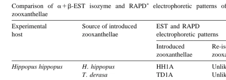
Table 1
a
Comparison of a1b-EST isozyme and RAPD electrophoretic patterns of introduced and re-isolated zooxanthellae
Experimental Source of introduced EST and RAPD
host zooxanthellae electrophoretic patterns
Introduced Re-isolated zooxanthellae zooxanthellae
b Hippopus hippopus H. hippopus HH1A Unlike HH1A
b
T. derasa TD1A Unlike TD1A
b
T. squamosa TS1A Unlike TS1A
b Tridacna gigas T. gigas TG1A Unlike TG1A
c
T. gigas, T. derasa, nd nd
H. hippopus and
H. porcellanus nd nd
a
RAPD primer[18 (GGG CCG TTT A) of primer set[1 from Nucleic Acid / Protein Service Unit of the
University of British Columbia.
b
All 200 re-isolated, then cultured zooxanthellae after 3, 6, and 9 months had patterns different from those of introduced zooxanthellae.
c
nd, no data since introduced zooxanthellae were a mixture of different clonal strains.
offshore, clams infected with homologous CZ remained significantly bigger (4160.8
mm, n5120) than those infected with homologous FIZ or heterologous zooxanthellae,
which by then had comparable sizes (38.060.6 mm, n5240) (Tukey, P,0.05).
3.2. Molecular genetic characterisation of introduced and re-isolated zooxanthellae
All zooxanthellae re-isolated at every stage of the experiments were found to have
a1b-EST isozyme and RAPD band patterns different from those of the clonal cultures
initially introduced to the clam larvae (Table 1). In contrast, the genetic patterns of the introduced zooxanthellae remained unchanged over many culture generations in the laboratory (Fig. 3). Also, the band patterns of the zooxanthellae re-isolates were different within individual hosts, within and among treatments (ca. 99% variation in 200 re-isolates).
4. Discussion
4.1. Genetically-diverse symbiont populations in giant clams
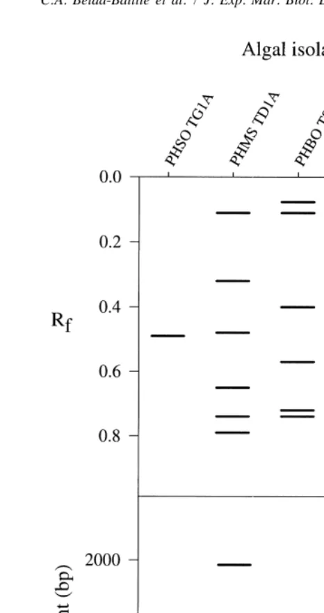
Fig. 3. (top) a1b-EST isozyme and (bottom) RAPD (UBC primer [18) electrophoretic patterns of the
original clonal stocks of zooxanthellae used for the infection trials. Cultured zooxanthellae were originally isolated from T. derasa (PHMS TD1A), T. squamosa (PHBO TS3A), H. hippopus (PHMS HH1A), T. gigas
] ] ]
(PHSO TG1A), and H. porcellanus (PHSU HP1A) (Rf, relative mobility; bp, base pairs).
] ]
of the giant clam family indicate that cultureable zooxanthellae, which belong to Clade A, comprise only a fraction of the total zooxanthellal population (Carlos et al., 1999). Furthermore, zooxanthellae from T. gigas have been previously observed to be predominantly type C with a lesser amount of type A based on RFLP analysis of the 18S rRNA gene (Rowan et al., 1996). This suggests that the symbiont diversity found in this study represents only a subset of the total genetic diversity present in clams and in the natural environment.
The potential sources of such symbiont genetic diversity in clam hosts have been discussed previously (Baillie et al., 1998), and may derive from a number of sources that include acquisition of new genotypes from the water column during filter feeding, mutation during clonal growth, and sexual reproduction. The fact that few, if any, of the zooxanthellal isolates were genetically indistinguishable strongly indicates that clonal growth is not the only process of reproduction in clam zooxanthellae. The high diversity of the zooxanthellae populations may be important in allowing the giant clam association to adapt to changing environmental conditions, including variations in solar radiation, temperature, and nutrients.
4.2. Molecular tools for monitoring of symbiont populations
This study has also demonstrated that allozyme and RAPD electrophoreses are very sensitive techniques that detect individual genetic differences in clam symbionts but not differences between algal taxa. On the other hand, ongoing analysis of the internal transcribed spacer (ITS) region of cultured zooxanthellae from giant clams shows that they all belong to Clade A, distributed into two subgroups that differ from one another by only one nucleotide (B.K. Baillie and T. Maruyama, unpublished data). RFLP and sequence analyses of the 18S rRNA gene have previously been unable to further resolve Clade A of the genus Symbiodinium from clams (Rowan and Powers, 1991a,b; Carlos et al., 1999).
The choice of isozyme and RAPD as genetic markers used in this study was dictated by the types of facilities available at the time. Use of these techniques, in turn, necessitated the culture of a large number of zooxanthellae samples under identical conditions and over several generations for a meaningful comparison of their band
patterns. For these reasons, only a couple of the most useful genetic markers [a1b-EST
and RAPD UBC primer [18 (Baillie et al., 1998)] were used to efficiently check the
4.3. Survival trends in clams versus introduced zooxanthellae
In view of the foregoing discussion, the observed differences in clam growth and survival cannot be attributed directly to the clonal strains of zooxanthellae initially introduced to the aposymbiotic clam larvae. The presence of such significant trends, however, suggests possible differences between subsequently-established dominant symbiont taxa, which were uncultureable or undetectable using the allozyme and RAPD techniques.
Symbiosis between giant clams and zooxanthellae starts soon after metamorphosis of the clam larvae (Fitt, 1985) at 1 to 2 weeks post-spawning, during which heavy mortalities occur (Lucas, 1994). In this study, all clams were maintained undisturbed in
serially-filtered sea water (50, 10, 1 mm) during the first month, after which flow of
unfiltered sea water into the experimental setups was commenced. Previously, Fitt (1985) convincingly demonstrated that all zooxanthellae ‘strains’ from giant clams, sea anemone, and jellyfish, which were offered to Hippopus hippopus and Tridacna
squamosa veliger larvae under controlled laboratory conditions, were observed to pass
through the clams’ digestive gland region, take up residence in the ‘‘haemal sinuses’’ of the metamorphosed clams, and divide for at least 1 week after metamorphosis, indicating healthy algae.
In this study, no attempt was made to re-isolate zooxanthellae from post-metamorphic clams prior to harvest (3 months post-spawning) in order to minimise clam stress and mortalities due to handling. This would ensure that early survival rates (a major variable under investigation) of juveniles that were amenable to harvest after 3 months (3–5 mm shell length) would be unaffected. It may be safe to assume that post-metamorphic clams initially established symbiosis with the introduced zooxanthellae during the first month when ‘wild’ zooxanthellae were excluded. This would be consistent with previous work on giant clams (Fitt, 1985), jellyfishes (Fitt, 1985), and sea anemones (Kinzie and Chee, 1979; Schoenberg and Trench, 1980b; Davy et al., 1997). However, given that the allozyme and RAPD techniques detected individual genetic differences, but not differences between algal taxa, we can only speculate on what happened immediately after initial establishment of symbiosis, and especially so after the clams were exposed to ‘wild’ zooxanthellae at 1-month post-spawning. Furthermore, inasmuch as the established symbionts in the harvested juveniles were found to be different and non-clonal, as opposed to the introduced clonal strains, it matters little that no symbiont re-isolation and re-characterisation were made earlier using the same molecular tools.
At 3, 6, and 9 months post-spawning, all the re-isolated zooxanthellae were found to have genetic patterns different from those of the introduced clonal strains of zoox-anthellae (Table 1). The original stocks of infecting zooxzoox-anthellae, on the other hand, maintained their genetic patterns over many culture generations in the laboratory (Fig. 3). Given that the cultureable zooxanthellae are only a fraction of the total symbiont populations in giant clams (Carlos et al., 1999), it is probably not surprising that the introduced genotypes could no longer be detected after exposure of the juveniles to an alternative source of zooxanthellae.
The significant trends in clam survival and growth are interesting, particularly for T.
higher survival rates and better growth rates than those infected with heterologous zooxanthellae (Fig. 1b). Comparable preliminary results were obtained in big raceways, where T. gigas infected with homologous CZ or FIZ had significantly higher survival and growth rates than those infected with a mixture of heterologous zooxanthellae from
Hippopus spp. (C.A. Belda-Baillie, unpublished data). Previously, it has been shown that
most re-isolated zooxanthellae from T. gigas clustered together as a group based on their allozyme patterns (Baillie et al., 1998) and RAPD patterns (B.K. Baillie, C.A. Belda-Baillie, E.D. Gomez, M. Sison, V. Silvestre and V. Monje, unpublished data). Based on these data, there appears to be some specificity in symbiosis between the largest and fastest-growing T. gigas species and a distinct group or taxon of symbionts in forming a successful symbiotic association. More manipulative studies on symbiosis specificity are clearly desirable.
5. Conclusions
The diversity of symbionts in giant clams raises the question of source of such genetic variation, and whether the same is true for most symbiotic reef organisms. The potential sources of such diversity (see Baillie et al., 1998) need to be elucidated to better understand the dynamics of zooxanthellae populations, and how these symbiotic associations adapt to their changing environment. Indeed, the increasing problem of widespread coral bleaching warrants more investigation on the extent to which zooxanthellae populations are involved in the bleaching phenomena. The giant clam– zooxanthellae symbiosis is a very useful system for investigating the mechanism of bleaching since they also suffer from it (Norton et al., 1995), host similar zooxanthellae taxa as corals (Rowan et al., 1996), are potentially sensitive indicators of disturbances in the environment (see Belda-Baillie et al., 1998), and can supply large quantities of both host tissue and zooxanthellae for analysis.
Acknowledgements
This study was funded by the Philippine Council for Advanced Science and Technology Research and Development, Philippine Council for Aquatic Marine Re-search and Development, and a UPMSI intramural grant (to C.A. Belda-Baillie). Various forms of assistance from the following people are also gratefully acknowledged: R. Pascua, D. Dumaran, R. Abcede, C. Diolazo, and J. Elefante. This publication is UPMSI contribution No. 295.
References
Baillie, B.K., Monje, V., Silvestre, V., Sison, M., Belda-Baillie, C.A., 1998. Allozyme electrophoresis as a tool for distinguishing different zooxanthellae symbiotic with giant clams. Proc. R. Soc. London B 265 (1409), 1949–1956.
Belda, C.A., 1994. Inorganic nitrogen and phosphorus nutrition in tridacnid clams and their algal symbionts. Ph.D. thesis, James Cook University of North Queensland, Australia, 182 pp.
Belda-Baillie, C.A., Leggat, W., Yellowlees, D., 1998. Growth and metabolic responses of the giant clam– zooxanthellae symbiosis in a reef-fertilization experiment. Mar. Ecol. Prog. Ser. 170, 131–141. Belda, C.A., Yellowlees, D., 1995. Phosphate acquisition in the giant clam–zooxanthellae symbiosis. Mar.
Biol. 124, 261–266.
Carlos, A.A., Baillie, B.K., Kawachi, M., Maruyama, T., 1999. Phylogenetic position of Symbiodinium isolates from tridacnids (Bivalvia), cardiids (Bivalvia), a sponge (Porifera), a soft coral (Anthozoa), and a free-living strain. J. Phycol. (in press).
Davy, S.K., Lucas, I.A.N., Turner, J.R., 1997. Uptake and persistence of homologous and heterologous zooxanthellae in the temperate sea anemone Cereus pedunculatus (Pennant). Biol. Bull. 192, 208–216. Fitt, W.K., 1985. Effect of different strains of the zooxanthella Symbiodinium microadriaticum on growth and
survival of their coelenterate and molluscan hosts. Proc. Int. Congr. Coral Reefs 6, 131–136.
Fitt, W.K., Warner, M.E., 1995. Bleaching patterns of four species of Caribbean reef corals. Biol. Bull. 189, 298–307.
Fitt, W.K., Trench, R.K., 1981. Spawning, development, and acquisition of zooxanthellae by Tridacna
squamosa (Mollusca: Bivalvia). Biol. Bull. 161, 213–235.
Freudenthal, H.D., 1962. Symbiodinium gen. nov. and S. microadriaticum sp. nov., a zooxanthella: taxonomy, life cycle, and morphology. J. Protozool. 9, 45–52.
Guillard, R.R.L., Ryther, J.H., 1962. Studies of marine planktonic diatoms. I. Cyclotella nana hustedt, and
Detonula confervacea (cleve) gran. Can. J. Microbiol. 8, 229–239.
Iglesias-Prieto, R., Trench, R.K., 1994. Acclimation and adaptation to irradiance in symbiotic dinoflagellates. I. Responses of the photosynthetic unit to changes in photon flux density. Mar. Ecol. Prog. Ser. 113, 163–175. Kinzie, R.A., Chee, G.S., 1979. The effect of different zooxanthellae on the growth of experimentally
reinfected hosts. Biol. Bull. 156, 315–327.
Laemmli, U.K., 1970. Cleavage of structural proteins during assembly of the head Bacteriophage T4. Nature (London) 227, 680–685.
Lucas, J.S., 1994. The biology, exploitation, and mariculture of giant clams (Tridacnidae). Rev. Fish. Sci. 2, 181–223.
Macaranas, J.M., 1991. A practical laboratory guide to the techniques and methodology of electrophoresis and its application to fisheries management. PCAMRD-DOST Fisheries Technology Manual. No.11, Philip-pines, 47 pp.
Macaranas, J.M., 1993. In: Munro, P. (Ed.), Genetic Aspects of Conservation and Cultivation of Giant Clams. ICLARM Workshop Report, Philippines, pp. 25–32.
Molea, T., Munro, P., 1994. Influence of symbiont strain on early growth of tridacnids. Asian Fish. Sci. 7, 91–102.
Norton, J.H., Prior, H.C., Baillie, B.K., Yellowlees, D., 1995. Atrophy of the zooxanthellal tubular system in bleached giant clams Tridacna gigas. J. Invert. Pathol. 66, 307–310.
Rowan, R., Powers, D.A., 1991a. A molecular genetic classification of zooxanthellae and the evolution of animal–algal symbioses. Science 251, 1384–1391.
Rowan, R., Powers, D.A., 1991b. Molecular genetic identification of symbiotic dinoflagellates (zooxanthellae). Mar. Ecol. Prog. Ser. 71, 65–73.
Rowan, R., Powers, D.A., 1992. Ribosomal RNA sequences and the diversity of symbiotic dinoflagellates (zooxanthellae). Proc. Natl. Acad. Sci. USA 89, 3639–3643.
Rowan, R., Knowlton, N., 1995. Intraspecific diversity and ecological zonation in coral–algal symbiosis. Proc. Natl. Acad. Sci. USA 92, 2850–2853.
Rowan, R., Whitney, S.M., Fowler, A., Yellowlees, D., 1996. Rubisco in marine symbiotic dinoflagellates: form II enzymes in eukaryotic oxygenic phototrophs encoded by a nuclear multigene family. Plant Cell 8, 539–553.
Schoenberg, D.A., Trench, R.K., 1980a. Genetic variation in Symbiodinium (5Gymnodinium) microadriaticum
Schoenberg, D.A., Trench, R.K., 1980b. Genetic variation in Symbiodinium (5Gymnodinium) microadriaticum
Freudenthal, and specificity in its symbionts with marine invertebrates. II. Morphological variation in
Symbiodinium microadriaticum. Proc. R. Soc. London B 207, 429–444.
Trench, R.K., 1987. Dinoflagellates in non-parasitic symbiosis. In: Taylor, F.J.R. (Ed.), The Biology of Dinoflagellates, Blackwell Scientific, Oxford, pp. 530–570.
Underwood, A.J., 1981. Techniques of analysis of variance in experimental marine biology and ecology. Oceanogr. Mar. Biol. A Rev. 19, 513–605.