Actions of Testosterone in Prepubertal and
Postpubertal Male Hamsters: Dissociation of
Effects on Reproductive Behavior and Brain
Androgen Receptor Immunoreactivity
Leslie R. Meek,
1Russell D. Romeo,
Colleen M. Novak, and Cheryl L. Sisk
2Department of Psychology, Neuroscience Program, Michigan State University, East Lansing, Michigan 48824
crease in the number of AR-ir cells per unit area was This study was conducted to determine whether there
found in both the MPN and MPNmag of intact male ham-is a increase in responsiveness to the activating effects
sters. These results indicate that a testosterone-depen-of testosterone on male reproductive behavior during
dent increase in brain AR during puberty may be neces-puberty in male golden hamsters and whether
respon-sary, but is not sufficient, to induce an increase in behav-siveness to behavioral actions of testosterone is
corre-ioral responsiveness to testosterone. q1997 Academic Press
lated with the ability of testosterone to upregulate brain androgen receptor immunoreactivity (AR-ir). Sexually naive male hamsters were castrated at 21 or 42 days of age and implanted subcutaneously with a pellet
con-taining 0, 2.5, or 5 mg of testosterone. One week later, One hallmark of puberty in males is the appearance males were given a 10-min mating test with a receptive of steroid-dependent reproductive behaviors. Clearly, female. Animals were euthanized 1 hr after the behav- one factor contributing to this behavioral maturation is ioral test, and blood samples and brains were collected. the pubertal rise in testosterone secretion from the tes-Plasma testosterone levels were equivalent in
prepuber-tes. However, an increase in responsiveness of the
ner-tal and adult males that had been administered the same
vous system to the actions of testosterone appears to be
dose of testosterone. However, adult males exhibited
a second mechanism involved in pubertal maturation of
more mounts, intromissions, and ejaculations than
pre-sexual behavior. Evidence for a pubertal increase in
pubertal males, demonstrating that postpubertal males
responsiveness to the behavioral actions of steroid
hor-are more responsive than prepubertal males to the
ef-mones comes from studies demonstrating that even
fects of testosterone on sexual behavior. In both age
groups, testosterone increased the number of AR-ir cells when juvenile males are treated with doses of testoster-per unit area in several brain regions involved in male one that activate male reproductive behavior in adults, sexual behavior, including the medial preoptic nucleus they do not express equivalent levels of male reproduc-(MPN), medial amygdala, posteromedial bed nucleus of tive behavior (Baum, 1972; Larsson, 1967; Sisk, Berg-the stria terminalis, and magnocellular preoptic nucleus
lund, Tang, and Venier, 1992; Sodersten, Damassa, and
(MPNmag). Surprisingly, testosterone increased AR-ir in
Smith, 1977). The cellular mechanisms contributing to
the latter three regions to a greater extent in prepubertal
the pubertal increase in behavioral responsiveness to
males than in adults. Thus, prepubertal males are more
testosterone are not known.
responsive to the effects of testosterone on AR-ir in
Although testosterone is the predominant circulating
these regions. In a separate experiment, a pubertal
in-steroid hormone in adult male rodents, activation of many components of male reproductive behavior is due 1Present address Division of Social Science, 109 Camden Bldg., 600
to the action of estradiol, which is formed locally in the
E. 4th St., Univ. Minnesota Morris, Morris, MN 56267.
brain by the aromatization of testosterone. However, 2To whom correspondence should be addressed at Department of
several lines of evidence indicate that testosterone or
Psychology, Neuroscience Program, Michigan State University, East
Lansing, MI 48824. Fax: (517) 353-1652. E-mail: [email protected]. an androgenic metabolite acts in concert with estrogen
75
0018-506X/97 $25.00
to stimulate the full repertoire of male reproductive whether there is a pubertal increase in brain AR-ir; in this experiment, the immunocytochemical protocol was behavior (reviewed in Meisel and Sachs, 1994). In
addi-tion, androgens play an indirect role in estrogenic acti- modified from that used in Experiment 1. vation of behavior, because activity of the aromatase
enzyme in the preoptic area, a known target site for
Subjects and Treatment
steroid activation of male reproductive behavior, is
reg-ulated by androgens (Roselli and Resko, 1984). Cellular Male golden hamsters (Mesocricetus auratus) were bred at Michigan State University (E. Lansing, MI). All responses to steroid hormones are mediated by specific
steroid receptors that, when activated by a ligand, alter animals were housed in clear polycarbonate cages (37.5 133117 cm) with wood chips (Aspen Chip Labora-transcription of steroid-regulated genes within target
cells. Thus, regulation of the abundance of androgen tory Bedding, Warrensburg, NY) and bedding (cotton upholstery batting). Following weaning at 21 days of receptor within the neural circuitry mediating male
re-productive behavior is a potential mechanism by which age, animals were housed in same-sex sibling groups until treatment, at which time they were singly housed. behavioral responsiveness to testosterone could be
modulated during puberty. Room temperature was maintained at 21{27C and the
light–dark schedule was 14 hr light/10 hr dark (lights In male ferrets, a pubertal increase in androgen
re-ceptor immunoreactivity (AR-ir) occurs within two on at 2300 hr EST). Food (Teklad Rodent Diet No. 8640, Harlan, Madison, WI) and water were available brain areas that are components of the neural circuit
mediating male reproductive behavior, the medial pre- ad libitum.
Experiment 1. Hamsters were castrated under me-optic area and the medial amygdala (Kashon and Sisk,
1995; Kashon and Sisk, 1994). Furthermore, administra- thoxyflurane anesthesia at either 21 or 42 days of age and were implanted with a 3-week timed-release pellet tion of a large dose of testosterone to prepubertal ferrets
increases AR-ir in these brain areas (Kashon, Hayes, (Innovative Research of America, Sarasota, FL) con-taining either 0, 2.5, or 5 mg of testosterone (nÅ 5–6 Shek, and Sisk, 1995). These findings raise the
interest-ing possibility that the pubertal rise in testosterone may per age and treatment group). Two additional groups of hamsters received a sham castration and a 0-mg pellet contribute to the pubertal increase in behavioral
re-sponsiveness to testosterone by upregulating androgen implant at either 21 (n Å6) or 42 (nÅ6) days of age. One week following treatment (either 28 or 49 days of receptor in relevant brain regions.
The present experiments were conducted to investi- age), all subjects were paired with a hormone-primed estrous adult female for a 10-min mating behavior test gate further the relationship between pubertal
matura-tion of male reproductive behaviors and pubertal regu- (see below). One hour after the onset of the mating test, hamsters were euthanized and perfused as described lation of androgen receptor by testosterone, using the
male golden hamster as an animal model. The experi- below.
Experiment 2. Twenty-one-day-old (nÅ3), 28-day-ments were designed to determine: (1) whether there
is a pubertal increase in responsiveness to behavioral old (nÅ6), and 49-day-old (nÅ6) intact male hamsters were used in this experiment. These animals received actions of testosterone in the golden hamster; (2) if there
is a pubertal increase in AR-ir within the neural circuit no experimental manipulation or behavioral test, but were sacrificed at approximately the same time of day known to mediate sexual behavior in male hamsters
and, if so, whether testosterone upregulates AR-ir in as were animals in Experiment 1 as described below. the same brain regions; and (3) whether responsiveness
to behavioral actions of testosterone is correlated with
Tests for Male Reproductive Behavior
the ability of testosterone to upregulate brain AR-ir in
(Experiment 1 only)
male hamsters.
Immediately prior to the onset of the dark portion of the light/dark schedule, hamsters were moved in their home cages to a testing room. All behavioral tests
oc-METHODS
curred between 10 min and 3 hr after lights out. The male was placed in a clean 10-gal. aquarium (51 126 Two experiments were conducted. In Experiment 1,
the effects of varying doses of testosterone on male 131.5 cm) with no bedding and was allowed 10 min to acclimate to the novel testing environment before the reproductive behavior and on brain AR-ir were
sequential injections of estradiol benzoate (10mg in 0.1 groups within an experiment was processed simultane-ously. In Experiment 1, the following protocol was ml sesame oil, sc, 48 hr prior to testing) and
progester-one (0.1 mg in 0.1 ml sesame oil, sc, 4 hr prior to testing). used. Sections were rinsed 101 in 0.1 M phosphate-buffered saline to remove the cryoprotectant. Sections The behavior tests were videotaped under red light
(three 25-W bulbs) with a Panasonic Color Video Cam- were then incubated sequentially in 0.1 M glycine in 0.1 M PBS (30 min), 0.3% H2O2 in PBS (10 min), 4%
era (WV 3250). The testing aquarium was mounted on
a stand over a slanted mirror, which provided a ventral normal goat serum (Vectastain ABC Elite kit, Burlin-game, CA) in 0.3% Triton X-100 in PBS (PBS-TX; 1 hr), view of the animals so that intromissions and
ejacula-tions were clearly visible. Videotapes were later scored and 0.5 mg/ml rabbit anti-AR in PBS-TX (PG-21-18A, obtained from G. S. Prins, Michael Reese Hospital, Chi-to determine the amount of time spent in anogenital
investigation of the female, the frequency of vaginally cago, IL; 48 hr). Sections were then incubated in second-ary antibody (goat anti-rabbit immunoglobulins, Vec-oriented mounts, intromissions, and ejaculations.
Anogenital investigation was scored when the male tastain ABC Elite kit, 1:200 in PBS-TX; 24 hr), followed by incubation in avidin–biotin–HRP complex (Vecta-investigated the perianal region of the female, including
the flanks. A vaginal mount was scored when the male stain ABC Elite kit, 1:50 in PBS-TX; 2 hr). For the chro-mogen reaction, sections were incubated for 6 min in mounted with an orientation that would permit
intro-mission. An intromission was scored if the male 1% 3,3*-diaminobenzadine (DAB) containing 2% b
-D-(/)-glucose, 0.04% NH4Cl, 0.038% imidazole, 50 ml/ml
achieved vaginal penetration. An ejaculation was
scored if, after a series of intromissions separated by 250 mM NiCl2, and 0.0075% H2O2in 0.05 M
Tris-buf-fered saline (TBS). Sections were rinsed 3 times in PBS short inter-intromission intervals, the male became
tem-porarily uninterested in the female (20 sec–1 min) and between incubations in each reagent. All incubations were at room temperature except for that with primary engaged in extensive genital grooming. We had
pre-viously determined that this pattern of behavior was antiserum, which was at 47C. After the DAB reaction, all
sections were rinsed 5 times in distilled water, mounted always correlated with sperm present in the vagina of
the female and that sperm were not present when this onto gelatin-coated slides, dried, dehydrated in increas-ing concentrations of alcohols, cleared in Hemo-De, and behavior pattern was not observed.
coverslipped. For Experiment 2, the
immunocytochem-Blood and Tissue Collection istry protocol was the same, except that the
concentra-tions for primary antiserum, NiCl2, and H2O2were
re-Hamsters were euthanized with an overdose of
Equithesin anesthetic (8 ml/kg, ip) and a 0.5-ml blood duced to 0.25 mg/ml, 125 mM,and 0.00375%, respec-tively. Processing tissue in the absence of the primary sample was obtained via cardiac puncture. Blood
sam-ples were centrifuged and plasma was removed and antiserum resulted in no detectable immunostaining by the secondary antibody.
stored at 0207C until radioimmunoassay was
per-formed. Body weight and flank gland length were re-corded, and the seminal vesicles were removed and
Analysis of Androgen Receptor Immunoreactivity
weighed after expression of the seminal fluid. Animals
were perfused intracardially with 150 ml of Sorenson’s The areal density (cells per unit area) of AR-ir cell profiles (cross sections) was quantified in the medial phosphate buffer (pH 7.4) containing 0.8% NaCl, 0.8%
sucrose, 0.4% b-D-(/)-glucose and 3 IU/ml heparin, preoptic nucleus (MPN), medial amygdala (MeAMY),
lateral septum (LSept), bed nucleus of the stria termi-followed by 250 ml of ice-cold fixative containing 4%
paraformaldehyde in 0.1Mphosphate buffer (pH 7.5). nalis, posteromedial subdivision (BNSTpm), and in the magnocellular region of the medial preoptic nucleus Brains were removed, postfixed in 4%
paraformalde-hyde for 4 hr, and then stored in 20% sucrose in phos- (MPNmag). These brain regions were selected for anal-ysis because they are components of the steroid-sensi-phate-buffered saline (PBS; 0.9% saline) at 47C until
sec-tioning approximately 48 hr later. Brains were cut into tive neural circuit controlling male reproductive behav-iors (Baum, Tobet, Starr, and Bradshaw, 1982; Lehman 40-mm-thick sections that were stored in a
polyethyl-ene-based cryoprotectant at 0207C until immunocyto- and Winans, 1982; Powers, Newman, and Bergondy,
1987; Wood and Newman, 1993b). Brain sections were chemistry was performed.
inspected under bright-field microscopy and regions
Androgen Receptor Immunocytochemistry were located according to Kollack and Newman (1992).
Two sections through each brain nucleus, separated by Every third section from each brain was used for
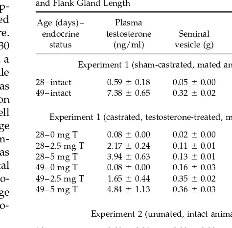
TABLE 1 were used for analysis. Each brain nucleus was centered
Mean Plasma Testosterone Concentration, Seminal Vesicle Weight,
in the field of view at 101, and magnification was then
and Flank Gland Length
increased to 601. The microscopic images were
cap-tured by a video camera (Sony XC-77) and displayed Age (days) – Plasma
on a computer monitor using NIH Image 1.56 software. endocrine testosterone Seminal Flank gland
status (ng/ml) vesicle (g) (mm)
These images, which represented an area of 100 by 130
mm at 60X, were then sharpened and printed out on a Experiment 1 (sham-castrated, mated animals) laser printer (Hewlett–Packard 5MP). All AR-ir profile
28 – intact 0.59{0.18 0.05{0.00 5.15{1.06
counts were made from these printouts. A profile was
49 – intact 7.38{0.65 0.32{0.02 8.00{0.22
considered immunopositive if blue–black reaction
product was visible and of the shape and size of a cell Experiment 1 (castrated, testosterone-treated, mated animals) nucleus. For each brain region examined, the average
28 – 0 mg T 0.08{0.00 0.02{0.00 3.64{0.95
number of immunopositive nuclear profiles in four
im-28 – 2.5 mg T 2.17{0.24 0.11{0.01 3.82{0.16
ages (bilateral counts in each of two tissue sections) was 28 – 5 mg T 3.94{0.63 0.13{0.01 3.22{1.48 computed for each animal, and data for experimental 49 – 0 mg T 0.08{0.00 0.16{0.03 6.97{0.89
49 – 2.5 mg T 1.65{0.44 0.35{0.02 8.46{0.52
groups were expressed as mean number of AR-ir
pro-49 – 5 mg T 4.84{1.13 0.36{0.03 7.94{0.23
files/13,000mm2. A single experimenter blind to the age
and treatment of the animals performed the
micro-Experiment 2 (unmated, intact animals)
scopic analysis.
21 – intact 0.11{0.01 0.01{0.00 Not measurable
28 – intact 0.34{0.17 0.03{0.00 6.82{0.52
49 – intact 5.54{0.89 0.37{0.03 8.42{0.45
Testosterone Radioimmunoassay
Plasma concentrations of testosterone were measured
RESULTS
with reagents in the Coat-A-Count Total Testosteronekit (Diagnostic Products, Los Angeles, CA) in two
dif-Experiment 1: Dose –Response Curves to ferent assays. The lower limits of detectability of the
Testosterone in Prepubertal and Adult Males assays were 0.1 and 0.08 ng/ml, and the intraassay
coef-ficients of variation (CV) were 5.5 and 6.7%,
respec-Behavior and Peripheral Measures
tively. The interassay CV was 6.1%.
Sham-castrated, mated animals. Sham-castrated adults had significantly higher levels of circulating T than the prepubertal sham castrates (tÅ100.188,Põ
Statistical Analysis 0.05, Table 1). Furthermore, seminal vesicle weights
were heavier (tÅ349.212,Põ0.05), and flank glands In Experiment 1,ttests were used to compare behav- were longer (t Å 6.877, P õ 0.05), in adults than in ioral, physiological, and anatomical measures in pre- prepubertal animals (Table 1).
vs postpubertal sham castrates. For testosterone-treated Prepubertal and adult sham-castrates did not differ castrated males, the effects of age and treatment on in the amount of time spent in anogenital investigation plasma testosterone concentrations and behavioral of the female (Table 2). Adult males exhibited signifi-variables were analyzed by two-way ANOVAs (dose cantly more vaginal mounts than their prepubertal by age). Significant interactions were further analyzed counterparts, who never displayed any mounting be-with Tukey HSD tests, and pairwise comparisons of havior (t Å 5.177, P õ 0.05, Table 2). There were no main effects were made with Fisher’s PLSD tests. Group significant differences between adult and juvenile differences in the percentage of animals displaying at males in the number of intromissions performed (Table least one occurrence of a behavior were analyzed byx2
2). However, no prepubertal males intromitted, tests. In Experiment 2, one-way ANOVAs orttests were whereas one-third of the adults displayed one or more used to examine the effect of age on physiological vari- intromissions (x2
Å 39.5, P õ 0.05). No ejaculations ables and AR-ir profile density in intact male hamsters. were observed in either group of animals.
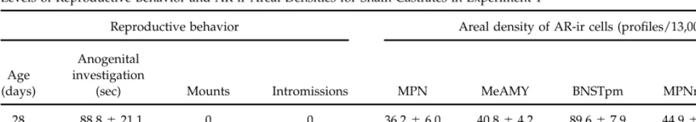
TABLE 2
Levels of Reproductive Behavior and AR-ir Areal Densities for Sham Castrates in Experiment 1
Reproductive behavior Areal density of AR-ir cells (profiles/13,000mm2)
Anogenital
Age investigation
(days) (sec) Mounts Intromissions MPN MeAMY BNSTpm MPNmag LSept
28 88.8{21.1 0 0 36.2{6.0 40.8{4.2 89.6{7.9 44.9{3.5 68.0{6.1
49 155.8{22.9 3.2{1.5* 1.0{0.68 39.1{5.3 46.8{6.1 81.9{4.2 51.1{2.4 59.8{5.9
* Significantly different from 28-day-old males.
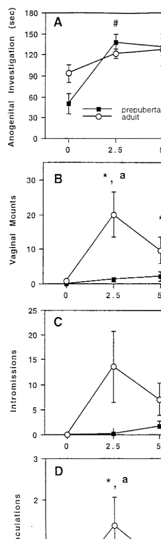
culating plasma testosterone levels (F Å 23.918, P õ between age and testosterone treatment on the number of ejaculations (FÅ3.295,Põ0.05, Fig. 1D). The adults 0.05; Table 1). Castrates that received the 2.5-mg pellet
had higher plasma testosterone levels than animals im- that were implanted with the 2.5-mg testosterone pellet engaged in a greater number of ejaculations than the planted with the blank pellet, in which plasma
testos-terone was undetectable. Similarly, castrates that re- adults that had received either the 0- or 5-mg testoster-one pellet (Põ0.05). This effect of testosterone on ejacu-ceived a 5-mg pellet had higher plasma testosterone
levels than groups that received either the blank or the lation was not observed in the juvenile animals. Adults displayed more ejaculations at the 2.5-mg testosterone 2.5-mg pellet. No interaction existed between age and
treatment on plasma testosterone; that is, testosterone dose than juvenile males receiving that dose of testos-terone.
pellets of a given dose produced similar levels of circu-lating testosterone in castrated males regardless of age. Testosterone treatment significantly affected the
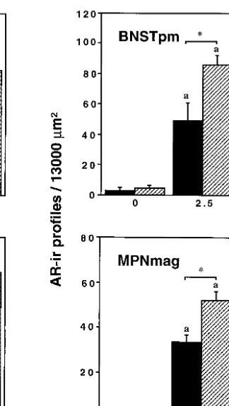
du-Androgen Receptor Immunoreactivity
ration of anogenital investigation (FÅ7.998,Põ0.05,
Fig. 1A), but there was neither an effect of age nor Sham-castrated, mated animals. There were no significant differences in the number of AR-ir profiles an interaction between testosterone treatment and age.
Fisher’s PLSD revealed that animals treated with either per unit area between juvenile and adult sham castrates in any of the brain regions examined (Table 2). the 2.5- or 5-mg testosterone pellet displayed
signifi-cantly more anogenital investigation than animals Testosterone-treated castrates. Two-way ANO-VAs revealed significant main effects of testosterone treated with the blank pellet, regardless of age.
There was a significant interaction between age and dose on the number of AR-ir profiles/13,000mm2in the
MPN and LSept (FÅ31.599,F Å31.814, respectively, testosterone treatment on the frequency of vaginal
mounts (F Å4.314,Põ 0.05, Fig. 1B). In adults, both bothPõ0.05), indicating similar effects of testosterone in both age groups. In both brain regions, the areal the 2.5- and 5-mg doses of testosterone elicited more
mounts compared with adults receiving a blank pellet. density of AR-ir profiles was higher in animals treated with either the 2.5- or 5-mg testosterone pellet than in This effect of testosterone on vaginal mounts was not
observed in the prepubertal animals. Adults engaged the groups treated with the blank pellet, and areal den-sity of AR-ir profiles did not differ between animals in more vaginal mounting at the 2.5-mg testosterone
dose compared with their juvenile counterparts. treated with either 2.5 or 5 mg of testosterone (Fig. 2). In the MeAMY, there were significant main effects of Age significantly affected the frequency of
intromis-sions (F Å6.218, Põ 0.05, Fig. 1C) such that overall, both age (F Å4.652, P õ 0.05) and testosterone (F Å 36.328,Põ0.05), but no interaction (Fig. 2). Thus, col-adults engaged in more intromissions than prepubertal
animals. Although there was no interaction between lapsed across doses of testosterone, the number of AR-ir profiles/13,000mm2was greater in juveniles than in
testosterone treatment and age on the frequency of
oc-currence of intromissions, only one prepubertal animal adults. Similarly, across both age groups, the areal den-sity of AR-ir profiles was significantly increased with receiving the 2.5-mg testosterone dose displayed one
intromission, whereas half of the adults treated with each increasing dose of tesoterone.
Two-way ANOVAs revealed significant interactions this dose displayed multiple intromissions (x2
Å26.12,
Põ0.05). between testosterone treatment and age on the number
of AR-ir profiles/13,000mm2in both the BNSTpm and
MPNmag (F Å 4.30 and F Å4.097, respectively, P õ 0.05). For both age groups, testosterone treatment in-creased the areal density of AR-ir profiles in both the BNSTpm and MPNmag (Fig. 2). However, the increase in the number of AR-ir profiles/13,000 mm2 was of a
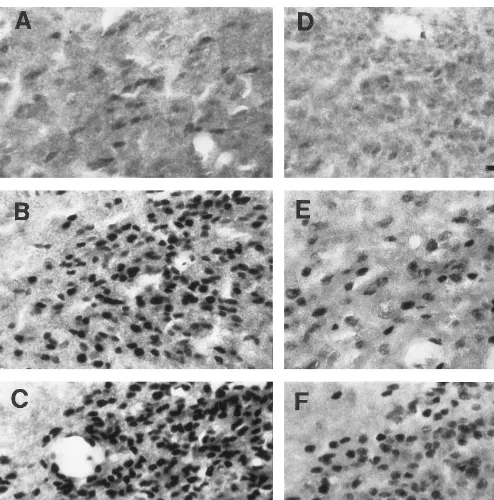
greater magnitude in juvenile males than in adults at both the 2.5- and 5-mg doses of testosterone. Photomi-crographs illustrating the effects of testosterone on the areal density of AR-ir profiles in the BNSTpm at the two ages are shown in Fig. 3.
Experiment 2: Pubertal Development of Brain Androgen Receptor Immunoreactivity
One-way ANOVAs and subsequent posthoc tests re-vealed that 49-day-old intact males had higher plasma testosterone levels (FÅ29.771,Põ0.05), seminal vesi-cle weights (F Å 123.22, P õ 0.05), and flank gland lengths (FÅ75.676,Põ0.05) than either the 28- or 21-day-old intact males (Table 1). In addition, 28-21-day-old males had significantly longer flank glands than 21-day-old males (Põ0.05).
Analysis of AR-ir profiles in BNSTpm and MPNmag could not be performed in 21-day-old males due to poor tissue quality of sections containing these regions. In all brain regions examined, there was a trend toward an increased number of AR-ir profiles/13,000mm2with
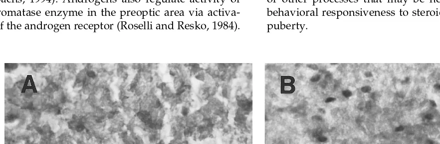
increasing age (Fig. 4). However, in just two brain re-gions was there a statistically significant effect of age. Within MPN, areal density of AR-ir profiles was greater in 49-day-old compared with either the 28- or 21-day-old animals (F Å 11.627, P õ 0.05). In MPNmag, the number of AR-ir profiles/13,000mm2was greater in
49-day-old males than in 28-49-day-old males (t Å 5.224, P
õ0.05). Figure 5 shows representative examples of AR immunopositive nuclei in the MPN of juvenile (28-day-old) and adult (49-day-(28-day-old) males.
DISCUSSION
Relationship between Behavioral Responsiveness and AR-ir
The behavioral dose response curves to testosterone for prepubertal and postpubertal hamsters in Experi-FIG. 1. Number of seconds engaged in anogenital investigation (A),
number of vaginal mounts (B), number of intromissions (C), and num-ber of ejaculations (D) in prepunum-bertal and adult male hamsters given a 10-min test with a sexually receptive female. Males were castrated
1 week prior to testing and were implanted with a pellet containing nificantly different from 0 mg testosterone for adults only. ‘‘a’’ indi-cates a significant difference between age groups for a given dose of 0, 2.5, or 5 mg of testosterone. ‘‘#’’ indicates significantly different
FIG. 2. AR-ir profiles/13000mm2in the MPN, LSept, MeAMY, BNSTpm, and MPNmag of prepubertal (28 days old) and adult (49 days old)
male hamsters that were castrated 1 week prior to sacrifice and received a pellet containing 0, 2.5, or 5 mg of testosterone. ‘‘a’’ indicates a significant difference from 0 mg testosterone within an age group. ‘‘b’’ indicates a significant difference from 2.5 mg testosterone within an age group. Asterisk indicates a significant difference between ages within a dose of testosterone. All values are means{SEM.
ment 1 provide clear evidence that prepubertal males mediating male sexual behavior (MPN and MPNmag; Experiment 2). In addition, testosterone can increase are less sensitive and less responsive to the effects of
testosterone on male reproductive behavior than are AR-ir in these same brain regions (Experiment 1), sug-gesting that the pubertal rise in testosterone upregu-postpubertal males. Doses of exogenous testosterone
that were effective in activating male reproductive be- lates AR within the behavioral circuitry and in this way may consequently contribute to the pubertal increase havior in adults elicited little or no behavior in
juve-niles, even though plasma concentrations of testoster- in responsiveness to behavioral actions of testosterone. However, Experiment 1 shows that an increase in AR-one were equivalent in the two age groups. In intact
FIG. 3. Photomicrographs of AR-ir cells in the BNSTpm of testosterone-treated prepubertal and postpubertal male hamsters. (A – C) Prepubertal males that received testosterone pellets of 0, 2.5, and 5 mg, respectively. (D – F) Adults that received testosterone pellets of 0, 2.5, and 5 mg, respectively. Note that the area of each of these photographs is larger than the area used for quantifying areal density (36,000 vs 13,000mm2
). Bar, 50mm.
behavioral responsiveness. In all brain regions exam- AR-ir responses to testosterone in the same individual, the present experiments extend earlier investigations of ined, the AR-ir response to testosterone in prepubertal
FIG. 4. AR-ir profiles/13000mm2
in the MPN, MeAmy, BNSTpm, MPNmag, and LSept of 21-day-old (prepubertal), 28-day-old (prepubertal), and 49-day-old (adult) male hamsters. Asterisk indicates a significant difference from other age groups for a given brain region. Tissue sections through BNSTpm and MPNmag were not available for 21-day-old males. All values are means{SEM.
Sisket al.,1992) and clearly do not support the hypothe- Thus, it is possible that testosterone is more effective in increasing aromatase activity in adults due to pubertal sis that lack of behavioral responsiveness to
testoster-one is due to a relative inability of testostertestoster-one to upreg- changes in mechanisms that couple AR with the aromatase enzyme. Alternatively, a pubertal increase ulate AR in the behavioral neural circuitry of
prepuber-tal males. in estrogen receptor may be an important component of
the neural mechanism underlying enhanced behavioral It appears, then, that other pubertal processes,
per-haps in concert with a testosterone-induced increase responsiveness in adult males. In any case, the pattern and/or duration of testosterone treatment given to pre-in AR, cause steroid hormones to be more effective pre-in
activating male reproductive behavior. Androgens act pubertal males in the present study apparently did not provide the appropriate stimulus for increased together with aromatized estrogenic metabolites to
acti-vate male reproductive behavior (reviewed in Meisel aromatase activity, upregulation of estrogen receptor, or other processes that may be necessary to increase and Sachs, 1994). Androgens also regulate activity of
the aromatase enzyme in the preoptic area via activa- behavioral responsiveness to steroid hormones during puberty.
tion of the androgen receptor (Roselli and Resko, 1984).
Testosterone-independent pubertal processes may 1983). The robust AR-ir response to testosterone within BNSTpm, MPNmag, and MeAMY in prepubertal males also contribute to increased behavioral responsiveness
to steroid hormones. Chemosensory cues from the fe- may therefore be related to the fact that anogenital in-vestigation was the only component of male sexual be-male are an important factor in the activation of be-male
reproductive behavior in hamsters. The salience of havior examined in this study that was activated equally by testosterone in prepubertal and postpubertal these cues or the way in which the information they
contain is integrated with steroid-dependent cellular males.
Alternatively, the underlying assumption that in-processes may change during pubertal maturation.
Thus, age-dependent changes in the processing of che- creases in steroid hormone receptor lead to increases in behavioral responses may need to be reevaluated. mosensory or somatosensory information may also be
an important component of the maturation of male re- For example, if AR-mediated cellular responses were in some way inhibitory to mounting, then enhanced productive behavior.
One obvious difference between juveniles and adults AR responsiveness to testosterone in BNSTpm, MPNmag, or MeAMY could underlie lack of activation in the present study was the length of time that their
nervous systems had been exposed to endogenous tes- of mounting by testosterone in prepubertal males. Fi-nally, it is worth noting that prepubertal hamsters are tosterone prior to treatment. However, in male rats,
1 week of treatment with subcutaneous capsules that more responsive to steroid inhibition of gonadotropin secretion compared with adult males (Sisk and Turek, produced circulating levels of 2–5 ng/ml was sufficient
to activate sexual behavior in adults that had been cas- 1983). If target cells for steroid-negative feedback regu-lation of gonadotropin secretion are intermingled with trated 3–4 weeks earlier (McGinnis, Mirth, Zebrowski,
and Dreifuss, 1989). Thus, 1 week of testosterone treat- target cells for steroid activation of behavior, it is possi-ble that the enhanced AR-ir response to testosterone in ment should have resulted in expression of male
repro-ductive behavior in juveniles if their nervous systems one or more of these three regions is related to en-hanced responsiveness to steroid negative feedback in were capable of adult-like responses. It also does not
appear that differential treatment of juvenile and adult juvenile males.
The dissociation between testosterone’s effects on males by the stimulus females can explain the age
dif-ference in responsiveness to testosterone. Regardless of sexual behavior and on AR-ir is also apparent in adult hamsters. In adults, the 5-mg dose of testosterone was the age of the male, the latency for the female to assume
the lordosis posture was less than 30 sec, and the aver- required to restore AR-ir to levels observed in age-matched sham castrates, yet full restoration of sexual age amount of time the female spent in lordosis during
the 10-min test was not different for juvenile and adult behavior was accomplished by the 2.5-mg dose in these same animals. In addition, the AR-ir response to testos-males. Adult female hamsters are generally larger than
adult males, and of course the size difference is exag- terone was generally graded in adults, i.e., AR-ir in-creased with increasing dose of testosterone, while a gerated when the female is paired with a juvenile male.
However, it seems unlikely that a body size difference maximal behavioral response was observed at the low-est dose administered. In these respects, the relation-was a major factor in the relative lack of occurrence of
mounts and intromissions in juvenile males because ship between testosterone and AR-ir in adult hamsters appears to be more like that between testosterone and some juveniles were able to perform these behaviors.
The fact that the AR-ir responses to testosterone in aggressive behavior rather than between testosterone and sexual behavior. Albert, Jonik, Watson, Gorzalka, BNSTpm, MPNmag, and MeAMY of prepubertal males
were of greater magnitude than those of postpubertal and Walsh (1990) compared dose response curves to testosterone for activation of sexual and aggressive be-males is surprising and intriguing. In all three of these
brain regions, the immediate early gene product fos is haviors in adult male rats and found that extremely low levels of testosterone were sufficient to fully acti-expressed after a male hamster is exposed to female
hamster vaginal pheromones (Fernandez-Fewell and vate sexual behavior, while aggressive behaviors were proportional to the dose of testosterone.
Meredith, 1994; Fiber, Adames, and Swann, 1993). In addition, interest in female odors is a behavior that appears to be regulated by testosterone directly (not
Methodological Considerations
via estrogen; Powers and Bergondy, 1983) and that can
be activated in both prepubertal males and adult fe- These experiments demonstrate that modification of immunocytochemical protocol can be used to reveal males by testosterone (unlike full copulatory behavior;
Experi-ment 1, no effect of age was found in the areal density The mechanism by which testosterone increases AR-of AR-ir prAR-ofiles in any brain region examined. This ir is not known, but it is probably a combination of at result was initially perplexing, because earlier pilot least two separate processes. First is a relatively rapid work had indicated that brain AR-ir in adult males was ligand-induced translocation of AR to the nucleus, fol-greater than that in prepubertal males, a finding that lowed by a second and more protracted response that in fact had provided the impetus to conduct Experiment results in a greater abundance of intracellular AR, 1. In the interim between the pilot experiment and Ex- through either an increase in AR protein synthesis or periment 1, however, immunocytochemical conditions an increase in the half-life of AR. Several laboratories had been changed to optimize AR immunostaining by have documented that testosterone induces an increase increasing the concentrations of primary antibody, in AR-ir within 15–30 min (Prins and Birch, 1993; Sar, NiCl2, and H2O2. Thus, this optimization could obscure Lubahn, French, and Wilson, 1990; Zhou, Blaustein, and
age differences in AR-ir cell numbers by increasing the De Vries, 1994), which is too rapid to reflect an increase sensitivity for detection of AR immunopositive nuclei. inde novosynthesis of AR. However, longer term treat-Experimental manipulation of primary antibody con- ment with testosterone increases AR-ir even further centration has been used to reveal changes in fos ex- (Kashon, Arbogast, and Sisk, 1996), and androgens ad-pression in neurons after steroid treatment (Auger and ministered to gonad-intact male rats increase AR-ir Blaustein, 1995). Therefore, in Experiment 2, lower con- (Menard and Harlan, 1993). This latter finding cannot centrations of primary antibody, as well as NiCl2 and be explained on the basis of ligand-induced
transloca-H2O2, were used. Under these conditions, a pubertal tion of AR to the nucleus, because endogenous
andro-increase in areal density of AR-ir profiles was observed gens are present and receptor is presumably already in certain brain areas. We conclude that the immunocy- within the nucleus. The ability of testosterone to in-tochemical conditions used in Experiment 1 were sensi- crease AR protein half-life (Kemppainen, Lane, Sar, and tive enough to stain cells that express even very low Wilson, 1992; Syms, Norris, Panko, and Smith, 1985) levels of AR so that the number of AR-ir profiles per or AR mRNA or protein (Gonzalez-Cadavid, Vernet, unit area was not different in pre- and postpubertal Navarro, Rodriguez, Swerdloff, and Rajfer, 1993; Kerr, males. In contrast, the conditions used in Experiment Allore, Beck, and Handa, 1995; Shan, Hardy, Catterall, 2 were such that cells expressing low levels of AR and Hardy, 1995) has been documented under certain would not be detectable, and thus only cells with a conditions in androgen-sensitive peripheral tissues and relatively high abundance of AR were stained. Thus, it brain. The immunocytochemical data indicating that appears that the pubertal increase in AR-ir in Experi- testosterone can upregulate the AR protein in brain are ment 2 does not reflect a pubertal change in cell
pheno-corroborated by androgen binding experiments, in type, but instead reflects an increase in AR expression
which nuclear AR concentration in brain homogenates within cells that, prior to puberty, expressed relatively
is increased by treatment with androgen at time points little AR.
later than that required for saturation of the androgen Another difference between the sham castrates in
Ex-receptor (Bittman and Krey, 1988; Krey and McGinnis, periment 1 and the intact males in Experiment 2 was
1990; Prins, Bartke, and Steger, 1990). Thus, we believe the occurrence of a mating test 1 hr prior to sacrifice
the effects of testosterone on brain AR-ir in the present (Experiment 1) or not (Experiment 2). It is possible that
study reflect true increases in intracellular levels of AR. some aspect of the mating experience, such as exposure
In all groups of males in Experiment 1, the frequency to the female or to her scent, may rapidly increase
tes-of occurrence tes-of sexual behaviors was relatively low, tosterone and consequently AR-ir. However, the
differ-and within-group variability on the behavioral mea-ence in plasma testosterone levels between gonad-intact
sures was relatively high. The most likely explanation prepubertal males in Experiments 1 and 2 was not large,
for these characteristics of the data is that all animals and it is more likely that the different
immunocyto-were sexually naive prior to the behavioral tests. Be-chemical conditions are responsible for the differences
cause the effect of sexual experience on responsiveness in the number of AR-ir profiles observed in the two
to testosterone is unclear, and experience could not be experiments. Finally, the more sensitive
immunocyto-easily equated in juvenile and adult males, we elected chemical protocol used in Experiment 1 did not
pre-to use sexually inexperienced animals for this study. clude an ability to detect group differences in the
num-Similarly, it is virtually impossible to equate general ber of AR-ir profiles, as there were clear effects of
social experience in juvenile and adult males because castration, testosterone, and age by testosterone
interac-adults by definition will have lived longer under partic-tions on the areal density of AR-ir profiles in
were housed alone for 1 week prior to behavioral test- ir in the preoptic area. The factors that contribute to brain region-specific effects of androgen on AR-ir under ing, but prior to that, prepubertal males were housed
with their mothers and littermates, whereas adults were physiological conditions are unknown, but undoubt-edly include parameters of hormone secretion and cell housed from weaning with same-sex siblings. Although
this difference in social experience could conceivably group differences in afferent input and neurochemical phenotype.
contribute to a difference in responsiveness to
testoster-one between the two age groups, other data from this Adult golden hamsters are seasonal breeders and re-productive competence is influenced by photoperiod. laboratory indicate that postweaning experience with
conspecifics per se does not result in an increase in Testicular regression occurs in males exposed to short daylengths, and along with the ensuing decline in tes-responsiveness to testosterone. Juvenile male ferrets
that had been housed with another male for 7 weeks tosterone production, there is a decrease in respon-siveness to behavioral effects of testosterone (Campbell, after weaning were still relatively unresponsive to the
effects of testosterone on activation of male reproduc- Finkelstein, and Turek, 1978; Morin and Zucker, 1978; Powers, Steel, Hutchison, Hastings, Herbert, and Walk-tive behavior, and the behavioral dose response curves
for prepubertal and postpubertal ferrets with similar ers, 1989). The decreased behavioral responsiveness in short days is correlated with a decrease in androgen postweaning housing conditions were remarkably
com-parable to those of the present study for hamsters (Sisk binding sites in the POA of male hamsters kept in short days (Bittman and Krey, 1988) and with decreased brain
et al.,1992). Thus we believe that some aspect of
puber-tal maturation, and not the amount of time housed with AR-ir (Wood and Newman, 1993a). If pubertal and sea-sonal changes in responsiveness to behavioral effects male conspecifics, is responsible for the differential
re-sponsiveness to testosterone in prepubertal and adult of testosterone are mediated by similar neural mecha-nisms, the present experiment would predict that sea-hamsters.
sonal changes in behavioral responsiveness cannot be explained solely on the basis of seasonal changes in the
Species Comparisons
ability of testosterone to upregulate brain AR-ir. The present data are in general agreement with
previ-ous studies in the rat (Freeman, Padgett, Prins, and
Summary
Breedlove, 1995; Menard and Harlan, 1993), hamster
(Wood and Newman, 1993a), ferret (Kashonet al.,1995; This set of experiments has documented a pubertal 1996), and opossum (Iqbal, Swanson, Prins, and Jacob- increase both in behavioral responsiveness to testoster-son, 1995), all of which used the PG-21 antibody to one and in AR-ir in brain regions important for male examine regulation of brain AR-ir by testosterone. In all reproductive behavior in male golden hamsters. Testos-of these reports, castration decreased and testosterone terone-induced increases in AR would presumably re-treatment restored brain AR-ir. It should be noted that sult in enhanced responsiveness to testosterone. How-when other antibodies to AR have been used to exam- ever, the lack of response of prepubertal males to the ine brain AR-ir, castration did not reduce AR immuno- behavioral actions of testosterone cannot be explained staining (Choate and Resko, 1992; Clancy, Whitman, on the basis of an inability of testosterone to upregulate Michael, and Albers, 1994), suggesting that AR confor- brain AR-ir. This finding requires us to reformulate our mation changes with the presence and absence of tes- assumptions about the relationship between abundance tosterone. In studies in which quantitative analysis of of AR and behavioral responses to testosterone, or to AR-ir cells was performed, the magnitude of regulation conclude that a testosterone-induced increase in AR is of AR-ir by androgen was brain region-dependent (Ka- not sufficient (although it may be necessary) to increase shon et al.,1995; 1996; Menard and Harlan, 1993). Al- behavioral responsiveness to testosterone in prepuber-though we did not observe dramatic regional differ- tal male hamsters, or both.
ences in the extent to which AR-ir is regulated by testos-terone in the present study, comparisons of results
across studies are difficult because of differences in hor-
ACKNOWLEDGMENTS
mone treatment paradigms and species. We did,how-ever, observe brain region differences in the extent to
This work was supported by NIH HD-26483 and NSF IBN
96-which pubertal changes in AR-ir occur, and presum- 02169. C.L.S. was supported by RCDA HD-00950; L.R.M. and R.D.R. ably, the pubertal increase in testosterone was one fac- were supported by Training Grant NS-07279; C.M.N. was supported by NRSA MH11232. We thank Dr. Gail S. Prins for the gift of the
AR-androgen receptor antiserum PG-21 and Jane Venier for expert techni- Kashon, M. L., Arbogast, J. A., and Sisk, C. L. (1996). Distribution and hormonal regulation of androgen receptor immunoreactivity in the cal assistance. Portions of this paper were presented at the 1996
meet-ing of the Society for Neuroscience, Washmeet-ington, DC. Tony Nunez, forebrain of the male European ferret.J. Comp. Neurol.376,567 – 586.
Laura Smale, and Juli Wade provided constructive comments on an
early version of the manuscript. Kashon, M. L., Hayes, M. J., Shek, P. P., and Sisk, C. L. (1995).
Regula-tion of brain androgen receptor immunoreactivity by androgen in prepubertal male ferrets.Biol. Reprod.52,1198 – 1205.
Kashon, M. L., and Sisk, C. (1995). Photoperiod times ontogeny of
REFERENCES
androgen receptor-immunoreactive cells in ferrets.NeuroReport6,337 – 341.
Kashon, M. L., and Sisk, C. L. (1994). Pubertal maturation is associated Albert, D. J., Jonik, R. H., Watson, N. V., Gorzalka, B. B., and Walsh,
with an increase in the number of androgen receptor-immunoreac-M. L. (1990). Hormone-dependent aggression in male rats is
pro-tive cells in the brains of male ferrets.Dev. Brain Res.78,237 – 242. portional to serum testosterone concentration but sexual behavior
Kemppainen, J. A., Lane, M. V., Sar, M., and Wilson, E. M. (1992). is not.Physiol. Behav.48,409 – 416.
Androgen receptor phosphorylation, turnover, nuclear transport, Auger, A. P., and Blaustein, J. D. (1995). Progesterone enhances an
and transcriptional activation. Specificity for steroids and antihor-estradiol-induced increase in Fos immunoreactivity in localized
re-mones.J. Biol. Chem.267,968 – 974. gions of female rat forebrain.J. Neurosci.15,2272 – 2279.
Kerr, J. E., Allore, R. J., Beck, S. G., and Handa, R. J. (1995). Distribu-Baum, M. J. (1972). Precocious mating in male rats following
treat-tion and hormonal regulatreat-tion of androgen receptor (AR) and AR ment with androgen or estrogen.J. Comp. Physiol. Psychol.78,356 –
messenger ribonucleic acid in the rat hippocampus.Endocrinology 367.
136,3213 – 3221. Baum, M. J., Tobet, S. A., Starr, M. S., and Bradshaw, W. G. (1982).
Kollack, S. S., and Newman, S. W. (1992). Mating behavior induces Implantation of dihydrotestosterone propionate into the lateral
sep-selective expression of Fos protein within the chemosensory path-tum or medial amygdala facilitates copulation in castrated male
ways of the male Syrian hamster brain.Neurosci. Lett.143,223 – 228. rats given estradiol systemically.Horm. Behav.16,208 – 223.
Krey, L. C., and McGinnis, M. Y. (1990). Time-courses of the appear-Bittman, E. L., and Krey, L. C. (1988). Influences of photoperiod on
ance/disappearance of nuclear androgen/receptor complexes in nuclear androgen receptor occupancy in neuroendocrine tissues of
the brain and adenohypophysis following testosterone administra-the golden hamster.Neuroendocrinology47,61 – 67.
tion/withdrawal to castrated male rats: Relationships with gonado-Campbell, C. S., Finkelstein, J. S., and Turek, F. W. (1978). The
interac-tropin secretion.J. Steroid Biochem.35,403 – 408. tion of photoperiod and testosterone on the development of
copula-Larsson, K. (1967). Testicular hormone and developmental changes tory behavior in castrated male hamsters.Physiol. Behav.21,409 –
in mating behavior of the male rat. J. Comp. Physiol. Psychol.63,
415.
223 – 230. Choate, J. V. A., and Resko, J. A. (1992). Androgen receptor
immuno-Lehman, M. N., and Winans, S. S. (1982). Vomeronasal and olfactory reactivity in intact and castrate guinea pig using antipeptide
anti-pathways to the amygdala controlling male hamster sexual behav-bodies.Brain Res.597,51 – 59.
ior: Autoradiographic and behavioral analyses.Brain Res.240,27 – Clancy, A. N., Whitman, C., Michael, R. P., and Albers, H. E. (1994).
41. Distribution of androgen receptor-like immunoreactivity in the
McGinnis, M. Y., Mirth, M. C., Zebrowski, A. F., and Dreifuss, R. M. brains of intact and castrated male hamsters. Brain Res. Bull.33,
(1989). Critical exposure time for androgen activation of male sex-325 – 332.
ual behavior in rats.Physiol. Behav.46,159 – 165. Fernandez-Fewell, G. D., and Meredith, M. (1994). c-fos expression in
Meisel, R. L., and Sachs, B. D. (1994). The physiology of male sexual vomeronasal pathways of mated or pheromone-stimulated male
behavior.InE. Knobil and J. D. Neill (Eds.),The Physiology of Repro-golden hamsters: Contributions from vomeronasal sensory input
duction,Vol. 2, pp. 3 – 105. Raven Press, New York. and expression related to mating performance.J. Neurosci.14,3643 –
Menard, C. S., and Harlan, R. E. (1993). Up-regulation of androgen 3654.
receptor immunoreactivity in the rat brain by androgenic-anabolic Fiber, J. M., Adames, P., and Swann, J. M. (1993). Pheromones induce
steroids.Brain Res.622,226 – 236. c-fos in limbic areas regulating male hamster mating behavior.
Neu-Morin, L. P., and Zucker, I. (1978). Photoperiodic regulation of copula-roReport4,871 – 874.
tory behaviour in the male hamster.J. Endocr.77,249 – 258. Freeman, L. M., Padgett, B. A., Prins, G. S., and Breedlove, S. M.
Powers, J. B., and Bergondy, M. L. (1983). Androgenic regulation of (1995). Distribution of androgen receptor immunoreactivity in the
chemoinvestigatory behaviors in male and female hamsters.Horm. spinal cord of wild-type, androgen-insensitive and gonadectomized
Behav.17,28 – 44. male rats.J. Neurobiol.27,51 – 59.
Powers, J. B., Newman, S. W., and Bergondy, M. L. (1987). MPOA Gonzalez-Cadavid, N. F., Vernet, D., Navarro, A. F., Rodriguez, J. A.,
and BNST lesions in male syrian hamsters:differential effects on Swerdloff, R. S., and Rajfer, J. (1993). Up-regulation of the levels of
copulatory and chemoinvestigatory behaviors.Behav. Brain Res.23,
androgen receptor and its messenger RNA by androgens in
181 – 195. smooth-muscle cells from rat penis.Mol. Cell. Endocrinol.90,219 –
Powers, J. B., Steel, E. A., Hutchison, J. B., Hastings, M. H., Herbert, 229.
J., and Walkers, A. P. (1989). Photoperiodic influences on sexual Iqbal, J., Swanson, J. J., Prins, G. S., and Jacobson, C. D. (1995).
Andro-behavior in male syrian hamsters.J. Biol. Rhythms4,61 – 78. gen receptor-like immunoreactivity in the Brazilian opossum brain
Prins, G. S., Bartke, A., and Steger, R. W. (1990). Influence of photoin-and pituitary: Distribution photoin-and effects of castration photoin-and testosterone
hibition, photostimulation and prolactin on pituitary and hypotha-replacement in the adult male.Brain Res.703,1 – 18.
lamic nuclear androgen receptors in the male hamster. Neuroendo-Johnston, R. E., and Coplin, B. (1979). Development of responses to
crinology52,511 – 516. vaginal secretion and other substances in golden hamsters.Behav.
androgen receptor along the ducts of the separate rat prostate lobes back on gonadotropin secretion in the golden hamster. Endocrinol-ogy112,1208 – 1216.
after androgen withdrawal and replacement. Endocrinology 132,
169 – 178. Sodersten, P., Damassa, D. A., and Smith, E. R. (1977). Sexual behavior
in developing male rats.Horm. Behav.8,320 – 341. Roselli, C. E., and Resko, J. A. (1984). Androgens regulate brain
aromatase activity in adult male rats through a receptor mecha- Syms, A. J., Norris, J. S., Panko, W. B., and Smith, R. G. (1985). Mecha-nism of androgen-receptor augmentation. Analysis of receptor syn-nism.Endocrinology114,2183 – 2189.
Sar, M., Lubahn, D. B., French, F. S., and Wilson, E. M. (1990). Immu- thesis and degradation by the density-shift technique.J. Biol. Chem.
260,455 – 461. nohistochemical localization of the androgen receptor in rat and
human tissues.Endocrinology127,3180 – 3186. Wood, R. I., and Newman, S. W. (1993a). Intracellular partitioning of androgen receptor immunoreactivity in the brain of the male Syrian Shan, L., Hardy, D. O., Catterall, J. F., and Hardy, M. P. (1995). Effects
of luteinizing hormone (LH) and androgen on steady state levels hamster: Effects of castration and steroid replacement.J. Neurobiol.
24,925 – 938. of messenger ribonucleic acid for LH receptors, androgen receptors,
and steroidogenic enzymes in rat Leydig cell progenitorsin vivo. Wood, R. I., and Newman, S. W. (1993b). Mating activates androgen receptor-containing neurons in chemosensory pathways of the male Endocrinology136,1686 – 1693.
Sisk, C. L., Berglund, L. A., Tang, Y. P., and Venier, J. E. (1992). Photo- Syrian hamster brain.Brain Res.614,65 – 77.
Zhou, L., Blaustein, J. D., and De Vries, G. J. (1994). Distribution of period modulates pubertal shifts in behavioral responsiveness to
testosterone.J. Biol. Rhythms7,329 – 339. androgen receptor immunoreactivity in vasopressin- and
oxytocin-immunoreactive neurons in the male rat.Endocrinology134,2622 – Sisk, C. L., and Turek, F. W. (1983). Developmental time course of