1538
T
he goal of this statement is to develop consensusrecom-mendations aimed at measuring and optimizing outcomes after in-hospital cardiac arrest (IHCA). For the purposes of this statement, IHCA is defined as a cardiac arrest that occurs in a hospital (whether the patient is admitted or not) and for which resuscitation is attempted with chest compressions, defibrillation, or both.
IHCA Consensus Process
Members of the writing group were selected for their expertise in cardiac resuscitation and post–cardiac arrest care. Monthly telephone conferences and “webinars” over a 10-month period were used to define the scope of the statement and to assign writing teams for each section. The first draft of each section was discussed and sent to the chair to be compiled into a single document. Revised versions were then sent to all writing group members until consensus was achieved. The final draft under-went 3 sets of independent peer review before publication.
IHCA Conflict of Interest or
Relationships With Industry
The American Heart Association (AHA) is committed to the highest ethical standards. The AHA believes that having
experts who have a relationship with industry or other rel-evant relationships on writing groups can strengthen the writing group effort when these relationships are transparent and managed.
The AHA conflict of interest policy requires each mem-ber to declare relevant and current conflicts of interest. The chair may not have any relationship with industry relevant to the topic. The majority of writing group members (defined as >50% +1) must be free of relevant relationships with industry. Every writing group member agrees to maintain his or her current status with respect to relationships with indus-try throughout the development of the manuscript to publica-tion. In addition, each member formally declares his or her conflict of interest or relationship with industry at the time of publication. All members of this writing group were compli-ant with this policy (“Writing Group Disclosures”).
Brief Overview
IHCA has not received the same level of focused research as out-of-hospital cardiac arrest (OHCA). There are many gaps in science, policy, and institutional application and account-ability for the care of these patients. There is variation across hospitals, regions, and nations in how IHCAs are defined and
(Circulation. 2013;127:1538-1563.)
© 2013 American Heart Association, Inc.
Circulation is available at http://circ.ahajournals.org DOI: 10.1161/CIR.0b013e31828b2770
The American Heart Association makes every effort to avoid any actual or potential conflicts of interest that may arise as a result of an outside relationship or a personal, professional, or business interest of a member of the writing panel. Specifically, all members of the writing group are required to complete and submit a Disclosure Questionnaire showing all such relationships that might be perceived as real or potential conflicts of interest.
This statement was approved by the American Heart Association Science Advisory and Coordinating Committee on January 3, 2013. A copy of the document is available at http://my.americanheart.org/statements by selecting either the “By Topic” link or the “By Publication Date” link. To purchase additional reprints, call 843-216-2533 or e-mail [email protected].
The American Heart Association requests that this document be cited as follows: Morrison LJ, Neumar RW, Zimmerman JL, Link MS, Newby LK, McMullan PW Jr, Vanden Hoek T, Halverson CC, Doering L, Peberdy MA, Edelson DP; on behalf of the American Heart Association Emergency Cardiovascular Care Committee, Council on Cardiopulmonary, Critical Care, Perioperative and Resuscitation, Council on Cardiovascular Nursing, Council on Clinical Cardiology, and Council on Peripheral Vascular Disease. Strategies for improving survival after in-hospital cardiac arrest in the United States: 2013 consensus recommendations: a consensus statement from the American Heart Association. Circulation. 2013;127:1538–1563.
Expert peer review of AHA Scientific Statements is conducted by the AHA Office of Science Operations. For more on AHA statements and guidelines development, visit http://my.americanheart.org/statements and select the “Policies and Development” link.
Permissions: Multiple copies, modification, alteration, enhancement, and/or distribution of this document are not permitted without the express permission of the American Heart Association. Instructions for obtaining permission are located at http://www.heart.org/HEARTORG/General/Copyright-Permission-Guidelines_UCM_300404_Article.jsp. A link to the “Copyright Permissions Request Form” appears on the right side of the page.
Strategies for Improving Survival After In-Hospital
Cardiac Arrest in the United States: 2013 Consensus
Recommendations
A Consensus Statement From the American Heart Association
Laurie J. Morrison, MD, MSc, Chair; Robert W. Neumar, MD, PhD; Janice L. Zimmerman, MD;
Mark S. Link, MD; L. Kristin Newby, MD, MHS, FAHA; Paul W. McMullan, Jr, MD, FAHA;
Terry Vanden Hoek, MD; Colleen C. Halverson, RN, MS; Lynn Doering, RN, DNSc, FAHA;
Mary Ann Peberdy, MD, FAHA; Dana P. Edelson, MD, MS, FAHA; on behalf of the American Heart
Association Emergency Cardiovascular Care Committee, Council on Cardiopulmonary, Critical Care,
Perioperative and Resuscitation, Council on Cardiovascular and Stroke Nursing, Council on Clinical
Cardiology, and Council on Peripheral Vascular Disease
by guest on November 16, 2017
http://circ.ahajournals.org/
counted and whether they are reported annually as an accredi-tation requirement or a metric of hospital performance. This scientific statement is organized into the following 4 sections to provide consensus recommendations based on scientific evidence from IHCA studies or reasonable extrapolation from the OHCA literature:
1. Epidemiology (incidence and outcome)
2. Best practices (institutional infrastructure, care path-ways, and process of care for each time interval [prear-rest, intra-ar[prear-rest, and postarrest])
3. Appeal for a culture change and standardized reporting and benchmarking
4. Conclusions and recommendations
This consensus statement on IHCA provides healthcare pro-viders, clinical leaders, administrators, regulators, and pol-icy makers with an overview of the various issues related to reporting, planning, and performing best practices as they relate to IHCA.
Epidemiology
Without a comparable data set composed of uniform defini-tions and reliable data abstraction across hospitals, it is chal-lenging to identify interventions that are effective and safe. It is also difficult to count and report the incidence and out-comes of IHCA without a standardized method of defining the denominator, which has led to confusion in the literature and affects the generalizability of study results. More impor-tant, there is a common assumption that scientific advances in OHCA are directly applicable to the epidemiology and treatment of IHCA, with no consideration given to the dif-ferent causes and burden of comorbidities that contribute to IHCA epidemiology.1 This assumption may be flawed,
but current guidelines lump the literature together to guide resuscitation.2
In most institutions, counting IHCAs is challenging. One method is to count the number of times the hospital’s emer-gency response team is activated. This may be a flawed mea-sure, because it can overcount (by including nonarrests) or undercount (by missing arrests in which victims were resus-citated by local staff without activation of the emergency response team, or missing arrests that occur in the emergency department [ED], operating rooms, cardiac procedure suites, and sometimes intensive care units [ICUs]).
The incidence of IHCA is not just a measure of the burden of illness; it is also a measure of the institutional response and system of care in the prevention of IHCA. Whereas IHCA out-comes may be a more refined measure of institutional readi-ness and effectivereadi-ness in the treatment of IHCA. The Joint Commission3 requires a common standard of care across the
inpatient and contiguous outpatient areas of the hospital, yet in practice, variability may exist in the institutional response based on the geography of the event (Table 1). Because all arrests that occur within the confines of a hospital test that hospital’s response and system of care, a strategy should be in place to ensure comprehensive monitoring and institutional reporting of outcomes for arrests in patients, employees, and
visitors in all areas, including the ED, diagnostic services, sur-gical suites, long-term care, and employee areas.
Hospitals that provide care for both acute and long-term patients may not consistently include or separate these patients when reporting incidence. Long-term care facilities and specialized facilities (eg, psychiatric care) may be physi-cally located within a hospital but operate under a separate license. Another important issue that must be addressed to ensure consistent reporting of institutional IHCA is how to count multiple arrests in the same patient during the same admission; each arrest in the same patient may be counted differently across institutions.
Finally, institutional variation in implementation of do-not-attempt-resuscitation (DNAR) orders for patients before or after IHCA and how DNAR patients are counted may skew reported incidence and survival rates.4 Hospitals that
fre-quently implement DNAR orders before IHCA may report lower incidences and higher survival rates than hospitals that infrequently implement DNAR orders. The institutional rate of survival will be dramatically affected if the institutional practice is to declare most patients DNAR after IHCA or to withdraw life-sustaining therapy. By one estimate from a reg-istry of 207 hospitals, as many as 63% of patients with IHCA who achieve return of spontaneous circulation (ROSC) may be declared DNAR, and 44% may have life support with-drawn.5 In 1 study, there was a significant increase (15%) in
the calculated survival-to-discharge rate when patients who were declared DNAR after an initial arrest were excluded.6
This suggests that DNAR rates can have a significant effect on reported outcome measures, and standard methods that account for the use of DNAR orders before or after IHCA must be implemented.
Published Estimates of Incidence
Given the lack of consistency in reporting, estimates of incidence and outcome should be reviewed and compared with caution. Single-institution studies using Utstein crite-ria have reported large vacrite-riations in hospital-wide incidence rates of adult IHCA, ranging from 3.8 to 13.1 per 1000 admissions.7,8
A systematic review and meta-analysis of rapid response systems within 41 hospitals (academic and community) involving >1 million admissions described an incidence of IHCA occurring outside of ICUs of 3.66 per 1000 adult admissions and 1.14 per 1000 pediatric admissions.9 Because
45% of adult arrests and 65% of pediatric arrests occur in ICUs,10 by extrapolation, the hospital-wide rate of
car-diac arrests is likely to be closer to 6.65 and 3.26 per 1000 admissions for adults and children, respectively. Given the estimated 32.2 million adult admissions and 1.8 mil-lion pediatric admissions (Healthcare Cost and Utilization Project data),11 extrapolation of the rapid response team
data9 yields ≈200 000 adult cardiac arrests and ≈6000
pedi-atric cardiac arrests in the United States each year (Table 2). The adult estimate was confirmed by a recently published extrapolation that used data from 150 hospitals participating in the Get With The Guidelines–Resuscitation registry.12 This
volunteer registry, funded by the AHA, was formerly known as the National Registry for Cardiopulmonary Resuscitation,
by guest on November 16, 2017
http://circ.ahajournals.org/
or the NRCPR.13 Remarkably, these estimates are similar to
those for emergency medical services–assessed (treated and untreated) OHCAs. On the basis of US census data and avail-able incidence data, it is estimated that each year ≈300 000 adult14 and 7000 pediatric15 OHCAs occur.16
Published estimates can be affected by rearrest rates as well. Ninety-two percent of admitted patients who have a car-diac arrest have only 1 arrest during the index hospitalization;
however, 7% have 2 arrests during the same admission, and, surprisingly, 1% have ≥3 arrests.5
Recommended Definition of Incidence
The incidence of IHCA in admitted patients should be cal-culated by dividing the total number of patients who receive chest compressions, defibrillation, or both by the number of patients admitted to the hospital. Admitted patients in the
Table 1. Variability of Institutional Response to In-Hospital Cardiac Arrest
Type of Patient/Arrest Potential Responders
OHCA
Arrives alive at ED with pulse ED staff
Arrives with ongoing active resuscitation attempt ED staff
OHCA rearrest in ED ED staff
Outpatient cardiac arrest
ED patient ED staff
ED patient admitted to hospital, waiting for inpatient bed
ED staff
Same-day surgery ED staff Operating staff Cardiac arrest team
Diagnostic tests and therapy ED staff Cardiac arrest team
IHCA
Inpatient ED staff* Cardiac arrest team
Operating room ED staff* Operating staff
Critical care unit ED staff* Critical care staff Cardiac arrest team
Recovery room ED staff* Critical care staff Cardiac arrest team
Nonpatient cardiac arrest
Staff with arrest anywhere ED staff* Cardiac arrest team
Visitors with arrest anywhere ED staff* Cardiac arrest team
Institutional response may differ across various locations where cardiac arrest occurs. This variability stresses the importance of good tracking of incidence and outcome to know how well the institution is performing in terms of prevention, response, and outcome.
ED indicates emergency department; IHCA, in-hospital cardiac arrest; and OHCA, out-of-hospital cardiac arrest.
*ED staff refers to institutions where the ED provides 24/7 (24 hours a day, 7 days a week) coverage for the hospital. This may occur in hospitals without 24/7 in-house support for critical care units.
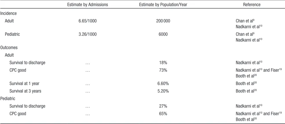
Table 2. Published Incidence and Outcome Estimates of ICHA (Adult and Pediatric)
Estimate by Admissions Estimate by Population/Year Reference
Incidence
Adult 6.65/1000 200 000 Chan et al9
Nadkarni et al10
Pediatric 3.26/1000 6000 Chan et al9
Nadkarni et al10 Outcomes
Adult
Survival to discharge … 18% Nadkarni et al10
CPC good … 73% Nadkarni et al10 and Fiser19
Booth et al20
Survival at 1 year … 6.60% Booth et al20
Survival at 3 years … 5.20% Booth et al20
Pediatric
Survival to discharge … 27% Nadkarni et al10
CPC good … 65% Nadkarni et al10 and Fiser19
Booth et al20 CPC indicates cerebral performance categories; and IHCA, in-hospital cardiac arrest.
by guest on November 16, 2017
http://circ.ahajournals.org/
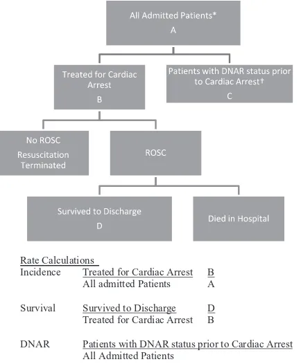
ICU and critical care units, recovery room, and operating room should be counted in the denominator, and the number of patients who experience an arrest in these areas should be included in the numerator. All patients with a DNAR order before the index cardiac event should be excluded. This seems intuitive, because the index event will be the patient’s last event; however, the presence of a DNAR order is often missed, which leads to activation of the emergency response team and initiation of resuscitative efforts only for hospital staff to find out about the DNAR order midarrest and then withdraw care. These patients should not be counted in IHCA incidence or outcome measures (Figure 1).
The incidence of IHCA in ED patients should include all patients who were registered in the ED and patients admitted to the hospital but who remained in the ED awaiting a bed before their index IHCA. This group of patients would exclude those with OHCA that occurred before their arrival in the ED and patients with OHCA who experienced another arrest on arrival in the ED. The incidence calculation should exclude all nonadmitted patients with OHCA or cardiac arrest that occurred outside the ED (outpatient settings) who were transferred to the ED after resuscitation while awaiting an in-hospital bed or were admitted directly to a hospital bed, because the true denominator of this type of patient is unknown (Figure 2). The incidence of IHCA among long-term care patients should be reported separately, using their
respective denominator in a fashion similar to that shown in Figure 1.
For patients with >1 IHCA during a single hospitalization, only the first IHCA is counted as the index cardiac arrest regardless of how many times the patient rearrests. If a patient has an arrest during >1 admission, then the first cardiac arrest that occurs in each separate admission is counted.
Published Estimates of Outcome
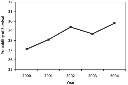
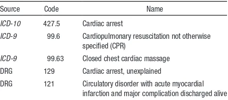
Although survival-to-discharge rates vary between studies, overall survival to hospital discharge has remained essentially unchanged for decades5 (Figure 3). In a retrospective
analy-sis of the data from ≈1000 US hospitals in the Nationwide Inpatient Sample, survival of post-IHCA patients was deter-mined by use of the International Statistical Classification of
Diseases and Related Health Problems(ICD)-9 code 427.5,
“cardiac arrest,” to identify patients with IHCA and patients who presented to the ED in cardiac arrest who were eventu-ally admitted to the hospital.17 The study suggested that there
was a 3% increase in in-hospital survival rates among IHCA patients between 2000 and 2004 (Figure 4). In a registry of 36 902 adults (≥18 years of age) and 880 children (<18 years old), survival to discharge after IHCA was higher in children than in adults for all rhythms (27% versus 18%); however, arrests that occurred in the delivery room and the ICU were
Rate Calculations
Incidence Treated for Cardiac Arrest B All admitted Patients
Survival Survived to Discharge Treated for Cardiac Arrest
A
D B
DNAR Patients with DNAR status prior to Cardiac Arrest C All Admitted Patients A
Figure 1. Reporting of incidence, survival, and do-not- attempt-resuscitation (DNAR) rate for patients admitted to an in-hospital bed with in-hospital cardiac arrest (IHCA). ROSC indicates return of spontaneous circulation. *All admitted patients includes all patients admitted in any in-hospital bed in any location of the hospital, including operating rooms, recov-ery, critical care units, procedural and diagnostic laboratories, and public areas. †Excludes admitted patients in the emergency department and patients designated DNAR after treatment for IHCA.
Rate Calculations
Incidence Treated for Cardiac Arrest All admitted Patients
Survival Survived to Discharge Treated for Cardiac Arrest
B A
D B
DNAR Patients with DNAR prior to Cardiac Arrest C All Admitted Patients A
Figure 2. Reporting of incidence, survival, and do-not-attempt-resuscitation (DNAR) rate for patients who have a cardiac arrest in the emergency department (ED). ROSC indicates return of spontaneous circulation. *All ED patients refers to all patients registered in the ED who have an arrest at any time in the ED before moving from the ED to an in-hospital ward. Patients who have had an out-of-hospital cardiac arrest, staff, and visitors are excluded, as are outpatients attending clinics who have an arrest and are transported to the ED for postresuscitation care until they are admitted to the hospital. †Patients with DNAR status before cardiac arrest and who were not treated or treated initially until the DNAR status was verified and resuscitation terminated.
by guest on November 16, 2017
http://circ.ahajournals.org/
excluded in this comparison10 (Table 3). One study compared
short- and long-term survival after IHCA and found 6.6% alive at discharge, 5.2% alive at 1 year, and 3% alive at 3 years.18 Good short-term neurological outcomes after IHCA,
as measured by cerebral performance category, were reported in 64% of children and 75% of adults who survived to dis-charge10,19,20 (Table 4).
Survival outcomes across different types of institutions, at different times, and of various subgroups have also been reported. A higher survival rate has been correlated with larger, teaching, and urban hospitals in some studies,5,17 but
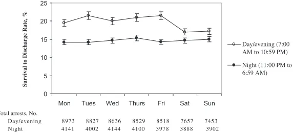
others report lower survival rates in metropolitan or teach-ing hospitals, perhaps related to the severity of underlyteach-ing illness18 (Table 5; Figure 5). In addition, survival rates after
IHCA have been reported to be lower at night and during weekends21 (Figure 6). In one report, survival-to-discharge
rates after IHCA among critical care patients were 15.9% overall but only 3.9% in patients who received vasopressors before the arrest.22 In a study of 433 985 elderly patients (≥65
years of age) who underwent in-hospital cardiopulmonary resuscitation (CPR), 18.3% survived to discharge (95% confi-dence interval, 18.2–18.5). There was no significant change in survival rates over time. Survival rates were lower in this sub-group of elderly patients when they were admitted to a skilled nursing facility and in patients who received care in a metro-politan or teaching hospital, and these findings were attributed to an overrepresentation of patients with more severe illness in these treatment facilities.23
Recommended Definition of Outcome
Survival to hospital discharge is the minimum standard, and survival to 30 days is preferred. In addition, a measure of functional survival (eg, cerebral performance category or Modified Rankin Scale at discharge or at 30 days). Out-comes should be reported for all patients who are admitted to the hospital who do not have a DNAR order before arrest who are treated with either chest compressions or defibril-lation (Figure 1). Arrests that occur in the ED should be reported separately and should not include patients whose initial arrest occurred out of hospital or people who were visitors, staff, or outpatients (Figure 2). DNAR rates should be defined by the number of patients with a DNAR status (before an index cardiac arrest) per 1000 admitted patients. The DNAR status of the patient after arrest is not included in the DNAR rate. The DNAR rate of patients before arrest should be reported separately for acute and long-term care inpatients. The rates of DNAR status assignment after arrest should be reported and compared with similar institutions to ensure that performance is in line with the standard of care.
Best Practices
The best practices are divided into 3 temporal sections: Prear-rest, intra-arPrear-rest, and postarrest. The discussion for each period includes (1) a brief introduction, (2) the structural aspects of the institutional response (personnel, training, equipment), (3) care pathways followed during the time interval (early identi-fication, focus on CPR and early defibrillation, comprehensive postarrest care), and (4) process issues related to how care is provided and quality improvement measures (real-time feed-back, automated equipment that can replace staff and deliver similar care, withdrawal of life-sustaining therapy).
Figure 3. Survival to hospital discharge after in-hospital car-diopulmonary resuscitation, according to year and race. Sur-vival rates are poorer for black and other nonwhite patients (P<0.001). There is no significant change in overall survival rate from 1992 to 2005 (P=0.57 with use of the likelihood-ratio test). From Ehlenbach et al.23 Copyright © 2009, Massachusetts
Medical Society. Reprinted with permission from Massachu-setts Medical Society.
Figure 4. US national data and adjusted probability of survival by year. Adjusted for age, sex, hospital location (urban/rural), hospital teaching status, hospital bed size (large, medium, small), median income for patient’s zip code, and region of the country in which the hospital is located. Modified from Carr et al17and
used with kind permission from Springer Science+Business Media. Copyright © 2008, Springer-Verlag.
Table 3. First Documented Rhythm in Pediatric and Adult Cardiac Arrests
First Documented Pulseless Rhythm
Pediatric Cardiac Arrest (n=880)
Adult Cardiac Arrest (n=36 902) P
Asystole 350 (40) 13 024 (35) 0.006
VF or pulseless VT 120 (14) 8361 (23) <0.001
VF 71 (8) 5170 (14) <0.001
Pulseless VT 49 (6) 3191 (9) 0.001
PEA 213 (24) 11 963 (32) <0.001
Unknown by documentation
197 (22) 3554 (10) <0.001
PEA indicates pulseless electrical activity; VF, ventricular fibrillation; and VT, ventricular tachycardia.
Adapted and used with permission from Nadkarni et al.10 Copyright © 2006 American Medical Association. All rights reserved.
by guest on November 16, 2017
http://circ.ahajournals.org/
Best Practices: Prearrest
Brief Introduction to Prearrest
In the pre-IHCA period, several aspects of preparation are important. These include placement of defibrillators and code carts (or crash carts); establishment of emergency response teams; training of IHCA code team personnel in clinical resus-citation care, as well as team leadership and resource man-agement; and development of a comprehensive performance review process, cardiac monitoring, and documentation in the medical record about the level of resuscitation appropriate for the patient (eg, DNAR status).
Structural Aspects of the Institutional Response
Defibrillators and Code Carts
Manual defibrillators or automated external defibrillators (AEDs) and code carts should be readily accessible to any patient area, and all staff should know the location of this
equipment and how to use it. In general, a defibrillator and code cart should be in close proximity to enable defibrillation of any patient in cardiac arrest within 2 minutes.24 This may be
achieved by staff carrying resources to the scene of the arrest rather than moving a cart; regardless of how staff access the defibrillator and code cart, it is most important that access be rapid. To minimize delays and confusion, it may be advisable to standardize defibrillator equipment across the institution.25
Ideally, staff should have the capacity to receive feedback on the quality of CPR at the point of care. This may include voice or visual cues on the quality of CPR (depth, interruptions or hands-off time, compression rate) that are measured and reported by the defibrillator, a handheld device, or alternative technology.26 Additionally, staff should have access to
physi-ological feedback about the quality of CPR at the point of care (eg, quantitative end-tidal CO2 or waveform capnography defined as continuous noninvasive measurement and graphic display of end-tidal CO2), at a minimum and intra-arterial pressure monitoring as outlined in the 2010 AHA guidelines
for CPR and ECC.27 There should be a process in place to
Table 5. Severity of Disease Predictors for Nonsurvivors and Survivors of Cardiac Arrest
Variable
Nonsurvivors (n=683)
Survivors (n=49) P *
Age at arrest, y 66±12 59±12 <0.01
Comorbidity score on discharge 3.0±1.5 2.6±1.6 0.03 Duration of resuscitation attempt, min 22.6±13 19.9±18 0.58
Ejection fraction, % 42±18 42±18 0.67
VT/VF 122 (18) 25 (52) <0.01
Medication use
ACE-I/ARB 270 (40) 30 (67) 0.01
β-Blocker 217 (31) 27 (56) <0.01
Antiarrhythmic 50 (8) 10 (20) 0.05
Calcium channel blocker 216 (32) 19 (39) 0.62
Data are presented as mean±SD or n (%).
ACE-I indicates angiotensin-converting enzyme inhibitor; ARB, angiotensin receptor blocker; VF, ventricular fibrillation; and VT, ventricular tachycardia.
*P value contrasts the 2 categories.
Reprinted from Bloom et al18 with permission from Elsevier. Copyright © 2007, Elsevier Inc.
age
comorbidity
antiarrhythmic
ACE-inhibitor
beta-blocker
VT/VF
0 1 1
1 . 0
Odds Ratio
Characteristi
c
Figure 5. Odds ratios for survival by significant clinical char-acteristics between survivors to discharge and nonsurvivors of in-hospital cardiac arrest. ACE indicates angiotensin-converting enzyme; and VT/VF, ventricular tachycardia/ventricular fibrilla-tion. Reprinted from Bloom et al18 with permission from Elsevier.
Copyright © 2007, Elsevier Inc.
Table 4. Outcomes of In-Hospital Pulseless Cardiac Arrest by First Documented Pulseless Arrest Rhythm*
VF or Pulseless VT Asystole PEA Unknown Rhythm
Pediatric (n=120)
Adult (n=8361)
Pediatric (n=350)
Adult (n=13 024)
Pediatric (n=213)
Adult (n=11 963)
Pediatric (n=197)
Adult (n=3554)
Any ROSC 80 (66.7) 5629 (67.3) 184 (52.6) 5858 (45.0) 123 (57.7) 6270 (52.4) 137 (69.5) 2062 (58.0)
ROSC >20 min 74 (61.7) 5185 (62.0) 157 (44.9) 4997 (38.4) 108 (50.7) 5135 (42.9) 120 (60.9) 1866 (52.5) Survival to discharge 35 (29.2) 3013 (36.0) 78 (22.3) 1379 (10.6) 57 (26.8) 1340 (11.2) 66 (33.5) 753 (21.2) Neurological outcome
Good 22 (62.9) 2268 (75.3) 43 (55.1) 841 (61.0) 36 (63.2) 834 (62.2) 35 (53.0) 447 (59.4)
Poor 1 (2.9) 264 (8.8) 16 (20.5) 243 (17.6) 13 (22.8) 222 (16.6) 11 (16.7) 111 (14.7)
Unknown 12 (34.3) 481 (16.0) 19 (24.4) 295 (21.4) 8 (14.0) 284 (21.2) 20 (30.3) 195 (25.9)
Values are number (%) of patients.
PEA indicates pulseless electrical activity; ROSC, return of spontaneous circulation; VF, ventricular fibrillation; and VT, ventricular tachycardia.
*First documented pulseless rhythm was defined as the first electrocardiographic rhythm documented at the time the patient became pulseless. Good neurological outcome was prospectively defined as cerebral performance category (CPC) 1 or 2 for adults; the comparable pediatric cerebral performance category (PCPC) of 1, 2, or 3 for children on hospital discharge; or no change from baseline CPC or PCPC.
Reprinted with permission from Nadkarni et al.10 Copyright © 2006 American Medical Association. All rights reserved.
by guest on November 16, 2017
http://circ.ahajournals.org/
collect and review the resuscitation data from the defibrillator and any other device or source documentation that captures data at the scene in a timely manner as a source of postevent feedback to the team.28 Code carts should be stocked with the
necessary ACLS medications and intubation and respiratory supplies, and, where applicable, specialty-specific supplies (eg, pediatric supplies, cesarean section tray).
Rapid Response Teams
Rapid response teams were established to prevent IHCA in patients whose condition is deteriorating.29,30 These teams
are usually composed of varying combinations of physicians, nurses, respiratory therapists, and pharmacists and can be summoned to the bedside of a patient who is noted to have an acute clinical decompensation or is thought to be at immedi-ate risk of IHCA and other immediimmedi-ate life-threimmedi-atening events. Although the theory is compelling, data on the effectiveness of these teams have actually been mixed.31,32 A recent
meta-anal-ysis suggests that although rapid response teams may decrease the incidence of IHCA outside the ICU, they have not convinc-ingly demonstrated significant improvements in survival rates.9
Possibilities for these counterintuitive results are (1) early identification and transfer of the patient to the ICU, where the patient subsequently experiences an IHCA, and (2) increased use of DNAR orders.33 Other possibilities include failure to
trigger the team when signs of deterioration are noted and poor surveillance methods for identifying clinical deterioration.34
Code Teams
The Joint Commission3 requires that resuscitation services
and equipment be provided to patients according to the hos-pital’s protocol and that resuscitation outcomes be collected and reviewed. In addition, The Joint Commission requires that evidence-based programs be used to train staff in the need for resuscitation and the use of resuscitation equipment and techniques. However, The Joint Commission does not man-date the composition of code teams. The American Board of Internal Medicine and the Accreditation Council for Graduate Medical Education likewise do not mandate composition or even training of code teams, although the new Accreditation Council for Graduate Medical Education common program requirements clearly emphasize the need for adequate super-vision and graded progressive responsibility as core tenets within graduate training in medicine.35 To fulfill the
require-ments of The Joint Commission, all hospital staff responsible for the care of patients should be trained in basic life support. This training should include how to recognize a patient whose
condition is deteriorating, call for help, start CPR, direct others to get the nearest AED, and use the AED. A designated emer-gency response team (eg, code blue in some hospitals) must be available at all times. Code team composition is mandated by individual hospitals and may consist of nurses, respiratory therapists, pharmacists, physicians, and clergy, as well as secu-rity personnel. Mechanisms for triggering a specialty-specific emergency response team for unique situations such as pediat-ric and maternal-fetal arrests should be available if such clinical situations are a possibility at a specific hospital. A code team leader is responsible for guiding the resuscitative efforts. Code team members must have ACLS Provider cards and be on duty in the hospital and available to respond to codes at all times.27
Education and Training
All hospital staff should know how to recognize cardiac arrest, call for help, perform chest compressions, and use an AED at the level of a bystander until staff with training in the care of patients with cardiac arrest respond to the event. Some hos-pitals have made this a minimum requirement for hiring, and others have mandated it as a minimum requirement for contin-ued employment, with annual retraining of all staff.
Education and training of IHCA code team staff are criti-cal to improved performance and better outcomes.36 IHCA
is a relatively low-frequency event, and IHCA code team members have reported feeling ill prepared to lead and par-ticipate as members of the team.37 IHCA treatment generally
relies on code teams whose personnel composition changes frequently, and members may not be focused solely on pro-viding emergency resuscitation care. Therefore, there may be aspects of training and skills retention related to providing intra-arrest care that are unique to the hospital setting and require frequent retraining of the team to maintain skills, minimize errors, and optimize outcome.38,39 Simulation
train-ing in addition to ACLS traintrain-ing of house staff at an academic hospital ICU was associated with greater adherence to the
AHA Guidelines for CPR and ECC.40
Very few studies have reported the effect of training on sur-vival from IHCA. In 1 study performed at a 550-bed tertiary care center, the survival rate of patients initially resuscitated by a nurse trained in ACLS was almost 4 times higher (37.5% versus 10.3%) than when resuscitation was initiated by a nurse without ACLS training.41 One study with some
method-ological concerns reported an increase in short- and long-term survival rates with ACLS-trained personnel.42
One of the more promising training strategies may involve the use of simulation-based mock codes. A recent study
0 5 10 15 20 25
Mon Tues Wed Thurs Fri Sat Sun
S
u
rv
iv
a
l
to
D
is
ch
a
rg
e
R
a
te
,
%
Day/evening (7:00 AM to 10:59 PM)
Night (11:00 PM to 6:59 AM)
Total arrests, No.
Day/evening 8973 8827 8636 8529 8518 7657 7453 Night 4141 4002 4144 4100 3978 3888 3902
Figure 6. Survival to discharge rate and total arrests by time category and day of week. Error bars represent 95% confidence intervals. Reprinted with permission from Peberdy et al.21 Copyright
© 2008, American Medical Association. All rights reserved.
by guest on November 16, 2017
http://circ.ahajournals.org/
conducted over the course of 48 months suggested that monthly random mock codes that used a simulator and occurred in various patient locations within the hospital were correlated with improved survival rates for pediatric arrest of >50%. These rates were higher than the 2008 national average.43 In
a simulation model evaluation of ventricular fibrillation (VF) IHCA resuscitation, patients were more likely to receive more defibrillations when the physician team arrived early (median arrival 50 seconds after onset). In all cases, there was a median delay of 85 seconds until CPR was started, and 100 seconds elapsed before the first defibrillation. These data suggest that gaps in knowledge, reluctance to act, and team work all need to be addressed through improved training.44
Care Pathways
Prevention Through Early Identification
IHCA is frequently preceded by clinical deterioration that is evident in symptoms and changes in vital signs that could be identified and treated by trained in-hospital staff.45 As a result,
greater emphasis has been placed on prevention of these events, based on the assumption that earlier identification and intervention to stabilize these patients can prevent IHCA.46,47
In 2008, The Joint Commission47 introduced patient safety
goals, in which goal 16 specifically targets improved recogni-tion of and response to changes in a patient’s condirecogni-tion.48
An observational study of surgical and medical wards reported that 1 of 5 patients developed abnormal vital signs, and >50% of these events went unnoticed by nursing staff. The patients with abnormal vital signs had a 3-fold higher 30-day mortality rate.49 A nested, controlled, in-hospital trial
compar-ing prearrest patients with control subjects at 48 hours before the event suggested that the Modified Early Warning Scores were different, but the authors noted that this scoring system does not include significant predictors such as diastolic and pulse pressures.50 A study that examined circadian variability and a
large registry study of >58 000 IHCAs both demonstrated lower survival rates during nights and weekends.8,21 Interventions to
address consistent and comprehensive staff training in monitor-ing vital signs, includmonitor-ing quantitative end-tidal CO
2 waveform
capnography and electrocardiographic (ECG) tracings, as well as anticipation of bad outcomes, initial response, and ACLS skills may enable earlier detection, better treatment, and better survival rates regardless of time of day or day of the week.
Very few high-quality evaluations of training interventions to improve the early identification of prearrest patients exist.51 The
more promising educational interventions are the Immediate Life Support52 and the Acute Life-Threatening Events Recognition
and Treatment53 courses; however, high-quality evaluations of
their efficacy are still pending. A longitudinal multicenter study54
of the Acute Life-Threatening Events Recognition and Treatment course suggested an increase in prearrest calls, a reduction in the number of IHCAs, and improved survival-to-discharge rate after IHCA. A before-and-after comparison of a 1-day course, based on a needs assessment, failed to show any difference; however, only 67% of the nursing staff were trained.55
ECG and Physiological Monitoring
It is important to keep in mind that many arrests are unmoni-tored and unwitnessed. Brady et al56 reported improved
survival to discharge and favorable neurological outcome with either monitoring or direct observation compared with unmonitored or unwitnessed IHCA. This retrospective look at registry data also suggested that there was no additional advantage of cardiac monitoring compared with staff obser-vation of the event, which reinforces that early identification and trained response are key.56 That said, it is very difficult to
predict who will experience a cardiac arrest. A tiered approach to the use of ECG monitoring may alert hospital personnel to a life-threatening arrhythmia before the clinical discovery of an unconscious patient; thus, it can save critical minutes from the onset of IHCA to the start of resuscitative efforts.
A 2004 AHA scientific statement provided some guidance on who should be monitored with electrocardiography.57 Class
I indications for monitoring include the following:
• Patients resuscitated from sudden cardiac death
• Patients in the early phase of acute coronary syndromes
• Patients with unstable coronary syndromes and newly diagnosed high-risk coronary lesions
• Adults and children who have undergone cardiac surgery
• Patients who have undergone nonurgent percutaneous coronary intervention (PCI) with complications
• Patients who have undergone implantation of an auto-mated defibrillator lead or a pacemaker lead and who are considered pacemaker dependent
• Patients with a temporary pacemaker or transcutaneous pacing pads
• Patients with atrioventricular block
• Patients with arrhythmias and Wolff-Parkinson-White syndrome
• Patients with long-QT syndrome and arrhythmias
• Patients with intra-aortic balloon pumps
• Patients with acute heart failure
• Patients with indications for intensive care
• Patients undergoing conscious sedation
• Patients with unstable arrhythmias
• Pediatric patients with symptoms of arrhythmia
Class II indications (may be beneficial in some patients) include the following:
• Patients with post–acute myocardial infarction (AMI)
• Patients with chest pain syndromes not thought to be acute coronary syndromes
• Patients who have undergone uncomplicated nonurgent coronary intervention
• Patients who have been administered antiarrhythmic drugs that are not potentially proarrhythmic
• Patients with implanted pacemakers who are not pace-maker dependent
• Patients with uncomplicated ablation of arrhythmia
• Patients with diagnostic coronary angiography
• Patients evaluated for syncope thought to be noncardiac
• Patients with DNAR orders who may have arrhythmias that cause discomfort
Selected low-risk patients admitted with chest pain may not need ECG monitoring.58,59 ECG monitoring must be of high
quality in capturing the trigger of true arrests (sensitivity) and
by guest on November 16, 2017
http://circ.ahajournals.org/
for avoidance of false alarms (specificity). Sufficient staffing is critical to allow a prompt and appropriate response to the alarms by a nurse or technician.
Other physiological monitoring is also necessary in disease-specific subgroups of patients. Arterial blood pressure monitoring may be performed noninvasively or invasively for patients at risk for hemodynamic instability.60 Respiratory
mon-itoring is of particular use in patients with sleep apnea. Pulse oximetry for monitoring of patients with pulmonary disease is quite valuable. Quantitative end-tidal CO
2 waveform
capnogra-phy is recommended for ventilation with a bag-mask, for intu-bated patients,27,61 and for those undergoing conscious sedation.
Process Issues
Plan for Routine Debriefing
It is important to put in place a process for postevent debriefing that best fits the culture of the institution, the resources, and the timing of data capture and analysis. It is important to define a priori who will lead the debriefing (preferably people trained as facilitators) and when it will occur. Debriefing is used to identify best practices unique to the institution, to optimize performance, and to address emotional responses related to the specific event. The impact of debriefing to date has been measured against per-formance and short- and long-term survival; however, other out-comes, such as factors related to team building, psychological responses, and retention, have not been studied. Debriefing ses-sions that review clinical and defibrillator-recorded information from a code may improve some but not all aspects of code team performance.28 In 1 study, debriefing with audiovisual
feed-back was associated with significantly improved rates of ROSC (59.6% versus 44.6%, P=0.03) but did not change survival-to– hospital discharge rates (7.4% versus 8.9%, P=0.69). Further study is needed to evaluate routine debriefing with respect to the capacity to build and retain teams, who should conduct the debriefing, when the debriefing should occur, and to define the cost-effectiveness of this intervention.
DNAR Orders
Resuscitation is not always desired by the individual, and in many cases it is medically futile. Advance directives, liv-ing wills, and durable power of attorney for health care and patient self-determination ensure that patient preferences will guide care even when the patient is unable to make decisions on his or her own. Advance planning by the patient or proxy decision maker is ultimately in the best interest of the patient, because studies have shown that these decisions are associated with better care, quality of life, and bereavement adjustment by caregivers.62 Advance directives should be discussed with and
documented for all patients admitted to the hospital. DNAR orders should be completed, signed, and dated by the physician after a documented discussion with the patient and/or family or legal representative. This will avoid unwanted or futile resus-citation and the subsequent need for early withdrawal of life-sustaining therapy. It is important to be frank with the patient and explain the probability of surviving IHCA, because most older patients readily understand prognostic information and can make decisions on whether they would like to receive CPR.63–65
The DNAR order should preferably state either full resuscita-tion or no attempt at resuscitaresuscita-tion; however, certain situaresuscita-tions or patient or family preferences may warrant explicit instructions
about which interventions to withhold or provide (eg, CPR with-out intubation, medications withwith-out CPR). This may include but is not limited to vasopressor agents, blood products, advanced airway interventions, nutrition, fluids, analgesia, sedation, anti-arrhythmic drugs, and defibrillation.66
Best Practices: Intra-arrest
Brief Introduction to Intra-arrest Care
High-quality CPR, with optimal chest compressions and ven-tilations, and early defibrillation are cornerstones of intra-arrest treatment that have improved survival from OHCA.67
There is growing evidence that optimizing these treatment cornerstones for IHCA could also improve outcomes in this setting.68,69 Periodic evaluation of residents trained in
pedi-atric advanced life support revealed that they did not meet performance standards specified by the 2010 AHA Guidelines
for CPR and ECC, which suggests that training is not enough
to ensure performance.70 Implementation strategies to ensure
timely access to equipment, visual reminders, regular testing, and point-of-care feedback may be required to optimize the translation of guidelines into practice during cardiac arrest.
Structural Aspects of the Institutional Response
Mechanical Chest Compressions
The use of mechanical chest compression devices in the in-hospital setting has been reported, particularly in settings where the performance of manual CPR is difficult, such as during in-hospital transport71 and PCI.72 Mechanical devices include
active compression-decompression and load-distributing band devices that automatically compress the chest. There are reports that mechanical chest compression devices improve coronary perfusion pressures during IHCA compared with manual chest compressions.73 IHCA studies of mechanical compression
devices have been limited to small case series involving a hand-ful of patients. As an example of the current literature on this subject, a recent case series of 28 patients with IHCA who pre-sented in pulseless electrical activity (PEA) and were treated with mechanical chest compressions demonstrated a 50% rate of survival to discharge and a 46% rate of good neurological outcome.74 If these devices are used, it is important to provide
training that minimizes interruptions in chest compressions dur-ing use of the device; however, there are limited data to support the routine use of these devices for IHCA.
Automated External Defibrillators
AEDs may play a role in improving early defibrillation times, particularly in less intensively monitored areas of the hospi-tal. Approximately half of all IHCAs occur outside the ICU.5
Implementation of a public access defibrillation program at a tertiary care hospital included targeted placement of AEDs in areas where time from arrest to arrival of a defibrillator would be >3 minutes, including time spent in parking garages and on walkways between buildings.75 In a study of 439 patients
with IHCA, a program to equip and train nurses outside of the ICU setting to use AEDs resulted in an 86% rate of ROSC for patients with pulseless ventricular tachycardia (VT)/VF and a 47% rate of survival to hospital discharge.76 In another study,
placement of AEDs in 14 locations that could be easily reached from all wards and diagnostic rooms within 30 seconds was
by guest on November 16, 2017
http://circ.ahajournals.org/
combined with a 2-hour AED training program for medical officers, nurses, and administrative and technical staff. In the 18 recorded cases of pulseless VT/VF, rates of ROSC and survival to hospital discharge were 88.9% and 55.6%, respectively.77
Although these studies did not compare AED resuscitation rates with prior non-AED rates, 1 study did show an improvement in outcomes of patients with pulseless VT/VF. After implementa-tion of a program that included educaimplementa-tion and encouraged use of manual biphasic defibrillators in AED mode, as well as place-ment of AEDs in all outpatient clinics and chronic care units, IHCA survival to discharge improved by 2.6 times from 4.9% to 12.8%.78 AEDs performed similarly to biphasic manual
defi-brillators in AED mode. A recent large registry study of IHCAs suggested that there was no association with increased survival and use of an AED with VF and pulseless VT and decreased survival with the nonshockable initial rhythms.79 The decrease
in survival from nonshockable rhythms could be attributed to the mandatory time off the chest to allow for analysis and shock delivery with an AED. Time to first contact by the cardiac arrest team was not compared in this study, and it is likely that AEDs were placed in areas less well served by the cardiac arrest team, representing a potential selection bias. In addition, AEDs were grouped with manual defibrillators that could be used in AED mode, but it was unknown whether the latter were used in manual or automatic mode, which makes it harder for the AED group to demonstrate superiority over the manual mode.79
Additional randomized clinical trials are required to evaluate and optimize use of AEDs in the hospital.
Automated External Cardioverter-Defibrillators
Automated external cardioverter-defibrillators (AECDs) may play a role in more intensively monitored areas of the hospital. These devices differ from AEDs in that they provide continu-ous cardiac monitoring with 2 pads placed on the patient’s chest and can automatically defibrillate shockable rhythms. In 1 pro-spective study of AECD monitoring of ED patients considered to be at risk for pulseless VT/VF (n=55), the average interval between onset of arrhythmia and first defibrillation was 33 sec-onds and resulted in a 94.4% rate of ROSC.80 A prospective
trial (Automatic External Defibrillation Monitoring in Cardiac Arrest; ClinicalTrials.gov, unique identifier NCT00382928) has completed enrollment of telemetry patients with IHCA randomly assigned to a cardiac arrest team or standard CPR plus AECD monitoring.81 The AECD in the study was
pro-grammed to deliver one 150-J biphasic shock to patients in sustained pulseless VT/VF. The primary end point was time to defibrillation, with secondary outcomes including neurological status and survival to discharge and 3-year follow-up. Prelimi-nary data demonstrated that 1 of 192 enrolled patients experi-enced sustained pulseless VT during AECD monitoring, and this patient was successfully defibrillated within 17 seconds. There were no events in the control group; however, during the same period, mean time to shock for pulseless VT/VF IHCA that occurred outside the telemetry ward was 230±50 seconds.
Care Pathways
Performance of CPR
A major opportunity for hospitals to improve patient care involves monitoring and improving CPR performance.26,28,82
Optimizing ventilations (a ratio of 30:2) and providing chest compressions at a rate of 100/min and a depth of at least 5 cm while minimizing pauses (hands-off time) will improve outcomes from IHCA.69 Despite the importance of chest
compressions in cardiac arrest outcome, they are rarely per-formed according to guideline recommendations.69 In studies
of IHCA, chest compression rates were too slow >30% of the time.68,69 In addition, 33% of compressions were too shallow,
and ≈20% of resuscitation time consisted of interruptions and no-flow time. Rescuer fatigue contributes to poor-quality CPR, and rescuers who provide ventilations and compressions should be replaced or should switch places after each 2-min-ute cycle.83 Strategies for improving the quality of each
com-ponent of CPR are reviewed below.
Decrease Interruptions in Chest Compressions
Bystander CPR and CPR provided by healthcare professionals improve outcomes in OHCA and IHCA, respectively.69,84
Inter-ruptions in chest compressions may decrease the compression fraction, which has been associated with decreased survival rates67 in OHCA. Some out-of-hospital strategies that include
continuous compressions without pauses for ventilations have been associated with improved outcomes.85,86 Interruptions for
even a few seconds can decrease coronary blood flow,87 and
are associated with worsened neurological outcome in animal models,88 and may decrease survival to discharge in OHCA.89
Pauses in chest compressions of ≥10 seconds’ duration have been associated with decreased success of defibrillation.90 A
correctly performed compression-to-ventilation ratio of 30:2 should be consistent, with 2 ventilations delivered within 2 seconds off the chest for each set of 30 compressions or 4 to 6 seconds off the chest per minute during the 2 minutes between rhythm analyses. To reduce hands-off time during analysis and charging, newer versions of defibrillator software enable interpretation of the ECG and continuous charging of the capacitor during chest compressions,91 which minimizes
the pause to a few seconds before the shock is delivered.
Avoid Hyperventilation
Excessive ventilation rates are often observed during CPR for OHCA92 and IHCA.69 Fast ventilation rates in the laboratory
are associated with increased intrathoracic pressures, lower coronary perfusion pressures, and decreased survival rates.92,93
Devices that prompt or time ventilation through timing lights or audio cue during CPR may be useful to prevent excessive ventilation. In addition to improving quality of chest compres-sions, code team debriefing with audiovisual feedback has been associated with a decrease in mean ventilation rates from 18/min to 13/min.28
Optimize Chest Compression Depth
Greater chest compression depth and a decreased preshock pause interval before defibrillation have been associated with increased defibrillation success and ROSC after IHCA.90
Ade-quate chest compression depth in OHCA has been associated with survival to hospital admission,94 but improved survival to
discharge as a function of adequate chest compression depth has not yet been demonstrated in either OHCA or IHCA. The
2010 AHA Guidelines for CPR and ECC have changed the
emphasis to a depth of at least 5 cm with each compression.95
by guest on November 16, 2017
http://circ.ahajournals.org/
The depth of compression in the IHCA setting has likely been overestimated because of movement of the patient’s mat-tress with manual chest compressions.96,97 Recent simulation
studies demonstrated that even with the use of a backboard, mattress compression can account for as much as 40% of mea-sured compression depth in patients with IHCA.96,97 When a
single accelerometer is applied to the sternum to measure chest compressions, as used in the majority of clinical studies, the actual compression depth is overestimated by as much as 4 to 13 mm, depending on mattress type.96,97 Thus, deeper chest
compressions in the IHCA setting may be needed to compen-sate for mattress movement if it cannot be neutralized by the use of a backboard.
Provide Early Defibrillation
Approximately 25% of patients with IHCA have a shockable rhythm of pulseless VT/VF.5,98 Despite proximity to advanced
health care, >30% of patients with IHCA have defibrillation times of >2 minutes after arrest.99 Defibrillation times longer
than 2 minutes after IHCA have been associated with decreased rates of survival to hospital discharge. Delays were also associ-ated with black race, noncardiac admitting diagnosis, time of arrest during evenings and weekends, and hospitals with <250 beds. When nonphysicians are allowed to perform defibrilla-tion, it can save critical seconds to minutes until the emergency response team arrives.76 Strategies used to increase early
defi-brillation after IHCA include use of hands-free pads (which decrease preshock pause),100 use of AEDs in non-ICU settings,
and use of AECDs.
Identify and Treat Underlying Causes
The most common causes of IHCA include cardiac arrhyth-mia, acute respiratory insufficiency, and hypotension.5 Studies
show that asystole and PEA are more common than VF in adult IHCA, with only 25% of patients having VF or pulseless VT as the initial rhythm,5,79 whereas children were more likely to
pres-ent with asystole (40% versus 35% in adults).10 The frequency
of PEA as the first presenting rhythm in adult IHCA is 30% and has remained unchanged over many years.5,10 Only 10% of
patients with IHCA who present with an initial rhythm of PEA or asystole have neurologically intact survival.5 Thus,
identifi-cation and treatment of the reversible causes that may present with PEA/asystole are important during IHCA. Very little has been published on the specific causes of PEA in this setting; however, expert opinion suggests that a substantial number of cases may be secondary to respiratory insufficiency and shock and may respond to targeted therapy. A number of special situ-ations may cause IHCA and require unique interventions that are disease specific. A detailed overview of these situations is provided in the sections on special considerations and pediatrics of the 2010 AHA Guidelines for CPR and ECC.101,102
Process Issues
Use Real-Time Feedback
Devices that prompt or time ventilation (eg, timing lights) and guide rhythm of chest compressions (eg, metronome) and quality of compressions (eg, quantitative end-tidal CO
2
wave-form capnography) during IHCA may be helpful. A cohort study with historical controls demonstrated improvements in chest compressions and ventilations with point-of-care
feedback, but no difference was found in either ROSC or survival to hospital discharge.26 Audio prompting of chest
compressions through use of technology as simple as a met-ronome has been found to improve blood flow during CPR both in animal models and during resuscitation attempts in humans.103,104 A recent randomized trial on OHCA
demon-strated that point-of-care feedback did not improve patient outcomes in well-trained services participating in randomized controlled trials,105 whereas Edelson et al demonstrated the
usefulness of employing quality of CPR measures during real IHCA to evaluate the efficacy of training,38 and this finding
was subsequently verified for both OHCA and IHCA in a sys-tematic review39 in 2009. The latter suggested that there was
good evidence to support the use of point-of-care feedback in training and that it may be beneficial in clinical application.39
Point-of-care feedback on CPR quality is generally thought to be helpful, and it makes sense in IHCA, because staff are accustomed to using technology to guide care.
Best Practices: Postarrest
Brief Introduction to Postarrest
For patients who achieve ROSC, variability in survival rates between hospitals exists and can range from 54% to 32%.106
Higher-volume hospitals and teaching hospitals have the high-est survival rate, which averages 38% for patients who have an arrest outside the ICU and 32% for patients who have an arrest in the ICU.10 Clinical investigation focused on improving
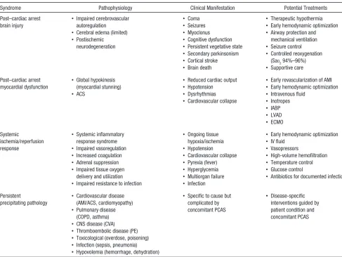
out-comes of patients who achieve ROSC after IHCA has been lim-ited and has made it necessary for practitioners to extrapolate from OHCA studies when developing diagnostic and treatment strategies. Patients with ROSC after cardiac arrest in any setting will suffer from a complex combination of pathophysiological processes previously described as the post–cardiac arrest syn-drome.107 Key components include (1) postarrest brain injury, (2)
postarrest myocardial dysfunction, (3) systemic ischemia/reper-fusion response, and (4) persistent acute and chronic pathology that precipitated cardiac arrest.107 Persistence of preexisting
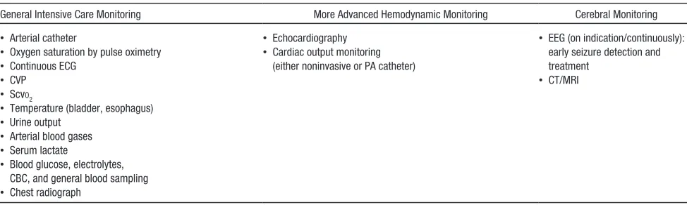
con-ditions and precipitating pathologies after ROSC provide sig-nificant challenges in management of patients resuscitated from IHCA (Table 6). Multisystem organ failure is a more common cause of death in the ICU after initial resuscitation from IHCA than after OHCA.108 Patient management is also affected by the
location of the arrest within the hospital (Table 1), the intensity of support (eg, mechanical ventilation and vasopressor therapy), and invasive monitoring in place at the time of arrest (Table 7). In all cases, optimal post-IHCA care requires a well-coordinated multidisciplinary team. Clinical trials evaluating treatment strategies in postarrest patients are lacking for both OHCA and IHCA. Much of the evidence is based on animal studies, cohort comparisons, and extrapolations from diseases that share similar characteristics, such as sepsis.
Structural Aspects of the Institutional Response
Comprehensive postarrest care requires access to and collabo-ration between a multidisciplinary team of providers, including emergency medicine (if the arrest occurs in the ED), cardiology, interventional cardiology, cardiac electrophysiology, intensive care, and neurology. If these services are not available, the insti-tution needs to have in place an interhospital transfer agreement
by guest on November 16, 2017
http://circ.ahajournals.org/
and a process to ensure access to these resources within the first 6 hours after arrest if therapeutic hypothermia cannot be initi-ated in the sending facility. The timing of transfers should take into consideration the time-sensitive nature of potential inter-ventions such as PCI and therapeutic hypothermia.
Care Pathways
Induction of Goal-Directed Mild Therapeutic Hypothermia Mild therapeutic hypothermia (32°C to 34°C) improves outcome of comatose survivors of witnessed OHCA when the initial rhythm is VF.109,110 Similar studies have not been
performed in patients who achieve ROSC after IHCA. The potential detrimental or beneficial effect of mild therapeutic hypothermia on active pathologies, comorbidities, and ongo-ing therapies must be considered. The role of therapeutic hypothermia in the management of IHCA and with initial rhythms other than VF in either the out-of-hospital or in-hospital setting is an important knowledge gap that needs to be addressed by future research. Despite this gap in research,
the 2010 AHA Guidelines for CPR and ECC recommend that
induced hypothermia may be considered for comatose adult patients with ROSC after IHCA of any initial rhythm.111
Coronary Reperfusion for ST-Segment Elevation Myocardial Infarction
PCI for patients resuscitated from IHCA is an important thera-peutic consideration. According to the Get With The Guide-lines–Resuscitation data, only 11% of treated IHCAs are caused by AMI.10 In a retrospective review of 110 survivors of IHCA
caused by VF, only 30 patients (27%) underwent cardiac cathe-terization on the day of the arrest, and of these, only 13 patients had an ECG with results consistent with ST-segment–elevation myocardial infarction or new left bundle-branch block. Patients who underwent cardiac catheterization were more likely to sur-vive than those who did not receive cardiac catheterization. Of those who underwent catheterization, 17 patients had a success-ful PCI.112 In post-IHCA patients, management of suspected
AMI should be similar to management of AMI in the nonarrest population; however, the extension of indications for immedi-ate PCI beyond ST-segment–elevation myocardial infarction or new left bundle-branch block remains controversial. A recent observational study of survivors of OHCA who were treated with therapeutic hypothermia and selected for cardiac catheter-ization demonstrated that at least 1 significant coronary lesion existed in 58% of patients without any ST-segment elevation.113
Table 6. Post–Cardiac Arrest Syndrome: Pathophysiology, Clinical Manifestations, and Potential Treatments
Syndrome Pathophysiology Clinical Manifestation Potential Treatments
Post–cardiac arrest brain injury
• Impaired cerebrovascular autoregulation
• Controlled reoxygenation (SaO
• Early revascularization of AMI
• Early hemodynamic optimization
• Systemic inflammatory response syndrome
• Impaired resistance to infection
• Ongoing tissue
• Antibiotics for documented infection
Persistent
precipitating pathology
• Cardiovascular disease (AMI/ACS, cardiomyopathy)
• Disease-specific interventions guided by patient condition and concomitant PCAS
ACS indicates acute coronary syndrome; AMI, acute myocardial infarction; CNS, central nervous system; COPD, chronic obstructive pulmonary disease; CVA, cerebrovascular accident; ECMO, extracorporeal membrane oxygenation; IABP, intra-aortic balloon pump; IV, intravenous; LVAD, left ventricular assist device; PCAS, post–cardiac arrest syndrome; and PE, pulmonary embolism.
Reprinted with permission from Neumar et al.107 Copyright © 2008, American Heart Association, Inc.
by guest on November 16, 2017
http://circ.ahajournals.org/