O R I G I N A L A R T I C L E
Dental Biomaterials
Effect of an extra layer of hydrophobic resin on the
microleakage of Cl V composite resin restorations with a
universal adhesive system
Mahmoud Bahari1,2, Narmin Mohammadi2, Parnian Alizadeh Oskoee2, Siavash Savadi Oskoee2& Farnaz Davoodi2
1 Dental and Periodontal Research Center, Faculty of Dentistry, Tabriz University of Medical Sciences, Tabriz, Iran 2 Department of Operative Dentistry, Faculty of Dentistry, Tabriz University of Medical Sciences, Tabriz, Iran
Keywords
etch-and-rinse, hydrophobic resin, microleakage, self-etch, universal adhesive.
Correspondence
Dr F. Davoodi, Department of Operative Dentistry, Faculty of Dentistry, Tabriz University of Medical Sciences, Tabriz 5166614711, Iran.
Tel: +98-41-33355965-9
Email: [email protected]
Received 9 March 2016; accepted 7 June 2016.
doi: 10.1111/jicd.12234
Abstract
Aim:The aim of the present study was to evaluate the effect of the application of a hydrophobic resin on the microleakage of Cl V composite resin restora-tions using a universal adhesive system applied in the self-etch (SE) and etch-and-rinse (ER) modes.
Method: Cl V cavities were prepared on the buccal surfaces of 64 human pre-molar teeth and divided into four groups (n =16): ER, ER with hydrophobic
resin (ERH), SE, and SE with hydrophobic resin (SEH). Microleakage at occlu-sal and gingival margins was evaluated using 0.5% methylene blue dye penetra-tion technique under stereomicroscope at 409 magnification. Data were
analyzed using Kruskal–Wallis and Mann–WhitneyU-tests (P<0.05).
Results: At the enamel margins, the ER group had significantly less microleak-age than the SE and SEH groups (P< 0.001), but had no significant difference
compared with the ERH group (P>0.05). There was also no
statistically-sig-nificant difference between the SE and SEH groups (P>0.05). At the dentinal
margins, the SE group had significantly less microleakage compared with the ER (P< 0.001) and ERH groups (P=0.005). Furthermore, microleakage of
the SEH group was significantly less than the SE group (P<0.001).
Conclusion: Applying an extra layer of hydrophobic resin significantly decreased microleakage in the SE mode at the dentinal margin, but had no sig-nificant effect in the ER mode.
Introduction
Traditionally, adhesive systems are divided into two groups: etch-and-rinse (ER) and self-etch SE.1 Consider-ing the controversy over professional and clinical judge-ments in the selection of an appropriate adhesive strategy, some manufacturers have introduced a new adhesive sys-tem that is much more flexible than the previous syssys-tems. This new system has been dubbed the universal adhesive (multipurpose or multimode) system, which could be used in the SE or ER mode based on the clinical judge-ment of the dentist.1
It has been reported that one-step SE systems result in water permeability of dentin and osmotic blistering of the enamel, finally affecting the clinical durability of the bond.2–5By considering the same water content in the SE
and universal systems, bond degradation might also take place in these systems.1 Mu~noz et al.6 showed that,
of phosphoric acid with the universal adhesive systems results in better bond quality in the enamel margin.7–9
An increased amount of solvents and hydrophilic monomers in the adhesive system has an adverse effect on the adhesive layer, which could hinder the formation of a high cross-linking polymer, decrease the DC, and increase the permeability of the adhesive layer.10To elimi-nate these drawbacks it is suggested to apply an addi-tional layer of a hydrophobic resin coat over the polymerized simplified adhesives. This resin layer increases the thickness and uniformity of the adhesive layer and decreases fluid flow across the adhesive layer. Excellent results have been seen after the placement of hydrophobic resin coat over one-step SE adhesives.10
Perdig~ao et al.1 demonstrated that the use of a hydrophobic resin in association with universal systems in the ER mode was effective for enamel bond strength. In addition, the use of a hydrophobic resin with a universal adhesive showed better dentinal bond strength in the SE mode.
Considering the ever-increasing clinical use of new uni-versal systems, in the present study, we investigated the effect of an extra layer of a hydrophobic resin coat on the microleakage of Cl V composite resin restorations using the universal adhesive systems in the SE and ER modes.
Materials and methods
In the present in vitro study, 64 sound human premolar teeth extracted for orthodontic reasons were used.9 The teeth were stored in 0.5% chloramine T solution until used for the purpose of the study. All the teeth were scaled and cleaned with a pumice and rubber cup.
Cavities measuring 3 mm mesiodistally, 3 mm occlu-sogingivally, and 2 mm in depth, with the occlusal and gingival walls placed 1.5 mm above and below the cemento–enamel junction, were prepared with a straight
fissure 008 (Diatech Dental AG, Heerbrugg, Switzerland) diamond bur using a high-speed handpiece under an air–
water spray.9 The burs were replaced with new ones after each five preparation rounds.8 All the margins were butt joined without any bevels.9 Then the samples were ran-domly divided into four groups.
Group 1
Group 1 used the universal adhesive system in the ER mode without a hydrophobic resin (ER group). After etching the tooth surface with 37% phosphoric acid (Kerr, Orange, CA, USA) for 15 s and irrigation for 10 s, a mild current of air was applied to the tooth surface for 2 s. Then a layer of the universal adhesive system (Bisco, Schaumburg, IL, USA) was applied to the cavity walls,
and the solvent was evaporated using a mild current of air. Thereafter, the adhesive was light cured for 10 s using Demetron A2 (Kerr/Sybron, Orange, CA, USA) at a light intensity of 1000 mw/cm2.6 In the next stage, Z250 (3M ESPE, St Paul, MN, USA) composite resin was placed in the cavity and light cured with the same light-curing unit for 20 s.6
Group 2
Group 2 used the universal adhesive system in the ER mode with a hydrophobic resin (ERH group). The proce-dural steps were similar to those in group 1, except that, before placing the composite resin, a very thin layer of a hydrophobic resin (Heliobond, Ivoclar Vivadent, Schaan, Liechtenstein) was placed in the cavity using a micro-brush, and air current was applied to achieve a very thin layer. Light curing was then carried out for 10 s using the same light-curing unit.1
Group 3
Group 3 used the universal adhesive system in the SE mode without a hydrophobic resin layer (SE group). One layer of the universal adhesive system was applied to the cavity surface for 20 s, and then the solvent was evapo-rated using a mild current of air. Light curing was then carried out for 10 s using the same light-curing unit.6 In the next stage, the cavities were restored in a manner sim-ilar to that in groups 1 and 2.6
Group 4
Group 4 used the universal adhesive system in the SE mode with a layer of hydrophobic resin (she group). All the procedural steps were similar to those in group 3, except that before placing the composite resin, a very thin layer of a hydrophobic resin (Heliobond, Ivoclar Viva-dent, Liechtenstein) was applied using a microbrush, and a current of air was used to achieve a very thin layer. Light curing was then carried out using the same light-curing unit.1
The characteristics of the applied materials are represented in Table 1
In the next stage, the samples were incubated in distilled water at 37°C for 24 h.11 The samples underwent a
ther-mocycling procedure consisting of 500 rounds in the tem-perature range of between 5 2 and 55 2°C, with a
varnish up to 1 mm away from the restoration margins. The root apices were sealed with wax.11 All the samples were immersed in 0.5% methylene blue solution for 8 h at room temperature and then stored in distilled water for 12 h.13 The samples were then sectioned in a buccol-ingual direction at the center of the restorations using a diamond disk (Diamant Gmbh, D&Z, Berlin, Germany). One disc was used for every four teeth. The samples were evaluated under a stereomicroscope (Nikon, Tokyo, Japan) at 940, and the depth of dye penetration at the
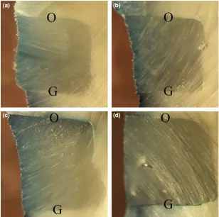
occlusal and gingival margins was investigated based on the following classification: (a) score 0: no dye penetra-tion; (b) score 1: dye penetration along the gingival/oc-clusal wall with no involvement of the axial wall; (c) score 2: dye penetration along the gingival/occlusal wall with involvement of the axial wall; and (d) score 3: dye penetration beyond the axial wall toward the pulp (Fig-ure 1).9
It should be pointed out that during the laboratory steps, the teeth were placed within a wet piece of gauze in the operator’s hand to prevent dehydration.
Statistical analysis was performed using Kruskal–Wallis
H-test. Pairwise comparisons were performed by the Mann–Whitney U-test for occlusal and gingival margins.
Data were analyzed at a significance level of 0.05 for all results.
Evaluation by scanning electron microscope
Samples for scanning electron microscope (SEM) were prepared based on the protocol reported by Korkmaz et al.14 Two teeth from each group were randomly selected for SEM evaluation of adhesive interfaces. The samples were sequentially polished with a series of sili-cone carbide abrasive papers, of 600, 800, 1200, 1500, and 2000 grit size, under running tap water as a lubricant to
Table 1. Compositions and manufacturers of study materials
Materials Composition Manufacturer
Hydrophobic resin Bis-GMA, TEGDMA Heliobond, Ivoclar Vivadent, Schaan, Liechtenstein Gel etchant 37.5% phosphoric acid, silica thickener Kerr, Orange, CA, USA
Composite resin Z250 UDMA, Bis-EMA, Bis-GMA, Zirconia glass, colloidal silica 3M ESPE, St Paul, MN, USA Bisco universal adhesive 10MDP,HEMA,Bis-GMA, ethanol, initiator, water Bisco, Schaumburg, IL, USA
(a) (b)
(c) (d)
Figure 1. Stereomicroscope images of dye penetration at occlusal (O) and gingival (G) margins of etch-and-rinse (a), etch-and-rinse with hydrophobic resin (b), self-etch (c), and self-etch with hydrophobic resin (d) groups at
smooth the surfaces. The samples were then brought into relief by being etched with 10% phosphoric acid for 10 s, followed by deproteinization in 5% sodium hypochlorite for 5 min. After being rinsed with distilled water, each sample was mounted on stubs and sputter coated with gold. The samples were carefully observed under an SEM (Tescan Vega II, Brno, Czech Republic) at the restorative material–tooth interfaces, and photographs were taken.
Results
Microleakage
Mean values standard deviations and test scores for microleakage measurements in the study groups are pre-sented in Table 2.
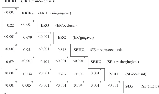
Kruskal–Wallis test showed statistically-significant
dif-ferences between test groups (P<0.001). Thus, pairwise
comparisons were done using the Mann–Whitney U-test
(Figure 2). Accordingly, the results are discussed sepa-rately for enamel and dentinal margins.
Enamel margin
The ER group had significantly less microleakage than the SE and SEH groups (P<0.001), but had no significant
difference compared with the ERH group (P>0.05). The
ERH group showed better results compared with the SEH and SE groups (P<0.001). However, there was no
statis-tically-significant difference between the SE and SEH groups (P>0.05).
Dentinal margin
The SE group had significantly less microleakage compared with the ER (P <0.001) and ERH groups (P=0.005).
Fur-thermore, microleakage of the SEH group was significantly less than the SE, ER, and ERH groups (P<0.001).
SEM observations
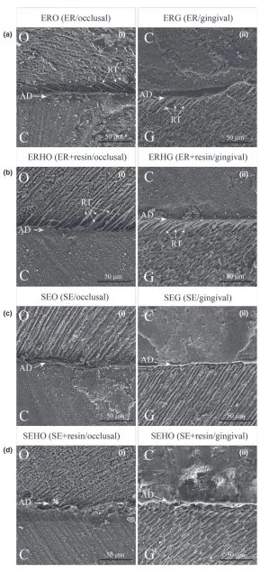
The ER samples at the enamel margin showed resin tags, microporosities, and a uniform adhesive layer that could
Table 2. MeanSD and scores of microleakage measurements
Score
Enamel margin Dentin margin
SE SEH ER ERH SE SEH ER ERH
0 0 2 10 13 0 12 0 1
1 2 1 5 3 3 4 0 3
2 5 5 1 0 5 0 9 4
3 9 8 0 0 8 0 7 8
MeanSD 2.430.18 2.180.26 0.430.15 0.180.10 1.310.11 0.250.11 2.430.12 2.180.24
ER, etch-and-rinse; ERH, etch-and-rinse with hydrophobic resin; SD, standard deviation; SE, self-etch; SEH, self-etch with hydrophobic resin.
ERHO (ER + resin/occlusal)
<0.001* ERHG (ER + resin/gingival)
0.22 <0.001* ERO (ER/occlusal)
<0.001* 0.679 <0.001* ERG (ER/gingival)
<0.001* 0.951 <0.001* 0.818 SEHO (SE + resin/occlusal)
0.674 <0.001* 0.401 <0.001* <0.001* SEHG (SE + resin/gingival)
<0.001* 0.534 <0.001* 0.767 0.603 0.001* SEO (SE/occlusal)
<0.001* 0.005* <0.001* <0.001* 0.004* 0.001* <0.001* SEG (SE/gingival)
* P< 0.05.
be seen in the enamel margin of SE samples. In the denti-nal margin, the SE samples showed a densely-packed hybrid layer compared to the ER samples, and the SEH samples had a more uniform and thicker hybrid layer compared to the SE samples (Figure 3).
Discussion
Investigation of microleakage is one of the most effective techniques for evaluating the quality of the tooth–
restora-tion interface.6 In general, this evaluation method is car-ried out byin vitrotests using dye penetration technique at the interface. The purpose of these tests is to evaluate the seal of restoration and clinical performance with regard to secondary caries and postoperative sensitivity.15 Despite some disadvantages and simplicity, this technique is both time and cost-effective, and is also considered a suitable technique for the evaluation of the performance of restorative materials.2
The last generation of dental adhesive systems, intro-duced recently, is the multimode one-bottle universal adhesive systems, which are more user-friendly. These sys-tems have a major advantage over previous syssys-tems, as they can be applied in both the SE and ER modes. This has attracted a lot of attention due to the possibility of decreasing chair time and greater flexibility with their application.6,16,17
The results of the present study showed less microleak-age with the ER and SE modes at the enamel and dentinal margins, respectively. It is obvious that the preparation of enamel for bonding purposes involves the use of acid etching, as shown in a large number of laboratory and clinical studies.18,19Acid etching of the enamel can create very fine porosities that increase micromechanical inter-locking. Furthermore, applying systems containing func-tional monomers, with the potential to form chemical bonds with hydroxyapatite, help the bonding mechanism beside the effects of porosities. Universal adhesive systems have been classified under the category of mild SE adhe-sive systems. The efficacy of mild SE systems on enamel has been evaluated in various studies.18,19 Peumans et al.18showed that if acid etching is not used, there will be more enamel defects compared to the situation when a separate pre-etching step is carried out at enamel mar-gins. Hanabusa et al.8 demonstrated that the universal adhesive systems exhibited higher bond quality with the ER mode at enamel margins and with the SE mode at dental margins. Furthermore, previous studies have shown that acid etching should be carried out on the enamel margins of the cavity only.7,20 It has also been
reported that etching the enamel alone without etching the dentin improves the marginal integrity of the adhesive systems, which is in agreement with the results of the
present study; however, this is associated with many problems.8 Limiting acid etching to enamel is difficult practically, if not impossible, especially when it is applied to the small cavities where the acid also contacts dentin. This significantly intensifies within the case of applying low-viscosity acids.21 Therefore, the use of acid etching
only on enamel (selective etching of enamel) is a major challenge for dentists. This necessitates further studies on the synthesis of materials using more controlled viscosities and techniques to apply such materials with further con-trol and selectively to enamel.
According to Alexandra et al., selective enamel etching did not improve the retention rate of two-step SE adhe-sives that can bond chemically to the tooth structure.17 However, After 8 years of clinical service, selective enamel etching resulted in minor positive effects on marginal integrity and less discoloration in the enamel margins when compared with the SE mode.18Similarly, in the case of applying the universal adhesive system, it has been reported that the long-term performance of these systems is not dependent on the adhesive strategy, except for mar-ginal adaptation.7,17
However, in the dentin substrate, the acid did not improve the bond strength due to structural complexities, the hydrophilic nature of dentin, and the presence of fluid-containing dentinal tubules.8 Marchesi et al.9 showed that the quality of the bond with the use of uni-versal adhesive systems in the SE mode was better with dentin. They showed that the ER mode yielded the same immediate bond strength compared to that with the SE mode in dentin; however, the bond strength decreased over time. The differences in the bond strength in such situations might be attributed to the presence of the 10-Methacryloyloxi-decyl-dihydrogen-phosphate (MDP) monomer in these adhesives. This monomer is responsi-ble for the chemical bond of the adhesive with the resid-ual hydroxyapatite on collagen fibers that might help the long-term stability and longevity of the bond.20In the ER mode, the application of acid removes the majority of dentinal hydroxyapatite, making it an inappropriate sub-strate for the chemical bonding of these systems. This supports the obtained results of the present study, as our applied adhesive system also contained this monomer. Of course, it should be pointed out that the use of these sys-tems in association with acid etching might increase the odds of postoperative sensitivity and degradation of colla-gen fibrils in dentin. The collapse of collacolla-gen fibers pre-vents the adhesive from penetrating the dentin surface, giving rise to the creation of an inefficient dentin bond.9
(i) (a)
(b)
(c)
(d)
(ii)
(i) (ii)
(i) (ii)
(i) (ii)
the prospecting long-term performance of adhesive sys-tems, their compositional formulation must be also con-sidered. For example, the presence of hydroxyethyl methacrylate in the adhesive could prevent self-assembled nanolayering. In fact, nanolayering of two 10-MDP mole-cules could result in an adhesive interface more resistant to degradation.7 It is has also been reported that the long-term performance of universal adhesives is not dependent on their using strategy in dentin.7,17
Another important finding of the present study was that the use of a layer of hydrophobic resin did not affect the ER mode in either enamel or dentinal mar-gins, but drastically improved the SE mode in the denti-nal margin. It should be noted that these findings are consistent with the results reported by Miguel et al.10 regarding the bonding efficacy of universal adhesives in dentin. They reported that applying an extra hydropho-bic resin coat increases DC of the SE mode at the denti-nal margin, but has no significant effect on the ER mode, which is in agreement with our results based on approximately the same microleakage in both the ER and ERH groups (Table 2). Moreover, we found that the resin coat did not improve the microleakage of the ER mode in the occlusal margin, which is attributed to the probable poor effects of the resin coat on DC. One-step SE systems lack a non-solvated resin layer, which makes them permeable membranes. This permeable membrane allows the rapid movement of fluids within the dentinal tubules through the adhesive layer. This fluid movement might jeopardize the DC, which finally results in its hydrolytic degradation.2–5,22 Studies have
shown that DC of the adhesives is inversely related to the inherent permeability of an adhesive layer.23 An adhesive layer that has not been cured completely is more permeable to fluids, such as water, which nega-tively affects bond strength.24 In addition, compared to a fully-cured adhesive layer, the presence of higher amounts of unreacted monomers in these layers can accelerate water sorption.25 An extra layer of hydropho-bic resin adds unsolvated hydrophohydropho-bic monomers to the interface leading to decreased amounts of relative con-centrations of retained solvents and uncured monomers in the adhesive layer.10 It has also been reported that applying an extra layer of a hydrophobic resin results in higher DC and lower adhesive permeability.10 Moreover, the presence of the resin layer decreases the degradation effects of the adhesive interface over time, which is attributed to the formation of a densely-packed hybrid layer in dentin.10
As illustrated in Figure 3, the dentin/composite inter-face of the SEH sample is somewhat thicker and more uniform compared to the interface of the SE sample. This could be responsible for the reduced microleakage in the
SEH group compared to the SE group (Table 2, Fig-ure 2). However, there were no detectable differences between the adhesive interface in the SE and SEH samples at the enamel margin, which verifies the corresponding microleakage results.
Resin tags can be seen in both ER and ERH samples, as expected.26The reduced microleakage in the ER and ERH groups compared to the SE and SEH groups was attribu-ted to the presence of resin tags at the enamel/composite interface (Figure 3). In the SE and SEH samples, there were some micro-irregularities at the enamel/composite interface, which could be the reason for the increased microleakage compared to that in the dentin/composite interface.
Generally,in vitro studies have limited significance for the prediction of clinical results, but they are necessary for the early screening of new materials, providing recom-mendation for handling, reducing the number of clinical trials, reducing risks in clinical trials, and analyzing fail-ures in detail for further improvements.15The
microleak-age and bond strength of the applied materials might be affected by the clinical performance of applied materials involved with a large number of variables, such as stresses resulting from occlusal forces, temperature and pH changes, and continuous presence in a moist environ-ment.21 In order to simulate an environment as close to the oral environment as possible the samples were ther-mocycled; however, this process cannot replace the long-term aging of the bond in the oral cavity.
Conclusion
Based on the findings of this study, it can be concluded that applying an extra layer of hydrophobic resin signifi-cantly decreases microleakage of the universal adhesive system in the SE mode at dentinal margin, while having no significant effect on the ER mode; the ER mode is more practical in the enamel margin compared to the dentinal margin; and the SE mode provides better results in the dentinal margin compared to the enamel margin.
Acknowledgments
References
1 Perdig~ao J, Mu~noz MA, Sezinando A
et al. Immediate adhesive properties to dentin and enamel of a universal adhesive associated with a hydropho-bic resin coat.Oper Dent2014;39: 489–99.
2 Tay FR, Pashley DH, Suh BI, Car-valho RM, Itthagarun A. Single-step adhesives are permeable membranes.J Dent2002;30: 371
–82.
3 Tay FR, Pashley DH, Suh B, Carvalho R, Miller M. Single-step, self-etch adhesives behave as perme- able membranes after polymerization. Part I. Bond strength and morphologic evidence American.Am J Dent2004; 17: 271
–8.
4 Tay FR, Pashley DH, Garcia-Godoy F, Yiu CK. Single-step, self-etch adhe-sives behave as permeable membranes after polymerization. Part II. Silver tracer penetration evidence.Am J Dent2004;17: 315
–22.
5 Tay FR, Lai CN, Chersoni Set al. Osmotic blistering in enamel bonded with one-step self-etch adhesives.J Dent Res2004;83: 290
–5.
6 Mu~noz MA, Luque I, Hass V, Reis A,
Loguercio AD, Bombarda NHC. Immediate bonding properties of uni-versal adhesives to dentine.J Dent 2013;41: 404
–11.
7 Perdig~ao J, Kose C, Mena-Serrano AP et al. A new universal simplified adhesive: 18 month clinical evalua-tion.Oper Dent2014;39: 113
–27.
8 Hanabusa M, Mine A, Kuboki Tet al. Bonding effectiveness of a new ‘multi-mode’ adhesive to enamel and den-tine.J Dent2012;40: 475
–84.
9 Marchesi G, Frasseto A, Mazzoni A et al. Adhesive performance of a
mul-ti-mode adhesive system:1-year in vitrostudy.J Dent2014;42: 603
–
12.
10 Miguel AM, Ana S, Issis LMet al. Influence of a hydrophobic resin coating on the bonding efficacy of three universal adhesives.J Dent2014; 42: 595
–602.
11 Santini A, Ivanovic V, Ibbetson R, Milia E. Influence of marginal bevels on microleakage around class V cavi-ties bonded with seven self-etching agents.Am J Dent2004;17: 257
–61.
12 Borges MA, Matos IC, Dias KR. Influence of two self-etching primer systems on enamel adhesion.Braz Dent J2007;18: 113
–8.
13 Crim GA, Chapman KW. Reducing microleakage in class II restorations: anin vitrostudy.Quintessence Int 1994;25: 781
–5.
14 Korkmaz Y, Ozel E, Attar N, Bicer CO, Firatli E. Microleakage and scan-ning electron microscopy evaluation of all-in-on self-etch adhesives and their respective nanocomposite pre-pared by erbium:yttrium-aluminum-garnet laser and bur.Laser Med Sci 2010;25: 493
–502.
15 Siegward DH. Systematic reviews: I. The correlation between laboratory tests on marginal quality and bond strength. II. The correlation between marginal quality and clinical out-come.J Adhes Dent2007;9: 77
–106.
16 van Dijkena J, Sunnegardh-Gronbergb K, Sorenssonb E. Clinical bonding of a single-step self-etching adhesive in noncarious cervical lesions.J Adhes Dent2007;9: 241
–3.
17 Mena-Serrano A, Kose C, De Paula EAet al. A new universal simplified adhesive: 6-month clinical evaluation. J Esthet Restor Dent2013;25: 55
–69.
18 Peumans M, De Munck J, Van Lan-duyt KL, Poitevin A, Lambrechts P, Van Meerbeek B. Eight-year clinical evaluation of a 2-step self-etch adhe-sive with and without selective enamel etching.Dent Mater2010;26: 1176–84.
19 Perdig~ao J, Carmo AR, Anauate-Netto Cet al. Clinical performance of a self-etching adhesive at 18 months. Am J Dent2005;18: 135
–40.
20 Perdig~ao J, Sezinando A, Monteiro
PC. Laboratory bonding ability of a multi-purpose dentin adhesive.Am J Dent2012;25: 153
–8.
21 Li H, Burrow MF, Tyas MJ. The effect of load cycling on the nanoleakage of dentin bonding sys-tems.Dent Mater2002;18: 111
–9.
22 Salz U, Zimmermann J, Zeuner F, Moszner N. Hydrolytic stability of self-etching adhesive systems.J Adhes Dent2005;7: 107
–16.
23 Cadenaro M, Antoniolli F, Sauro S et al. Degree of conversion and per-meability of dental adhesives.Eur J Oral Sci2005;113: 525
–30.
24 Tay FR, Pashley DH, Hiraishi Net al. Tubular occlusion prevents water-treeing and through-and-through fluid movement in a single-bottle, one-step self-etch adhesive model.J Dent Res2005;84: 891
–6.
25 Takahashi A, Inoue S, Kawamoto C et al.In vivolong-term durability of the bond to dentin using two adhe-sive systems.J Adhes Dent2002;4: 151–9.
26 Bertrand MF, Hessleyer D, Muller-Bolla M, Nammour S, Rocca JP. Scanning electron microscopic evalua-tion of resin-dentin interface after Er: YAG laser preparation.Lasers Surg Med2004;35: 51