International Medical Journal Vol. 21, No. 1, pp. 101 - 105 , February 2014
SHORT COMMUNICATIONS
The Effect of Microleakage on Composite Resin Restorations
Cured by Different Light Curing Units (LCU)
Sri Ram Kumar
1), Norhayati Luddin
2), Ziyad Kamal Mahmoud Mohammad
3),
Mohammad Khursheed Alam
2)ABSTRACT
Objective: This study aims to compare the microleakage of Class V composite resin (CR) restorations; (i) cured using differ-ent LCU, (ii) restored by differdiffer-ent CR and cured with differdiffer-ent LCU, (iii) restored by differdiffer-ent CR and cured with differdiffer-ent LCU at the occusal and gingival margin.
Materials and Methods: Sixty (60) permanent upper premolars were used. Two class V cavities (3 millimeter (mm) x 2 mm) with the occlusal and gingival margin ended 1 mm above and below cemento-enamel junction were prepared on the buccal and lingual surface of each tooth. The 120 cavities were divided randomly into four groups (n=30). Cavities in group one and three were restored with nanocomposite while cavities in group two and four were restored with microhybrid. Cavities in group one and two were cured using LED LCU while cavities in group three and four were cured using Halogen LCU. The samples were then immersed in 0.5% methylene blue dye for 24 hours and sectioned longitudinally. Microleakage at the occlusal and gingival margin was quantified in mm using stereomicroscope at 40x magnification. Data were analyzed using Mann-Whitney test and results with p < 0.05 were considered significant.
Results: No significant differences in microleakage score were observed between use of different LCUs and different CRs. Both types of CRs cured using Halogen LCU showed statistically significant difference in microleakage score at the occlusal and gingival margin (p < 0.05).
Conclusion: Microleakage was still present in both types of CRs cured using both LCUs. However, nanocomposite cured using LED LCU showed the least microleakage score.
KEY WORDS
microleakage, Halogen, LED, nanocomposite, microhybrid
Received on October 1, 2012 and accepted on January 16, 2013 1) Ministry of Health, Malaysia
2) School of Dental Sciences, Universiti Sains Malaysia Kubang Kerian, 16150, Kelantan, Malaysia 3) Arab American University
Jenin, Palestine
Correspondence to: Norhayati Luddin (e-mail: norhayati @ kck.usm.my)
101
INTRODUCTION
The demand for esthetic dentistry has grown dramatically and there has been rapid development of new restorative materials that can restore the color and characteristic of natural teeth. Among vari-ous restorative materials, the light activated dental material, namely composite resin has been widely used due to its low cost and conser-vative technique when compared with indirect restorations. Light cured composite resin (CR) materials have several advantages such as control of the contour during restoration placement, better color stability and a more complete polymerization as compared to chemi-cally activated materials (Burgess et al., 2002). Yet, a major draw-back with CR is the formation of microleakage. Microleakage is a phenomenon of the diffusion of organic or inorganic substances into a tooth through the interface between the restorative material and the tooth structure due to gap formation (de Almeida et al., 2003). This leads to a variety of clinical conditions such as discoloration, pulpal irritation, postoperative sensitivity, and eventual failure of the restoration (Hofmann et al., 2002). After decades of research, several
causes have been identified as contributing factors to microleakage. Among them, there are polymerization shrinkage, dissolution of lin-ear or smlin-ear layers, and varying coefficients of thermal expansion for restorations (Oilo et al., 1992, Fortin et al., 1994, Suzuki et al., 1985). It has been well established that polymerization shrinkage plays a major role in the outcome of the CR restorations as compared to other factors (Yap et al., 2000). Nurray et al (2007) cited that, Pradelle et al (2003) and Feilzer et al (1987) reported that shrinkage depends on the configuration of the restoration (C factor), type and shade of the CR, viscoelastic properties of the dentin bonding system used in adhesive procedure and the restorative technique used. Apart from that, the light curing units can significantly influence the degree of polymerization of the light activated CRs (Rahiotis et al., 2004, Knobloch et al., 2004). The effectiveness of light curing itself seems to be affected by the intensity of light emitted, the spectral output of the light source and the curing mode. In conjunction with this, differ-ent light curing units have been evaluated with regard to their effec-tiveness on light curing CR, and some controversies still existed in the literature (Powell et al., 2000).
Light activated CR has been polymerized by UV light since their first introduction in the world of dentistry. Later on, it has been replaced by blue light commonly found in halogen bulbs. This is pos-sible due to the usage of camphorquinone as the photoinitiator in CR, which is sensitive to this blue light. Yet, it has been noted that this light curing had many inherent limitations. Among them is the degra-dation of bulb, due to limited life span (40-100 hours). Such degrada-tion occurs over time due to high operating temperature. As the degradation takes place, it affects the intensity of light. Thus, an aged light curing unit (LCU) is unable to polymerize CR, which in turn causes a decrease in favorable properties of composite material, due to limited depth of cure and relatively long exposure time needed to light cure the material (Rahiotis et al., 2004, Nomoto et al., 1994). In the meantime, the vitality of pulp can be affected by rising tempera-ture during polymerization (Hannig et al., 1999). As a result, new technologies such as the light emitting diode (LED) have been intro-duced to the dental profession as an alternative to the conventional curing units. Innovative LED technology for light curing dental mate-rials has been improved in order to overcome the inadequacies of halogen LCU. LED LCU creates a narrow bandwidth of light spectral output. This easily falls within the absorption spectrum of the cham-porquinone photoinitiator (400-500 nm). That is present in the light activated dental materials, so no filters are required in LED LCUs (Haitz et al., 1995). LEDs also have a lifetime of several thousand hours without significant degradation of light emission over time (Haitz et al., 1995). They are inexpensive, very compact low voltage devices, have better resistance to shock and vibration, and they are also portable and safe (Mills et al., 2002). Although they have lower light emission, LED LCU has the ability to cure like other light sources or slightly less (Dunn et al., 2002). In addition, the tempera-ture increase is significantly less and it does not pose any threat to the pulpal tissue (Hofmann et al., 2002, Soh et al., 2003). Saving time during the light curing process is one of the most important aspects when using LED LCU especially to clinicians who use incre-mental filling technique.
Some studies have evaluated the use of these different light sources for composite resin polymerization, and it has been reported that the LED (Rahiotis et al., 2004) and Halogen (Powell et al., 2000) can be considered as an alternative tools in clinical practice to cure CR. Even though LED LCU has less than half of irradiance as com-pared to Halogen LCU, it displayed significantly less microleakage at the gingival interface (Oberholzer et al., 2004). However, no signifi-cant difference was found in microleakage between the use of two
different LCUs at the enamel interface.
Light emitting diode has gained vast popularity among dentist and is currently the light curing unit of choice in clinical practice. Even though numerous studies have been carried out to prove the capability of LED LCU, it is still not clear whether the LED LCU can cure newer generation of composite resin, which is different in its composition and filler particle size and yet still obtain the desired or acceptable microleakage level.
Therefore, the aims of this study are:
I. To compare the microleakage of Class V composite resin restorations cured using different LCUs (Halogen vs. LED). II. To compare the microleakage of Class V composite resin
restoration restored with different composite resins (microhy-brid vs. nanocomposite) and cured using different LCUs (Halogen vs. LED).
III. To compare the microleakage of Class V composite resin restoration restored with different composite resins (microhy-brid vs. nanocomposite) and cured using different LCUs (Halogen vs. LED), between the occlusal and gingival mar-gin.
MATERIALS AND METHODS
A total of 60 extracted, sound, human permanent upper premolars were used in this study. The tooth samples were collected, calculi and residual soft tissues were carefully removed using ultrasonic scaler, and then the tooth were stored in 10% formalin at room temperature (27 ) within one month after extraction. The tooth samples were checked for cracks under 10X magnification with a stereomicroscope (Material Testing System, LEICA, Germany).
Two Class V cavities were made on the buccal and lingual sur-face of each tooth with rounded internal angles. The cavities were prepared 1mm above and below the cemento-enamel junction (CEJ). CEJ was the reference point for the cavity preparation, whereby the occlusal margin was finished in enamel and the gingival margin was in dentin/cementum. No enamel was left on the gingival margin. The cavities were approximately 3 mm in width, and 2 mm in depth. All cavity preparations were performed with a high-speed hand piece with water spray using a #1090-diamond fissure bur (Diatech Dental AG, Heerbrugg Switerland) with a diameter of 0.5mm. Dimensions
Table 1. Experimental group in the study
Groups Composite Light Curing Unit Sample Size
1 Filtek Z350 LED 15 tooth (30 cavities)
2 Filtek Z250 LED 15 tooth (30 cavities)
3 Filtek Z350 Halogen 15 tooth (30 cavities)
4 Filtek Z250 Halogen 15 tooth (30 cavities)
Table 2. Composition of composite resins and adhesive system used in this study
MATERIAL BASIC COMPOSITION MANUFACTURER
Nanocomposite Filler particle: (filler loading 59.5%) 3M, ESPE, Dental
(Filtek Z350) Aggregated Zirconia/silica filler (5-20nm) Product, St. Paul,
Non-agglomerated silica (20-25nm) MN, USA
Resin:
Bis-GMA, UDMA, TEGDMA, bis-EMA
resins
Microhybrid Filler particle: (filler loading 60%) 3M ESPE Dental
(Filtek Z250) Zirconia/silica filler (0.01-3.5 µ m ) Product, St. Paul,
Resin: MN, USA
Bis-GMA, UDMA, bis-EMA resins
G-Bond Phosphoric acid ester monomer, 4-META GC, Japan
monomer, Nano-filled particles, Acetone
and Water Solvent
Table 3. Types of LCUs that were used in this study
LC MANUFACTURER COUNTRY POWER SOURCE
Smartlite (LED) Denstlpy UK Mains
LC Astralis 3 Ivolar Schaan Mains
were checked with the periodontal probe. The prepared teeth were stored in distilled water until restored. Block randomization method were used to divide the prepared cavities into four groups (www.ran-domization.com), the groups were arranged and filled as shown in Table 1 (n = 30). The compositions of adhesive system and compos-ite resins used in this study are described in Table 2. The LCU used in this study is described in Table 3.
The entire 120 cavities were conditioned with G-bond (GC, Japan). A microbrush was used to coat the entire cavities in a group (n = 30), then after 10 seconds, the cavities were air thinned with high-pressure syringe for 5 seconds in the presence of vacuum air suction and cured according to the following groups:
Group 1 (G1):
The G-Bond was cured using LED LCU for 10 seconds. The cav-ities were filled with nanocomposite (Filtek Z350) in one horizontal increment. It was then cured with LED LCU for 40 seconds from the buccal or lingual aspect.
Group 2 (G2):
The G-Bond was cured using LED LCU for 10 seconds. The cav-ities were filled with microhybrid (Filtek Z250) in one horizontal increment. It was then cured with LED LCU for 40 seconds from the buccal or lingual aspect
Group 3 (G3):
The G-Bond was cured using Halogen LCU for 10 seconds. The cavities were filled with nanocomposite (Filtek Z350) in one horizon-tal increment. It was then cured with Halogen LCU for 40 seconds from the buccal or lingual aspect.
Group 4 (G4):
The G-Bond was cured using Halogen LCU for 10 seconds. The cavities were filled with microhybrid (Filtek Z250) in one horizontal increment. It was then cured with Halogen LCU for 40 seconds from the buccal or lingual aspect.
For all groups, cellulose strip was used to reestablish the contour of the tooth. The cellulose strip was held by finger pressure against the margin of the cavity so that the preparations were not overfilled at both the occlusal and gingival margin. Prior to curing each new restoration, the light source irradiance intensity was measured with a
radiometer CURE RITE (Dentsply, USA). The light intensities mea-sured was between 480 and 520 mW per square centimeter and no decrease in the output could be observed. The exit window of the LCU then was placed opposing the center of the restored cavity and cured at a constant irradiation distance of 4 mm. Immediately, the restorations were polished with a graded series of Sof-lex disc sys-tems (3M ESPE, USA). Specimens were then dried at room tempera-ture and the apices were sealed with sticky wax. Then, two coats of fingernail varnish were applied on the tooth 1mm short of the mar-gins of each restoration. Nail varnish was then left to dry at room temperature. Teeth were then kept in distilled water for 24 hours to rehydrate as dehydration might have occurred during nail varnish application and drying.
Finally, all the teeth were then immersed in 0.5% methylene blue solution for 24 hours. Samples were subsequently rinsed under run-ning tap water to remove dye for 5 seconds and then dried at room temperature. Then samples were sectioned buccal-lingually through the center of the restoration and teeth under copious of water using a diamond disc (EXAKT hard material cutting, Germany). Each section was labeled by the group name (G1, G2, G3 and G4), sample number (1, 2, 3...120), the section name (A, B) and the side measured were labeled by ( ).
The degree of dye penetration was observed under a 40X magni-fication with a stereomicroscope (Material Work Station, LEICA, Germany). Section showing greater leakage was used for microleak-age measurements. Dye penetration was quantified for occlusal and gingival margins separately. The depth measurements of microleak-age through the tooth-restoration interface at occlusal and gingival wall of the cavity were analyzed under calibrated image analyzer (Material Work Station, LEICA, Germany). Specimens showing dye penetration from the lateral and pulpal floor of the restoration were not recorded to exclude other sources of dye penetration.
The data was statistically analyzed via SPSS Software version 12.01. Non-parametric Mann-Whitney test was used to compare lin-ear dye penetration between the light curing units at 95% confidence interval. The test was performed at level of significance of P < 0.05.
Microleakage on Composite Resin Restorations
103
Table 4. Comparison of microleakage of Class V composite resin restorations cured using Halogen (n = 60) and LED LCUs (n = 60)
Variable Halogen LED Z P
Median (IQR) Median (IQR) statistica valuea
Microleakage at
occlusal margin 0.12 (0.26) 0.06 (0.21) 1.074 0.28
Microleakage at
gingival margin 0.25 (0.35) 0.15 (0.27) -1.594 0.11
aMann-Whitney
Table 5. Comparison of microleakage of Class V composite resin restoration restored with microhybrid (n = 60) and nanocom-posite (n = 60) and cured using different LCUs
Variable Microhybrid Nanocomposite Z P
Median (IQR) Median (IQR) statistica valuea
LED LCU 0.12 (0.20) 0.12 (0.30) -0.151 0.88
Halogen LCU 0.21 (0.36) 0.18 (0.35) -0.726 0.47
aMann-Whitney
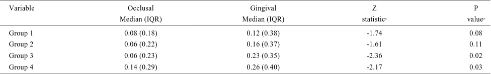
Table 6. Comparison of microleakage of Class V composite resin restoration restored with microhybrid and nanocomposite and cured using different Halogen and LED LCUs, between the occlusal and gingival margin
Variable Occlusal Gingival Z P
Median (IQR) Median (IQR) statistica valuea
Group 1 0.08 (0.18) 0.12 (0.38) -1.74 0.08
Group 2 0.06 (0.22) 0.16 (0.37) -1.61 0.11
Group 3 0.06 (0.23) 0.23 (0.35) -2.36 0.02
Group 4 0.14 (0.29) 0.26 (0.40) -2.17 0.03
RESULTS
Comparison of microleakage of Class V composite resin restorations cured using different LCUs (Halogen vs. LED).
This study compares the microleakage of Class V composite resin restorations cured using different LCUs (Halogen vs. LED). Cavities in group one and two were compared with cavities in group three and four for the occlusal and gingival margin separately. As a result, the measurements of 60 samples of Halogen cured composite resin were compared with the measurements of 60 samples of LED cured com-posite resins for both the occlusal and gingival margin. The statistical analysis of microleakage of Class V composite resin restoration cured using Halogen and LED LCUs showed no significant differences between the two groups at either of the margins. Table 2, represents the results of the statistical analysis of the first objective of the study with P > 0.05.
Comparison of microleakage of Class V composite resin restoration restored with different composite resin (micro-hybrid vs. nanocomposite) and cured using different LCUs (Halogen vs. LED).
This study compares the microleakage of Class V composite resin restoration restored with different composite resin (microhybrid vs. nanocomposite) and cured using different LCUs (Halogen vs. LED). Cavities in group one and three were compared with cavities in group two and four. As a result, the measurements of 60 samples of micro-hybrid were compared with the measurements of 60 samples of nanocomposite for both the occlusal and gingival margin.
The statistical analysis of microleakage of Class V composite resin restoration restored with microhybrid (Filtek Z250) and nanocomposite (Filtek Z350) and cured using different LCUs, showed no significant differences in the two groups. Table 3, repre-sents
Comparison of microleakage of Class V composite resin restoration restored with different composite resin (micro-hybrid vs. nanocomposite) and cured using different LCUs (Halogen vs. LED), between the occlusal and gingival margin.
This study compares the microleakage of Class V composite resin restoration restored with different composite resin (microhybrid vs. nanocomposite) and cured using different LCUs (Halogen vs. LED), between the occlusal and gingival margin. The linear dye penetration in each group is compared between the occlusal and gingival margin. The statistical analysis of microleakage of Class V composite resin restoration restored with different composite resin (microhybrid vs. nanocomposite) and cured using LED LCU showed no significant differences at occlusal and gingival margin. The statistical analysis of microleakage of Class V composite resin restoration restored with different composite resin (microhybrid vs. nanocomposite) and cured using Halogen LCU showed significant differences at occlusal and gingival margin in both the groups. Table 4, represents the results of the statistical analysis of the third objective of the study with P < 0.05.
DISCUSSION
Microleakage is an important parameter that has a bearing on the behavior of composite resin restorations in the oral environment. Thus, it is imperative to prevent or improve the causes of microleak-age (Cheong et al., 2012). Among the causes that are mainly dis-cussed by dental practitioners is the "polymerization shrinkage". Adequate polymerization is required for clinically successful restora-tions. The quality of polymerization of composite is one of the important factors affecting the longevity of any composite resin restorations. Thus, it is important in order to have adequate polymer-ization, proper LCU must be used. As such, the main purpose of this study was to determine the curing performance of the commercially used LED and Halogen light curing units in curing different types of
composite restorative material in Class V restorations. LED LCUs are gaining vast popularity among dental practitioners, thus it is impor-tant from a practical and fundamental point of view that this type of LCUs are used to achieve maximum and adequate polymerization of composite resins. However, conflicting results are often reported in the literature when different LCUs were used on composite restora-tive materials. This might be due to the fact there is a difference between the irradiation protocols used, especially in the intensity of light used. Due to this, a standard clinically used Halogen LCU was used in this study for comparison with LED LCU. The methylene blue immersion test is a method, attempted to stimulate the possibili-ty of a gap existing between a restoration and tooth structure that causes microleakage.
The first hypothesis tested in this experiment was that the posite resins cured using LED LCU has lower microleakage com-pared to Halogen LCU. This study found that the median microleak-age score of LED LCU cured composite material at the occlusal (0.06 mm) and gingival (0.15 mm) margin, was lower than Halogen LCU cured composite material at the occlusal (0.12 mm) and gingival (0.25 mm) margin. However, the results showed no statistical signifi-cant difference between the two different LCUs (p > 0.05). This result is in consistent with a study done by Oberholzer et al., (2004) which concluded that significantly less microleakage occurred at the gingival margin when restorations were cured with an LED LCU compared to curing with standard Halogen LCU. And they also found that there is no significant difference in microleakage between LCUs at the occlusal margin.
In order to prevent microleakage it seems clear that all the mar-gins of the cavity should be kept within the enamel margin. However, root caries, abfraction, and abrasion lesions have their margins in dentin or cementum (Georges et al., 2002). Hence, the effect of dif-ferent LCU on composite resin at the gingival margin provides a good clinical view on the effectiveness of preventing microleakage. Based on this study, LED LCU has shown lesser microleakage score compared to Halogen LCU at the occlusal and gingival margin in both types of the composites even though it is not statistically signifi-cant (p > 0.05). The result of this study however, might be clinically relevant.
The second hypothesis tested in this experiment was that nanocomposite (Filtek Z350) has lower microleakage compared to microhybrid (Filtek Z250) composite resin if cured using LED LCU. This study found that the median microleakage score of nanocompos-ite (0.12 mm) and microhybrid (0.12 mm) cured using LED LCU was not different. Meanwhile the mean microleakage of nanocomposite (0.18) was lower than microhybrid (0.21 mm) cured using Halogen LCU. The results were not of statistical significant difference (p > 0.05). This result is in consistent with a study done by Ernst et al., (2006) whom concluded that there are no significant differences between both types of dental composite.
Composite resins referred to as "nanofilled composite" are pro-duced with nanofiller technology and formulated with nanomer and nanocluster filler particles. The combination of nanomer sized parti-cles and nanocluster formulations reduces the interstitial spacing of the filler particles providing increased filler loading, better physical properties, and improved retention of a polished surface (3M Inc., 2002). Regardless of any current significant improvements in the either the type of LCUs used, the material used for filling and finally the bonding systems, no system is currently able to completely pre-vent the formation of polymerization shrinkage gaps between the interface of the restoration and the cavity margin, especially at the gingival margin.
The third hypothesis tested in this experiment was that gingival margin has greater microleakage as compared to occusal margin in both types of the composite materials cured using Halogen and LED LCUs. This study, found that the median microleakage score of nanocomposite cured using LED LCU at the occlusal (0.08 mm) was lower than the gingival (0.12 mm) margin, similar to the median microleakage of microhybrid cured using LED LCU at the occlusal (0.06 mm) was lower than the gingival (0.16 mm) margin. However, the results showed no statistical significant difference between the occlusal and gingival margin in both group 1 and 2 respectively (p > 0.05).
and 4 (p < 0.05).
With latest invention and research, there has been an increase in usage of adhesive system and dental composites, thus newer materi-als with simpler and better properties are being introduced, which in turn help to improve dental services and prolong the restorations life. Self-ecth adhesives have recently become available and combine the functions of primer and adhesive components that have eliminated the need for separate acid etching and rinsing steps (Sensi et al., 2005). The difference in result between the occlusal and gingival margin is because the etching of the cavity wall will remove smear layer and clearing the dentinal tubules thus enhances formation of hybrid layer. However, there is a mark difference of the number and size of dentinal tubules present between a gingival and occlusal mar-gin within the same cavity. Pronounced microleakage at the mar-gingival than occlusal margin had appeared in other study (Oberholzer et al., 2004). Resin bonding of the gingival margin was less predictable due to the oblique orientation (Mixson et al., 1995) and the lower density of tubules than in deep dentine (Ferrari et al., 2001). The absence of an outward flow of dentinal fluid and a completely altered dentinal surface, leads to a poor correlation between in vivo and in vitro con-ditions.
CONCLUSION
It can be concluded that nanocomposite cured using LED LCU showed the least microleakage score. However, microleakage was still present even though with the adhesive systems, nanofilled com-posite resins, and the LCUs used in this study. Both LED and Halogen LCU did not eliminate microleakage in curing both types of the composite resin materials, but there was lesser microleakage score in nanocomposite cured using LED LCU.
REFERENCES
Burgess JO, Walker RS, Porche CJ, Rappold AJ. (2002). Light curing an update. Compend Contin Educ Dent, 23, 889-892.
de Almeida JB, Platt JA, Oshida Y, Moore BK, Cochran MA, Eckert GJ. (2003). Three dif-ferent methods to evaluate microleakage of packable composites in Class II restora-tions. J Op Dent, 28, 453-460.
Cheong Ian AT, Abdul Muttlib NA, Wan Bakar WZ, Alam MK. (2013). Comparison
between microleakage of composite and porcelain in class V restoration: an in vitro study. International Medical Journal, 20(3), 359-362.
Dunn WJ, Bush AC. (2002). A comparison of polymerization by light-emitting diode and halogen-based light curing units. J Am Dent Assoc, 133, 335-341.
Hofmann N, Hugo B, Klaiber B. (2002). Effect of irradiation type (LED or QTH) on photo-activated composite shrinkage strain kinetics, temperature rise, and hardness. J Oral Sci, 110, 471-479.
Ferrari M, Cagidiaco MC, Vichi A, Mannocci F, Mason PN, Mjor IA. (2001). Bonding of all-porcelain crown: structural characteristics of the substrate. J Dent Mat, 17, 156-164.
Fortin D, Swift EJ Jr, Denehy GE, Reinhardt JW. (1994). Bond strength and microleakage of current dentine adhesives. J Dent Mat, 10, 253-258.
Hannig M, Bott B. (1999). In-vitro pulp chamber temperature rise during composite resin polymerization with various light curing sources. J Dent Mat, 15, 275-281. Haitz RH, Craford MG, Weissman RH. (1995). Light emitting diodes. In Bass M, (Ed.),
Handbooks of optics. 2nd ed. New York: Mc Graw Hill Inc.
Knobloch LA, Kerby RE, Clelland N, Lee J. (2004). Hardness and degree of conversion of posterior packable composites. J Op Dent, 29, 642-649.
Mills RW, Uhl A, Jandt KD. (2002). Optical power outputs, spectra and dental composite depths of cure, obtained with blue light emitting diode (LED) and halogen light curing units (LCUs). Br Dent J, 193, 459-463.
Mixson JM, Spencer P, Moore DL, Chappel RP, Adams S. (1995). Surface morphology and chemical characterization of abrasion/erosion lesions. Am J Dent, 8, 5-9.
Nomoto R, Uchida K, Hirasawa T. (1994). Effect of light intensity on polymerization of light cured composite resins. J Dent Mat, 12, 198-205.
Nuray A, Yonca K. (2007). The effect of two light-emittting diode (LED) and one Halogen curing light on the microleakage of class V flowable composite restorations. J Contem Dent Practice, 8(2), 80-8.
Oberholzer TG, Schunemann M, Kidd M. (2004). Effect of LED curing on microleakage and microhardness of Class V resin-based composite restorations. Am J Dent, 54, 15-20.
Oilo G. (1992) Biodegradation of dental composites/glass ionomer cements. Advanc Dent Restoration, 6, 50-54.
Powell GL, Blankenau RJ. (2000). Laser curing of dental materials. Dent Clin North, 44, 923-930.
Rahiotis C, Kakaboura A, Loukidis M, Vougiouklakis G. (2004). Curing efficiency of vari-ous types of light curing units. J Oral Sci, 112, 89-94.
Soh MS, Yap AU, Siow KS. (2003). The effectiveness of cure of LED and halogen curing lights at varying cavity depths. J Op Dent, 28, 707-715.
St Georges AJ, Wilder AD Jr, Perdigao J, Swift EJ Jr. (2002). Microleakage of Class V composites using different placement and curing techniques: an in vitro study. Am J Dent, 15, 244-247.
Suzuki M, Jordan RE, Boksman L. (1985). Posterior composite resin restorative-clinical consideration. J Dent Mat, 455.
Microleakage on Composite Resin Restorations