Research Report
Clinicopathologic Factor, Endostatin Serum Level, and Vascular Endothelial
Growth Factor-C (VEGF-C) Serum Level as Predictors of Lymph
Nodes Metastasis in Early Stage Cervical Cancer Patients
Faktor Kliniko-Patologik, Kadar Serum Endostatin, dan Vascular Endothelial Growth Factor-C sebagai Prediktor Metastasis Kelenjar Getah Bening dan Kanker Serviks Stadium Awal
Ong Tjandra, Andrijono, Bambang Sutrisna
Oncology Division Department of Obstetrics and Gynecology Faculty of Medicine University of Indonesia/
Dr. Cipto Mangunkusumo Hospital Jakarta
INTRODUCTION
Cervical cancer is the most common cancer that oc-curs in developing countries. There are estimated around 500.000 newly diagnosed cases every year in the world, with 250.000 cases resulted in death. In USA, cervical cancer ranked as the third gynecology malignancy with estimated 12.800 new cases in year 2000 with 4600 death.1,2 Whereas in different part of
the world, Indonesia, cervical cancer is reported to be the number one female malignancy (17.8%). In one of Indonesian government hospitals, named Dr. Cipto Mangunkusomo Hospital, the statistic of year 1989-1992 showed that cervical cancer took a high percent-age of total cancer cases, which was 76.2% out of 1,717 gynecology cancer cases with life expectancy in 5 years between 56.7% - 72%.3
Abstract
Objective: To analyze endostatin serum level and VEGF-C se-rum level as predictors of lymph nodes metastasis and lymph nodes metastasis in early stadium of cervical cancers.
Method: This research was done in Oncology Division, Depart-ment of Obstetrics and Gynecology Faculty of Medicine University of Indonesia, Dr. Cipto Mangunkusumo Hospital, Jakarta. The de-sign of this research was nested case control and consecutive sam-pling. The subjects were all of the early stadium cervical cancer pa-tients in Oncology clinic, Dr. Cipto Mangunkusumo hospital that had qualified according to inclusion criteria and exclusion criteria. The bloods were taken, the serums were separated, VEGF-C serums were examined by ELISA method, and endostatin serums were ex-amined by Immunoassay method. Patients went through radical hys-terectomy surgeries, and then pathology anatomy examinations.
Result: The samples were 47 patient, consisted of 33 patients (70.21%) without lymph nodes metastasis and 14 patients (29.79%) with lymph nodes metastasis. By using ROC cut off point method, the obtained cut off point of VEGF-C level was 10,066.9 pg/ml,with sensitivity 78.57%, and specificity 96.97%. It was obtained the in-creasing risk of lymph nodes metastasis in patients with VEGF-C level > 10,066.9 pg/ml compare to patients with VEGF-C level ≤
10,066.9 pg/ml, with OR 80, 95% CI (7.99; 800.71), and p < 0.001. Based on cut off point, 184.5ng/ml, with sensitivity 64.3%, and spe-cificity 63.4%, the endostatin level was divided into two groups, which were the < 184.5ng/ml group and the ≥ 184.5ng/ml group. Pa-tients in ≥ 184.5 ng/ml group had the risk of lymph nodes metasta-sis 3.15 times more compare to patients in < 184.5 ng/ml group, but not statistically significant (p=0.075, 95% CI (0.856;11.595)).
Conclusion: VEGF-C serum levels can be used as prognosis factor or predictor of lymph node metastasis in early stage cervical cancer patients. Endostatin serum levels as the predictor of lymph node metastasis still need further research by adding sample quan-tity.
[Indones J Obstet Gynecol 2011; 35-4:225-9]
Keywords: endostatin, VEGF-C, lymph node metastasis, cervi-cal cancer
Abstrak
Tujuan: Menganalisa peran kadar endostatin serum dan VEGF-C serum sebagai prediktor metastasis Kelenjar Getah Bening (KGB) dan invasi limfo-vaskuler pada penderita kanker serviks uteri stadium awal.
Metode: Penelitian ini dilakukan di Divisi Onkologi, Departe-men Obstetri-Ginekologi Fakultas Kedokteran Universitas Indone-sia - Rumah Sakit Umum Pusat Nasional Dr. Cipto Mangunkusumo - Jakarta. Desain penelitian ini adalah nested case control dan kon-sekutif sampling. Subjek penelitian ini adalah semua pasien kanker serviks uteri stadium awal di Poliklinik Onkologi RSUPN Dr. Cipto Mangunkusumo yang memenuhi kriteria inklusi dan kriteria eks-klusi. Pasien diambil darahnya, dipisahkan serumnya, dan diperik-sa kadar VEGF-C serum dengan metode ELISA, dan kadar endos-tatin serum dengan metode Immunoassay. Pasien dioperasi histe-rektomi radikal, dan diperiksa oleh patologi anatomi.
Hasil: Diperoleh jumlah subjek penelitian adalah 47 pasien, dengan 33 pasien (70,21%) tanpa metastasis KGB, dan 14 pasien (29,79%) dengan metastasis KGB. Dengan menggunakan metode ti-tik potong ROC, diperoleh cut off point kadar VEGF-C adalah 10.066,9 pg/ml, dengan sensitivitas 78,57%, dan spesifisitas 96,97%. Didapatkan peningkatan risiko metastasis KGB pada pasien dengan kadar VEGF-C > 10.066,9 pg/ml dibandingkan pasien dengan ka-dar VEGF-C ≤ 10.066,9 pg/ml, dengan OR 80, 95% CI 7,99;800,71, dan p < 0,001. Kadar endostatin dibagi menjadi dua kelompok ber-dasarkan cut off point, 184,5 ng/ml, dengan sensitivitas 64,3%, dan spesifisitas 63,4%, yaitu kelompok < 184,5 ng/ml, dan kelompok ≥ 184,5 ng/ml. Kelompok pasien ≥ 184,5 ng/ml berisiko metastasis KGB 3,15 kali dibandingkan kelompok pasien < 184,5 ng/ml, tetapi tidak bermakna secara statistik (p = 0,075, 95% CI (0,856;11,595)).
Kesimpulan: Kadar VEGF-C serum dapat digunakan sebagai faktor prognosis atau prediktor risiko metastasis KGB pada pasien kanker serviks uteri stadium awal. Kadar endostatin serum sebagai prediktor risiko metastasis KGB masih memerlukan penelitian lebih lanjut dengan menambah jumlah sampel.
[Maj Obstet Ginekol Indones 2011; 35-4:225-9]
Kata kunci: endostatin, VEGF-C, metastasis KGB, kanker ser-viks
Correspondence: Ong Tjandra. Oncology Division, Department of Obstetrics and Gynecology, Faculty of Medicine University, Dr. Cipto Mangunkusumo Hospital, Jakarta, Indonesia. Telp.: 021-3162722, Fax.:021-3162722, E-mail: [email protected]
In patient with early-stage cervical cancer that has undergo radical hysterectomy and lymphadenectomy, adjuvant therapy is given based on the evaluation of various prognostic factors. Clinical-pathological fac-tors (stage, lesion size, histological type, degree of differentiation, deep cervical stromal invasion, lym-phovascular invasion, lymph node metastasis), cell adhesion molecules (E-cadherin, katenin), the enzyme protease (matrix metalloproteinase (MMP), kaptensin D, Heparanase), DNA index, tumor suppressor genes p53 and HPV type are the prognostic factors used in the therapy of cervical cancer, and new prognostic factors still needed.4-7
Tumor development, neovascularization, and me-tastasis depend on the ability of cancer cells to invade tissue that consist of extracellular matrix degradation and membrane structures basal.8,9 Angiogenesis, the
formation of new blood vessels, is suspected to take role in the development and growth of primary tumor or metastases.10
Endostatin which is a 20-kDa proteolytic fragment of collagen XVIII, has been found to be a potent in-hibitor of angiogenesis process, and can inhibit tu-mor growth and metastatic process in animal sub-ject.11 In addition, endostatin therapy that repeated in
several cycles of extension of time shows the effect of tumor dormancy without any resistance to endo-statin itself.12 At the cellular level, endostatin inhibits
endothelial cell proliferation and migration and indu-ces cell endothelial apoptosis.11,13,14
In a study of endostatin levels in patients with thy-roid cancer, result showed that the concentration of endostatin was significantly higher in patients with distant metastases than in patients with remission and healthy patients. During endogenous TSH stimulation, levels of endostatin significantly decreased.15
Vascular endothelial growth factor (VEGF), a high-potential mitogen on endothelial cells, is an im-portant mediator of angiogenesis and also involves in endothelial cell differentiation and development vas-cular system.16-18 Therefore, VEGF is considered to
be the most important angiogenesis factor closely re-lated to the induction and maintenance of the neo-vasculature in tumors. VEGF shows its works by bin-ding two receptor tyrosine kinases, KDR/Flk-1 and FLT-1, which expressed on the endotel cell.19-21
The goal of this study is to analyze endostatin se-rum and VEGF-C sese-rum as the predictor of lymph nodes metastasis and lymph nodes metastasis in early stadium of cervical cancers.
METHODS
A nested case-control study was conducted at the nur-sery and surgery room in Department of Gynecology Oncology of Dr. Cipto Mangunkusumo Hospital. Tar-get populations of this research were all patients with cervical cancer who came to Dr. Cipto Mangunku-sumo Hospital and met the inclusion and exclusion criteria. The inclusion criteria were: patient with cer-vical uteri cancer stage IB1, IB2, and IIA, obesities index ≤ 0.65, has not received any treatment for the
cancer, and willing to go for surgery. The exclusion criteria were: patient who has medical or systemic
dis-ease, have other tumor beside the cervical cancer, and unwilling to go for surgery or choosing radiation treatment.
The number of initial cases was 69 patients who suffered from early stage cervical cancer, i.e. stage IB1, IB2, and IIA. Out of 69 cases, 11 cases were excluded from the study, because they did not meet the inclusion and exclusion criteria. The details of 11 excluded cases were 6 cases that had undergone neo-adjuvant chemotherapy previously, 2 cases of cervical cancer with ovarian tumors, and 3 cases with systemic disease (2 cases of hepatitis and 1 case of pulmonary tuberculosis).
Another 11 cases were also excluded from research despite they previously had meet the criteria of in-clusion and exin-clusion. Eight cases were upstaging into stage IIB, 6 out of the 8 were observed during the surgery and the next 2 cases were observed just before the surgery when the examination was under the in-fluence of anesthesia. Another excluded case was a patient who had another tumor during surgery which was ovarian tumor. Lastly, 2 cases were examined and resulted to suffer from histopathological, which was not cervical cancer, but carcoma and condylo-mata acuminata. Thus, the total number of cases which can be assessed was as many as 47 cases.
Patients who came to the polyclinic of gynecologic oncology had to go through a clinical examination, cervical lesions measurement, radiology, rectoscopy, cystoscopy, thorax x-ray, and BNO-IVP to determine the cancer stage.
Patients with cervical cancer stage IB/ IIA who met the criteria of inclusion and exclusion were examined, given informed consent, and 5cc of blood sample was obtained from the patients to check the serum levels of endostatin and VEGF-C serum for surgery prepa-ration. Serum test was conducted by the Prodia La-boratory using immunoassay technique. All blood was evaluated in the same laboratory and with same tec-hnique. However, duplo was not done in the labora-tory due to the limited amount of blood. After the results of the test have been obtained, the results were categorized into two groups using the ROC method. Cut off point selected was the optimal result between sensitivity and specificity.
Histopathologic test was performed to assess the lymph node metastasis, histological type, degree of differentiation, lymphovascular infiltration, invasion of parametrium, and vaginal incision margin. It was conducted in the anatomic pathology of Dr. Cipto Ma-ngunkusumo Hospital using the same standard me-thod. Faults that might occured include error in test procedure, inaccurate reading, and the number of sli-des. However, they were all minimized through the same inspection standards. The test was done by com-petent personnel who examined and read all tissues and the lymph node samples.
Important note on this research is that 2 out of 47 cases (4.25%) had histological type and degree of dif-ferentiation which were different from the results of anatomic pathology and tissue biopsy. Likewise, there were 7 cases that did not get a description of the lym-phovascular invasion on biopsy results. There-fore, as the basic data study, the results of histopatho-logical test were used.
All data obtained from above mentioned tests de-termined the correlation between each variable. Data were analyzed with analysis of univariate, bivariate, and multivariate logistic regression
RESULT
In this case-control study, 14 were the case group with positive metastasis, and 33 were the control group with negative metastases. In accordance with the problem formulation, which is metastatic nodes in cervical cancer is one of the strong prognostic deter-minant, the study aimed to identify factors that can predict lymph nodes metastasis. Once these factors can be identified, arranged or designed scoring sys-tems to predict whether patients with these factors have a risk of lymph nodes metastasis. The probabi-lity of occurrence of metastases can also be calculated First hypothesis. The risk of lymphatic metastases is influenced by clinical pathology factors (stage, lesion size, histological type, degree of differentiation), cha-nges in serum levels of endostatin, and VEGF-C se-rum levels. To prove the hypothesis, bivariate analysis between categories of each variable was performed, and then the ratio of equilibrium metastasis and its p value were calculated. Patients with stage IB2 (lesion size > 40 mm) had OR 12.5 occurrence of lymph node metatstasis OR 12,5 than patients with stage IB1 (le-sion size ≤ 40 mm) (95% CI: 1.60, 97.64, p = 0.016).
Patients with lesion size > 40 mm had occurrence of metastasis OR times more than patients with a lesion size ≤ 40 mm (95% CI: 2.05, 48.69, p = 0.004).
Pa-tients with poorly differentiated metastases had occu-rrence of lymph node metastasis OR 2.76 compared to patients with well differentiated (95% CI: 1.03, 7.42, p = 0.043). Patients with positive vaginal inci-sion had OR 5.55 lymphatic metastases occurrence compared to patients with negative vaginal incision line (95% CI: 1.10, 27.89, p = 0.037). Patients with positive lymphatic invasion had OR 20 lymphatic me-tastases occurrence compared to patients with nega-tive lymphatic invasion (95% CI: 2.32; 171.77, p = 0.006). Patients with positive lymphatic vascular in-vasion had OR 20 lymph metastases occurrence com-pared to patients with negative lymphatic invasion (95% CI: 2.32; 171.77, p = 0.006). Patients with lev-els of VEGF-C > 10066.90 had OR 80 metastases oc-currence compared to patients with levels of VEGF-C
≤ 10066.90 (95% CI: 7.99; 800.71, p <0.001).
Second hypothesis: clinical pathology factors (sta-ge, lesion size, histological type, degree of differen-tiation), endostatin serum levels, and VEGF-C serum levels are predicting factors of lymph node metasta-sis. To prove the above hypothesis, multivariate lo-gistic regression analysis was performed with the lymphatic metastases being determined as the depend-ent variable, while the factors that were tested as in-dependent variables. From the above results of biva-riate analysis, factors that had a high OR with the value of p ≤ 0.25 was chosen for subsequent inclusion into the multivariate analysis model. The results of multivariate analysis are as follows. Endostatin levels
≥ 184.5 was a risk factor, OR 2.086 (95% CI: 0.402, 10.821, p = 0.381). Parametrium invasion risk
lym-phatic metastases with OR 2.534 (95% CI: 0.406, 15.829, p = 0.320). Positive vaginal incision margin had OR 3.492 (95% CI: 0.424, 28.764, p = 0.245). Stage IIA had OR 2.146 (95% CI: 0.231, 19.912, p = 0.502). Parity> 4 had OR 3.539 (95% CI: 0.602, 20.792, p = 0.162). Poor differentiation had OR 1.822 (95% CI: 0.340, 9.768, p = 0.484). Size of primary lesion > 40 mm had OR 5.550 (95% CI: 0.678, 45.457, p = 0,110). Based on this analysis, model to predict the occurrence of lymphatic metastasis can be made. The probability of occurrence of lymphatic me-tastasis = -3.471 + 0.735 (endostatin levels ≥ 184.5)
+ 0.93 (positive parametrium invasion) + 1,25 (posi-tive vaginal incision margin) + 0.764 (stage IIA) + 1.264 (parity> 4) + 0.600 (poor differentiation) + 1.714 (primary lesion size > 40 mm). The equation had a good calibration, and robust quality based on the parameters of discrimination.
Third hypothesis: The risk of lymphovascular in-vasion is influenced by clinical pathology factors (sta-ge, lesion size, histological type, degree of differen-tiation), endostatin serum levels, and VEGF-C serum levels.To prove the hypothesis, bivariate analysis was performed between categories of each variable, and then the ratio of metastases equilibrium and its p va-lue were calculated. From these calculations, there were several variables that significantly arised, which were: Patients with primary lesion size > 40 mm had 10 times lymphovascular invasion risk compared to patients with primary lesion size ≤ 40 mm (95% CI: 1.21, 92.25, p = 0.033). Patients with medium differ-entiation had OR 13.5 lymphovascular invasion oc-currence more than patients with well differentiation (95% CI: 1.42; 128.25, p = 0.023). Patients with poor differentiation had OR 29.25 lymphovascular invasion occurrence more than patients with well differentia-tion (95% CI: 2.78; 306.80, p = 0.005). Patients with positive parametrium invasion had OR 4.4 lympho-vascular invasion occurrence more than patients with negative parametrium invasion (95% CI: 1.03, 18.73, p = 0,045). Patients with high levels of VEGF-C > 10066.9 had OR 12,5 lymphovascular invasion occur-rence more than patients with levels of VEGF-C ≤
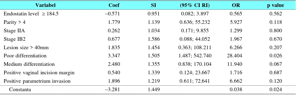
10066.90 (95% CI: 1.44; 108.18, p = 0.022). Fourth hypothesis: clinical pathology factors (sta-ge, lesion size, histological type, degree of differen-tiation), endostatin serum levels, and VEGF-C serum levels were predicting factors of lymphovascular in-vasion. To prove the above hypothesis, multivariate logistic regression analysis was performed with lym-phatic metastases being determined as the dependent variable, while the factors that were tested as inde-pendent variables. From the results of bivariate ana-lysis above, factors that have a high OR with the value of p ≤ 0.25 were selected for subsequent
inclu-sion into the multivariate analysis model. The results of multivariate analysis were as follows. Endostatin levels ≥ 184.5 ng/ml was a protective factor of
108.211, p = 0.207). Poor differentiation had OR 28.404 (95% CI: 1.487; 542.740, p = 0.026). Medium differentiation had OR 11.940 (95% CI: 0.838; 170.104, p = 0.067). Positive vaginal incision margin had OR 1.716 (95% CI: 0.124, 23.667, p = 0.687). Positive parametrium invasion had OR 6.662 (95% CI: 0.611, 72.641, p = 0.120). Based on this analysis, a model to predict the occurrence of lymphovascular invasion can be made as follows. The probability of occurrence of lymphovascular invasion = 3.281 + -0.571 (endostatin levels ≥ 184.5) + 1.779 (parity > 4)
+ 0.262 (stage IIA) + 0.677 (stage IB2) + 1.835 (pri-mary lesion size > 40 mm) + 3.347 (poor differentia-tion) + 2.480 (differentiation medium) + 0.540 (limit of positive vaginal incision) + 1.896 (positive para-metrium invasion). The equation had a good ca-libra-tion, and robust quality based on the parameters of discrimination.
Fifth hypothesis: clinical pathology factors (stage, lesion size, histological type, degree of differentia-tion), endostatin serum levels. To prove the hypothe-sis, bivariate analysis between categories of each va-riable was performed, and then ratio of metastases equilibrium and its p value were calculated. From these calculations, there were several variables that significantly arised, which were: Patients with prima-ry lesion size > 40 mm had the risk of endostatin levels ≥ 184.5 ng/ml 2.222 times compared to patients
with primary lesions ≤ 40 mm (95% CI: 1.369, 3.608,
p = 0.046).
Sixth hypothesis: clinical pathology factors (stage, lesion size, histological type, degree of
differentia-tion) are associated with serum levels of VEGF-C. To prove the hypothesis, bivariate analysis between ca-tegories of each variable was performed, and then the ratio of metastases equilibrium and its p value were calculated. From these calculations, there are several variables that significantly arised, which were: Pa-tients with primary lesion size > 40 mm had the risk of VEGF-C levels > 10066.90 pg/ml of 5.16 times compared to patients with primary lesions ≤ 40 mm
(95% CI: 1.13, 23.54, p = 0.034) .
Seventh hypothesis: endostatin serum levels are as-sociated with higher levels of VEGF-C serum. To test this hypothesis, bivariate analysis between the two va-riables was performed. Therefore, the obtained results were as follows. The risk for having VEGF-C levels > 10066.90 was 9 times higher in patients with endo-statin levels ≥ 184.5 than in patients with higher levels of endostatin < 184.5 (95% CI: 1.675, 48.367, p = 0.006).
DISCUSSION
Up until now, there are not any satisfactory theories to explain the role of endostatin in lymphatic metas-tasis, and as anti-angiogenesis therapy. Clinically, en-dostatin increases the risk of lymphatic metastasis in bivariate analysis (OR = 3.15, p = 0.075), as well as multivariate analysis (OR = 2.086, p = 0.381), but not statistically significant. Endostatin is produced by the body as a reaction to the presence of a malignant tu-mor through basement membrane degradation. With
Table 1. Prediction of Lymphatic Metastases Occurence in Patients with Early Stage Cervical Cancer.
Variabel Coef SI (95% CI RI) OR p value
Endostatin level ≥ 184.5 0.735 0.840 0.402; 10.821 2.086 0.381 Positive parametrium invasion 0.930 0.935 0.406; 15.829 2.534 0.320 Positive vaginal incision margin 1.250 1.076 0.424; 28.764 3.492 0.245
Stage IIA 0.764 1.137 0.231; 19.912 2.146 0.502
Parity > 4 1.264 0.903 0.602; 20.792 3.539 0.162
Poor differentiation 0.600 0.857 0.340; 9.768 1.822 0.484 Primer lesion size > 40mm 1.714 1.073 0.678; 45.457 5.550 0.110
Constanta –3.471 1.179 0.031 0.003
Table 2. Prediction of Lymphovascular Invasion Occurence in Patients with Early Stage Cervical Cancer.
Variabel Coef SI (95% CI RI) OR p value
Endostatin level ≥ 184.5 –0.571 0.951 0.082; 3.897 0.565 0.562
Parity > 4 1.779 1.139 0.636; 55.232 5.927 0.118
Stage IIA 0.262 1.034 0.171; 9.855 1.299 0.800
Stage IB2 0.677 1.586 0.088; 44.052 1.967 0.670
Lesion size > 40mm 1.835 1.454 0.363; 108.211 6.266 0.207 Poor differentiation 3.347 1.505 1.487; 542.740 28.404 0.026 Medium differentiation 2.480 1.355 0.838; 170.104 11.940 0.067 Positive vaginal incision margin 0.540 1.339 0.124; 23.667 1.716 0.687 Positive parametrium invasion 1.896 1.219 0.611; 72.641 6.662 0.120
Constanta –3.281 1.449 0.038 0.024
more advanced od tumors metastases, endostatin le-vels are higher. When metastasis occurs, there is also degradation of basal membrane, so that more endo-statin is produced. In theory, endoendo-statin on angioge-nesis switch chart is one of the angiogeangioge-nesis inhibitor. Endostatin is produced in line with the process of angiogenesis, which is described by an increase in VEGF-C. Thus, high levels of endostatin serum in this study in accordance with the high VEGF-C as proven by the significant difference between serum levels of VEGF-C and serum levels of endostatin, (OR = 9; and the value of p = 0.006).
In theory, it is said that the endostatin is angioge-nesis inhibitor. The increasing levels of endostatin in tumors is a by product of the increasing VEGF-C. If removal of the primary tumor was done, it could ac-celerate the metastases more rapidly.22 Primary
tu-mors have a role to suppress metastasis.23 This has
accordance with the endostatin being produced by the primary tumor to slightly suppress angiogenesis. After the primary tumor is removed, endostatin production will decrease and VEGF-C will increase and then me-tastases will occur. If created a table based on levels of VEGF-C and endostatin levels of lymphatic nodes metastatic, could be seen the relationship of positive lymphatic nodes metastasis. High VEGF-C and low endostatin (lymphatic node metastasis 100%), High VEGF-C and high endostatin (lymphatic node metas-tasis 88.89%); Low VEGF-C and low endostatin (12.5% lymphatic node metastasis), low VEGF-C and high endostatin (lymphatic node metastasis 8.33%). Percentage view above explained that VEGF-C acted as stimulator of angiogenesis, and endostatin was functioning to inhibit VEGF-C also metastasis.
In this study, group obtained low VEGF-C and high endostatin is as much as 25.5% (12/47) with lymphatic nodes metastasis in 1 case. In this popula-tion, role of endostatin for lymphatic nodes metastasis as angiogenesis inhibitors was more visible. There-fore, it may take more samples to investigate the ef-fect of endostatin against the lymphatic nodes me-tas-tasis.
CONCLUSION
Stage, lesion size > 40 mm, differentiation, vaginal incision margin, lymphatic invasion, lymphovascular invasion, levels of VEGF-C > 10066.90 were risk fac-tor for lymph node metastasis, and can be used as predictors. The size of lesions > 40 mm, differentia-tion, invasion of parametrium, levels of VEGF-C > 10066.90 were risk factor of lymphovascular inva-sion. Clinically, endostatin was increasing the risk of lymphatic metastatic, but not statistically proven. VEGF-C was proven as risk factors and predictors of lymphatic metastasis.
REFERENCES
1. Basil JB, Horowitz IR. Cervical carcinoma Contemporary management. Obstet Gynecol Clin 2001; 28(4): 727-42 2. Benedet JL, Pecorelli S. FIGO Special Report on
Gyneco-logic Cancer 2000. Int J Gynecol Obstet 2000; 70: 209-62
3. Aziz MF. Faktor kliniko-patologik, molekul adhesi sel E-kadherin, katenin-A, dan enzim proteolitik matriks ekstra-selular kathepsin-D sebagai prediktor metastasis kelenjar getah bening dan prognosis kanker serviks stadium awal [disertasi]. Jakarta: Program studi ilmu kedokteran S3 Fa-kultas Kedokteran Universitas Indonesia; 2004.
4. Takeda N, Sakuragi N, Takeda M, Okamoto K, Kuwabara M, Negishi H. Multivariate analysis of histopathologic prognostic factors for invasive cervical cancer treated with radical hysterectomy and systematic retroperitoneal lym-phadenectomy. Acta Obstet Gynecol Scand. 2002 Dec; 81(12): 1144-51
5. Thompson JD. Cancer of the cervix. In: Thompson JD, Rock JA, editors. Te linde’s operative gynecology. 7th ed.
Philadelphia: JB Lippincott Company; 1992: 1161-247 6. Menon U, Jacobs IJ. Tumor markers. In: Hoskins WJ, Perez
CA, Young RC, editors. Principles and practice of gyne-cologic oncology. 3rd ed. Philadelphia: Lippincott Williams
& Wilkins; 2000: 165-76
7. Skates SJ. Tumor markers in the diagnosis and management of gynecologic cancers. In: Gershenson DM, Mcguire WP, Gore M, Quinn MA, Thomas G, editors. Gynecologic can-cer (controversies in management). Philadelphia: Elsevier Ltd; 2004: 847-53
8. Zachary I. VEGF signalling: integration and multi-tasking in endothelial cell biology. Biochem Soc Trans. 2003; 31: 1171-7
9. Kodama J, Shinyo Y, Hasengaowa, Kusumoto T, Seki N, Nakamura K. Loss of basement membrane heparan sulfate expression is associated with pelvic lymph node metastasis in invasive cervical cancer. Oncology Reports. 2005; 14: 89-92
10. Weidner N. VEGF and metastasis in breast cancer. N Eng J Med 1991; 324: 1-8
11. O’Reilly MS. Endostatin: an endogenous inhibitor of angio-genesis and tumor growth. Cell 1997; 88: 277-85 12. Boehm T, Folkman J, Browder T, O’Reilly MS.
Antiangio-genic therapy of experimental cancer does not induce ac-quired drug resistance. Nature 1997; 390: 404-7
13. Yamaguchi N. Endostatin inhibits VEGF-induced endothe-lial cell migration and tumor growth independently of zinc binding. EMBO J. 1990; 18: 4414-23
14. Dhanabal M. Endostatin Induces Endothelial Cell Apopto-sis. J. Biol. Chem. 1999; 274: 11721-6
15. Gwiezdzinska JK, Junik R, Kopczynska E. Serum Endo-statin Levels in Patients with Metastatic and Non-metastatic Well-differentiated Thyroid Cancer. Endocrynol Pol. 2010; 61: 7-12
16. Ferrara N. Vascular endothelial growth factor. Eur. J. Can-cer. 1996; 32A: 2413-22
17. Ferrara N, Davis-Smyth T. The biology of vascular en-dothelial growth factor. Endocine Rev 1997; 18: 4-25 18. Veikkola T, Alitalo K. VEGFs, receptors and angiogenesis.
Semin Cancer Biol 1999; 9: 211-20
19. McMahon G. VEGF-receptor signalling in tumour angio-genesis. Oncologist. 2000; 5: 3-10
20. Bernatchez PN, Soker S, Sirois MG. Vascular Endothelial Growth Factor Effect on Endothelial Cell Proliferation, Mi-gration, and Platelet-activating Factor Synthesis is Flk-1-dependent. J Biol Chem 1999; 274: 31047-54
21. Davis-Smyth T, Chen H, Park J, Presta LG, Ferrara N. The Second immunoglobulin-like domain of the VEGF tyrosine kinase receptor Flt-1 determines ligand binding and may initiate a signal transduction cascade. EMBO J. 1996; 15: 4919-27
22. Woodruff M. The Walter Hubert lecture. Interaction of can-cer and host. Br J Cancan-cer. 1982; 46: 313-22
23. Camphausen K, Moses MA, Beecken W-D. Radiation the-rapy to a primary tumor accelerates metastatic growth in mice. Cancer Res. 2001; 61: 2207-11