Approach to acute or subacute myelopathy
William F. Schmalstieg, MD
Brian G. Weinshenker, MD
Neurology®Clinical Practice 2010;75(Suppl 1):S2–S8
I
mproved understanding of the differential diagnosis and improved investigative tech-niques, particularly neuroimaging and sero-logic testing, have facilitated the diagnosis of patients with acute and subacute myelopathy and reduced the proportion of patients who are labeled as having “idiopathic transverse myelitis.” Addi-tionally, these advances have identified subgroups of patients in whom progression of deficit or fu-ture relapses are anticipated, allowing intervention and prophylaxis as appropriate. However, early management remains empiric and consists of high-dose corticosteroids for most patients. In the event of an inadequate response to corticosteroids or a subsequent atypical course, further investigations to detect diagnoses other than “transverse myelitis” should be considered and additional treatments, such as plasmapheresis, may be appropriate. Indi-vidualized diagnosis and treatment is more feasible now than in the past.IS IT AN ACUTE MYELOPATHY?Localizing an acute neurologic process to the spinal cord is often, but not always, straightforward (table 1). The as-cending sensory symptoms of acute inflammatory demyelinating polyradiculoneuropathy (AIDP) may confuse the diagnosis, as this complaint is also highly associated with acute myelopathy. Un-equivocal upper motor neuron signs exclude AIDP, but are often not apparent in the early stages of a spinal cord insult. Although not invariably present, an unequivocal sensory level on the torso, sensory loss indicating involvement of spinal tracts (e.g., spinothalamic modality impairment contralateral to motor findings), or urinary retention localize to the spinal cord. Myopathy or neuromuscular junction disor-ders may be mistaken for myelopathy, particularly if the lower limbs are predominantly affected, but the absence of any sensory abnormality should suggest the correct localization. Bilateral mesial frontal lobe lesions (e.g., bilateral anterior cerebral artery distribution infarcts) could mimic a myelopathy, although abulia or other signs of frontal lobe dysfunction typically coexist. Autoimmune or paraneoplastic muscle stiffness syn-dromes, such as stiff-person syndrome associated with glutamic acid decarboxylase or amphiphysin autoanti-bodies, may be confused with spasticity and erroneously lead one to suspect myelopathy.
Occasionally patients with chronic myelopathy may present a history that mistakenly suggests an acute process. For example, patients with primary progressive multiple sclerosis (MS) may experience acute, transient worsening (pseudoexacerbation) in the setting of an underlying infection or heat exposure. In such cases, careful history will uncover symptoms that have been insidiously progressive over a longer interval than first
From the Department of Neurology, Mayo Clinic College of Medicine, Rochester, MN.
Disclosure:Author disclosures are provided at the end of the article. Illustration by Amy P. Collins
suspected. Patients with myelopathy who have no clear lesion on spinal MRI or multiple chronic-appearing lesions should be questioned to uncover subtle previous symptoms of chronic myelopathy and examined to detect cognitive or bulbar impair-ment localizing elsewhere in the nervous system.
WHEN IT IS AN ACUTE MYELOPATHY, WHAT CAUSES SHOULD BE CONSIDERED? In pa-tients with recent onset symptoms, particularly ones that evolve rapidly, the initial priority is to exclude a surgical emergency such as epidural metastasis or ab-scess. When the index of suspicion for an acute com-pressive lesion is high, immediate imaging is required, ideally with MRI of the entire spine. If im-aging demonstrates spinal cord compression due to an acute lesion such as epidural metastasis, definitive management (i.e., surgery) should be pursued with-out delay to prevent rapid and irreversible worsening. Often the cause of an acute or subacute myelopa-thy is inapparent after an initial evaluation. In a re-cent French series of patients presenting with acute noncompressive myelopathy, 101/170 (59.4%) were of uncertain cause initially, although 55/101 (54.4%) patients were ultimately diagnosed with a demyelinating or inflammatory disorder. After aver-age follow-up of 73.2 months, 49/170 (28.8%) had a final diagnosis of myelopathy of uncertain etiology. The most commonly identified causes were demyeli-nating disorders (MS and neuromyelitis optica), spi-nal cord infarction, parainfectious myelitis, and
systemic inflammatory disorders (e.g., Sjo¨gren syn-drome and lupus).1
Transverse myelitis is the default diagnosis for an unexplained myelopathy evolving over the course of days to 3 weeks with subsequent stabilization or im-provement. In practice, there are no satisfactory ways to distinguish among idiopathic transverse myelitis, parainfectious myelitis, and postvaccinial myelitis. When a viral illness occurs in close temporal associa-tion, parainfectious myelitis is often diagnosed, but the causal role of the associated infection is difficult to determine for an individual patient. One can fidently link the two only when myelitis occurs con-currently or within days of an infection known to be associated with myelitis (e.g., zoster) or when investi-gation such as CSF PCR demonstrates unequivocal evidence of CNS infection.
Nevertheless, serologic evidence of recent infection with pathogens known to be associated with myelopa-thy (e.g., enteroviruses, Chlamydia, Mycoplasma) may limit the need for further diagnostic investigations in an otherwise unexplained myelopathy. Features suggesting an infectious etiology include fever, rash (zoster, entero-virus, Lyme disease), meningismus, a history of recent travel (tuberculosis, parasitic infections such as schisto-somiasis with travel to endemic regions), suspected ra-bies exposure, or immunosuppression (herpes zoster, cytomegalovirus). It is particularly important to con-sider treatable infections such as syphilis, HIV, tubercu-losis, Lyme disease, and herpesviruses.
Occasionally patients with
chronic myelopathy may
present a history that
mistakenly suggests an acute
process
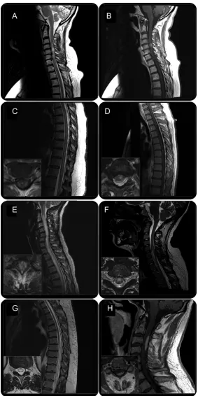
Other diagnoses that may be made confidently in most instances include cord compression, vascular disorders, toxic/metabolic syndromes, neoplasm, paraneoplastic syndromes, and sarcoidosis. Although compression is often obvious as the cause of myelop-athy on MRI, spinal stenosis may cause impressive and occasionally longitudinally extensive T2 signal abnormalities (ⱖ3 vertebral segments) on spinal MRI that may lead one to suspect an inflammatory myelopathy. Circumscribed gadolinium enhance-ment at the point of maximal stenosis and a history of progressive symptoms over many weeks to months are consistent findings in such cases (figure 1, A and B).2Vascular myelopathies include those due to
in-farction resulting from arterial embolism or hypo-perfusion, hemorrhage, or vascular malformations associated with venous hypertension. Dural
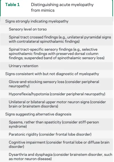
arterio-Table 1 Distinguishing acute myelopathy from mimics
Signs strongly indicating myelopathy
Sensory level on torso
Spinal tract crossed findings (e.g., unilateral pyramidal signs with contralateral spinothalamic findings)
Spinal tract-specific sensory findings (e.g., selective spinothalamic findings with preserved dorsal column findings; suspended band of spinothalamic sensory loss)
Urinary retention
Signs consistent with but not diagnostic of myelopathy
Glove-and-stocking sensory loss (consider peripheral neuropathy)
Hyporeflexia/hypotonia (consider peripheral neuropathy)
Unilateral or bilateral upper motor neuron signs (consider brain or brainstem disorders)
Signs suggesting alternative diagnosis
Spasms, rather than spasticity (consider stiff-person syndrome)
Paratonic rigidity (consider frontal lobe disorder)
Cognitive impairment (consider frontal lobe or diffuse brain disorder)
Figure 1 MRI of representative cases of acute and subacute myelopathies
venous fistula (DAVF)–associated myelopathies may be distinguished by the distinctive history of stepwise progression or transient worsening precipitated by walking or prolonged standing. This myelopathy can be successfully treated by obliteration of the fistula and should not be missed. Toxic myelopathy due to nitrous oxide abuse is a consideration in younger pa-tients and medical professionals; papa-tients with under-lying vitamin B12 deficiency are particularly vulnerable. Vitamin and trace metal deficiencies usu-ally cause chronic myelopathies, but as these etiolo-gies are readily treatable, vitamin B12 and copper levels should be tested in unexplained steroid nonre-sponsive myelopathy. Spinal cord tumors may present subacutely or with apoplectic onset in the case of hemorrhage into tumor and are usually readily identifiable on MRI, although rarely they may be mistaken radiologically for myelitis. Screen-ing for serum paraneoplastic autoantibodies includ-ing collapsin response mediator protein-5 (CRMP-5) antibody3 should be considered in patients with
known cancer, constitutional symptoms, smoking history, or suggestive neuroradiology with isolated,
tract-specific involvement. Sarcoidosis may present as isolated myelopathy. Definitive diagnosis requires biopsy evidence of noncaseating granulomatous inflam-mation, either from the nervous system or other in-volved organs. A high serum angiotensin-converting enzyme level is suggestive but nonspecific. A diagnosis of isolated CNS sarcoidosis should be suspected when subacute myelopathy is accompanied by patchy, asym-metric, slowly evolving, and persistently enhancing gad-olinium cord lesions. A satisfactory therapeutic response to empiric, long-term (months to years) corticosteroid treatment provides a reasonable basis for a tentative but usually correct diagnosis.
WHAT CLINICAL FEATURES SUGGEST A PAR-TICULAR DIAGNOSIS? The time course (figure 2), specific spinal cord syndrome, and symptoms other than those referable to the spinal cord may pro-vide useful clues as to the diagnosis. Apoplectic onset suggests a cord infarct or spinal hemorrhage, both of which may worsen over hours to days. Parainfectious or idiopathic myelitis, myelitis related to inflamma-tory demyelinating diseases, and some paraneoplastic
Figure 2 Differential diagnosis of acute myelopathy: Time course and MRI findings
*Relapses upon withdrawal of corticosteroids/immunosuppression. **MRI may be normal. ADEM⫽acute disseminated
en-cephalomyelitis; DAVF⫽dural arteriovenous fistula; HTLV⫽human T-lymphotropic virus; MS⫽multiple sclerosis; NMO⫽
syndromes evolve over days to weeks, but generally reach a nadir within 3 weeks, after which there is either improvement or stability.4When a
myelopa-thy develops insidiously or continues to progress af-ter 3 weeks, transverse myelitis becomes unlikely and the differential diagnosis includes an intrinsic cord tumor, compressive lesion, DAVF, metabolic de-rangement, sarcoidosis, or a degenerative process.
The clinical syndrome of spinal cord involvement may suggest a particular etiology, although none are specific. Incomplete Brown-Se´quard syndrome (loss of pain and temperature sensation contralateral to weakness) may be associated with either compression or an intrinsic cord lesion such as demyelination. An anterior spinal cord syndrome with bilateral cortico-spinal and spinothalamic involvement sparing dorsal column function is typical of anterior spinal artery distribution infarction, but may also occur in MS. A complete spinal cord syndrome with bilateral in-volvement of all spinal tracts is rarely caused by an MS relapse or infarct, but may occur in idiopathic or neuromyelitis optica (NMO)–associated transverse myelitis or cord compression. NMO-associated my-elitis more commonly presents with clinical and
im-aging signs of central cord involvement than does MS-associated myelitis, which more commonly af-fects the periphery of the cord. Highly selective tract involvement (e.g., pure corticospinal tract involve-ment), especially when confirmed by MRI evidence of highly localized enhancing tractopathy, is charac-teristic of a paraneoplastic disorder (figure 1C).
Neurologic or constitutional symptoms not refer-able to the spinal cord focus the differential diagno-sis, but may be irrelevant and distract one from the true diagnosis. Optic neuritis or a prior diagnosis of inter-mediate uveitis may suggest MS. Severe optic neuri-tis and an episode of unexplained intractable nausea or hiccoughs are characteristic of NMO.5
Coexist-ing peripheral neuropathy can occur in sarcoid, Sjo¨gren syndrome, lupus, metabolic disorders (e.g., subacute combined degeneration), and para-neoplastic syndromes.
WHAT INVESTIGATIONS SHOULD BE PER-FORMED? MRI scan of the spinal cord with and without gadolinium contrast is the initial investiga-tion of choice in the evaluainvestiga-tion of acute myelopathy. Contraindications are limited to MRI-incompatible ferromagnetic medical devices or foreign bodies and incompatibility with the scanner due to habitus. With careful coordination between cardiologists and radiologists, MRI can be performed in selected pa-tients with cardiac pacemakers who are not entirely pacemaker-dependent. For patients unable to un-dergo MRI, CT myelography may be considered when cord compression is suspected. CSF evaluation including cell count, glucose, protein, oligoclonal bands, immunoglobulin G (IgG) index, and cytology is appropriate unless imaging, history, and examina-tion already suggest a clear diagnosis. The results of spinal MRI and clinical suspicion should guide the selection of additional investigations (table 2).
For noncompressive myelopathy, the results of MRI can be broadly subdivided into 3 categories:
1. Short T2 hyperintensity (⬍3 vertebral segments in length).
Focal, discrete lesions that do not occupy the en-tire cord in axial cross-section are highly sugges-tive of MS (figure 1D), although remote, sometimes forgotten trauma can occasionally pro-duce such lesions. MRI scan of the brain may help to clarify the cause; detection of 1 or more brain lesions typical of MS (discrete periventricular, juxtacortical, or infratentorial T2 hyperintense foci) correlates with at least an 85%–90% future risk of developing MS.6 Oligoclonal bands and
elevated CSF IgG index help to confirm a sus-pected MS diagnosis, but CSF analysis may be
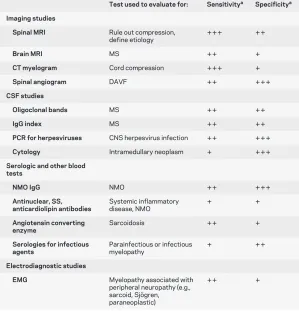
Table 2 Utility of diagnostic tests in evaluation of myelopathy
Test used to evaluate for: Sensitivitya Specificitya
Imaging studies
Spinal MRI Rule out compression,
define etiology ⴙⴙⴙ ⴙⴙ
Brain MRI MS ⴙⴙ ⴙ
CT myelogram Cord compression ⴙⴙⴙ ⴙ
Spinal angiogram DAVF ⴙⴙ ⴙⴙⴙ
CSF studies
Oligoclonal bands MS ⴙⴙ ⴙⴙ
IgG index MS ⴙⴙ ⴙⴙ
PCR for herpesviruses CNS herpesvirus infection ⴙⴙ ⴙⴙⴙ
Cytology Intramedullary neoplasm ⴙ ⴙⴙⴙ
Serologic and other blood tests
EMG Myelopathy associated with peripheral neuropathy (e.g., sarcoid, Sjögren, paraneoplastic)
ⴙⴙ ⴙ
Abbreviations: DAVF⫽dural arteriovenous fistula; IgG⫽immunoglobulin G; MS⫽multiple sclerosis; NMO⫽neuromyelitis optica; SS⫽Sjögren syndrome.
aThe symbols
superfluous if other clinical and radiographic fea-tures are highly suggestive.
2. Longitudinally extensive T2 hyperintensity (ⱖ3 vertebral segments in length).
Longitudinally extensive transverse myelitis oc-curs in idiopathic transverse myelitis, NMO (fig-ure 1E), acute disseminated encephalomyelitis, cord infarction, and myelitis associated with sys-temic diseases such as syssys-temic lupus erythemato-sus. Serum NMO IgG testing is indicated before assigning a diagnosis of idiopathic transverse my-elitis.7Brain lesions on MRI eventually occur in
the majority of patients with NMO, but usually NMO does not lead to the discrete Dawson finger pattern of periventricular lesions characteristic of MS. However, confluent and linear lesions encir-cling the ventricles may occur in NMO. CSF oligo-clonal bands are usually absent in NMO.
Certain patterns of signal abnormality on MRI predict a vascular disorder. Anterior and central cord signal change and swelling with sparing of the posterior columns suggest infarct, particularly in patients with a suggestive history (figure 1F). Posterior flow voids on spinal MRI representing dilation of the epidural venous plexus are a fairly specific but less sensitive indicator of DAVF, whereas longitudinally extensive gadolinium en-hancement and T2 hyperintensity often extend-ing to the conus are typical but nonspecific findings (figure 1G). Magnetic resonance angiog-raphy may help to visualize a DAVF, but spinal angiography is required for definitive diagnosis and treatment.
If symptoms suggestive of recent infection or CSF pleocytosis (⬎50 leukocytes/L) are present, CSF PCR testing for herpesviruses (e.g., herpes simplex, cytomegalovirus, varicella zoster) and se-rologic testing for HIV, syphilis, and Lyme dis-ease should be considered. Prominent CSF pleocytosis and occasionally neutrophilic pleocy-tosis may occur in myelitis associated with NMO. Symptoms and signs of systemic inflammatory disease such as polyarthritis should prompt auto-immune serologic testing (i.e., antinuclear anti-bodies, SS-A, SS-B antibodies). In the absence of clinical indications of these diseases, positive sero-logic tests may be unimportant, although they may indicate NMO; a quarter of NMO spectrum disor-der patients have nonspecific serologic evidence of autoimmunity, usually in the absence of clinical signs of other autoimmune disorders.8
Indiscrimi-nate use of autoantibody testing in all patients with myelitis is not recommended. MRI find-ings including nodular and persisting (⬎2
months) gadolinium enhancement or menin-geal and nerve root enhancement suggest sar-coidosis (figure 1H) or, rarely, lymphoma.
3. Normal MRI.
Patients with suspected myelopathy and appar-ently normal MRI should undergo careful review of the images for subtle findings of cord signal change, atrophy, or extrinsic compression by un-common causes (e.g., epidural lipomatosis). If ex-amination demonstrates unequivocal evidence of a spinal cord process and the MRI is normal, con-sider and test for degenerative, infectious, and metabolic causes of myelopathy. EMG and nerve conduction studies occasionally help to identify a primary peripheral process (e.g., AIDP) or my-elopathy associated with concomitant peripheral neuropathy as can be seen in sarcoidosis and sub-acute combined degeneration.
HOW SHOULD AN ACUTE MYELOPATHY BE TREATED?Controlled studies of treatment of acute myelitis are lacking. In myelitis due to demyelinat-ing, inflammatory, or undetermined cause, expert consensus favors high-dose IV corticosteroids, typi-cally 1 gram of IV methylprednisolone daily for 5 days. This treatment should not be withheld in the case of suspected recent viral infection; the role of ste-roid treatment in patients with definitive evidence for direct viral infection of the cord (e.g., myelitis occurring simultaneously with or within days of a zoster eruption) is unclear. Plasmapheresis should be considered in pa-tients who continue to have significant impairment af-ter high-dose corticosaf-teroids. In a sham-controlled trial of plasma exchange in patients with an acute relapse of demyelinating disease unresponsive to corticosteroid treatment, many of whom had acute myelitis, 8 of 19 (42.1%) treated patients experienced moderate to marked improvement vs 1 of 17 (5.9%) who received sham treatment.9There are no established treatments
for patients with cord infarction.
Ongoing clinical observation is an important part of the care of patients with an unexplained myelopa-thy. Subsequent appearance of new neurologic or systemic symptoms may reveal a demyelinating or systemic inflammatory disorder. Patients with relent-lessly progressive symptoms despite appropriate em-piric treatment may require spinal cord biopsy for definitive diagnosis, particularly when follow-up im-aging demonstrates worsening.
DISCUSSIONAcute and subacute myelopathies re-quire urgent medical evaluation. Imaging, preferably by MRI, should be performed without delay to ex-clude a compressive lesion. Subsequently, history and physical examination should guide subsequent inves-tigations to reach a definitive diagnosis. As the etiol-ogy is often unclear at initial presentation, empiric treatment should be provided while conducting fur-ther investigations to determine the etiology of my-elopathy. A thorough evaluation often reveals evidence of a treatable disorder or one that may re-lapse without preventive treatment.
DISCLOSURE
Dr. Schmalstieg reports no disclosures. Dr. Weinshenker has served on data safety monitoring boards for Novartis and Biogen Idec; serves on the editorial boards ofMultiple Sclerosis, theCanadian Journal of Neurological Sciences, and theTurkish Journal of Neurology; receives research support from the Guthy-Jackson Charitable Foundation; and receives license roy-alties from RSR Ltd. and may receive royroy-alties from Mayo Medical Ven-tures for a patent/intellectual property re: Aquaporin-4 associated antibodies for diagnosis of neuromyelitis optica.
Received July 14, 2010. Accepted in final form September 2, 2010.
REFERENCES
1. Debette S, de Seze J, Pruvo JP, et al. Long-term outcome of acute and subacute myelopathies. J Neurol 2009;256: 980 –988.
2. Kelley BJ, Erickson BJ, Weinshenker BG. Compressive myelopathy mimicking transverse myelitis. Neurologist 2010;16:120 –122.
3. Keegan BM, Pittock SJ, Lennon VA. Autoimmune my-elopathy associated with collapsin response-mediator protein-5 immunoglobulin G. Ann Neurol 2008;63:531– 534.
4. Proposed diagnostic criteria and nosology of acute trans-verse myelitis. Neurology 2002;59:499 –505.
5. Takahashi T, Miyazawa I, Misu T, et al. Intractable hiccup and nausea in neuromyelitis optica with anti-aquaporin-4 antibody: a herald of acute exacerbations. J Neurol Neuro-surg Psychiatry 2008;79:1075–1078.
6. Brex PA, Ciccarelli O, O’Riordan JI, Sailer M, Thompson AJ, Miller DH. A longitudinal study of abnormalities on MRI and disability from multiple sclerosis. N Engl J Med 2002;346:158 –164.
7. Weinshenker BG, Wingerchuk DM, Vukusic S, et al. Neuromyelitis optica IgG predicts relapse after longitudi-nally extensive transverse myelitis. Ann Neurol 2006;59: 566 –569.
8. Pittock SJ, Lennon VA, de Seze J, et al. Neuromyelitis optica and non organ-specific autoimmunity. Arch Neurol 2008;65:78 – 83.
9. Weinshenker BG, O’Brien PC, Petterson TM, et al. A randomized trial of plasma exchange in acute central nervous system inflammatory demyelinating disease. Ann Neurol 1999;46:878 – 886.
If you liked this article, you may be interested in. . .
Continuum
Spinal Cord, Root, and Plexus Disorders. June 2008;www.aan.com/go/elibrary/continuum
Neurology
Nathan P. Staff et al. Hypertrophic nerves producing myelopathy in fulminant CIDP. August 24, 2010;www.neurology.org
Jeremy D. Isaacs et al. Noncompressive myelopathy associated with violent axial tics of Tourette syndrome. February 23, 2010;www.neurology.org
David Roshal et al. Pearls & Oy-sters: Fibrocartilaginous embolism myelopathy. February 16, 2010;www.neurology.org
DOI 10.1212/WNL.0b013e3181fb3638
2010;75;S2-S8
Neurology
William F. Schmalstieg and Brian G. Weinshenker
Approach to acute or subacute myelopathy
This information is current as of November 1, 2010
Services
Updated Information &
http://www.neurology.org/content/75/18_Supplement_1/S2.full.html
including high resolution figures, can be found at:
References
ref-list-1
http://www.neurology.org/content/75/18_Supplement_1/S2.full.html##
This article cites 9 articles, 2 of which you can access for free at:
Citations
otherarticles
http://www.neurology.org/content/75/18_Supplement_1/S2.full.html##
This article has been cited by 2 HighWire-hosted articles:
Subspecialty Collections
http://www.neurology.org//cgi/collection/transverse_myelitis
Transverse myelitis
http://www.neurology.org//cgi/collection/mri
MRI
http://www.neurology.org//cgi/collection/all_spinal_cord
All Spinal Cord
s
http://www.neurology.org//cgi/collection/all_demyelinating_disease_cn
All Demyelinating disease (CNS) following collection(s):
This article, along with others on similar topics, appears in the
Permissions & Licensing
http://www.neurology.org/misc/about.xhtml#permissions
its entirety can be found online at:
Information about reproducing this article in parts (figures,tables) or in
Reprints
http://www.neurology.org/misc/addir.xhtml#reprintsus
Information about ordering reprints can be found online:
rights reserved. Print ISSN: 0028-3878. Online ISSN: 1526-632X.