Effect of the interval of serial sections of ovarian
tissue in the tissue chopper on the number of
isolated caprine preantral follicles
C.M. Lucci
a,), C.A. Amorim
a, S.N. Bao
b, J.R. Figueiredo
a,
´
A.P.R. Rodrigues
a, J.R.V. Silva
a, P.B.D. Gonc
¸
alves
ca
Faculty of Veterinary, UniÕersity of Ceara, Fortaleza, CE, Brazil´
b
UniÕersity of Brasilia, Brasılia, DF, Brazil´
c
UniÕersity of Santa Maria, Santa Maria, RS, Brazil
Accepted 20 March 1999
Abstract
The present work investigated the effect of the interval of serial sections of ovarian tissue on the number of isolated preantral follicles in the goat. Goat ovaries were cut in the tissue chopper at eight different intervals. The quality of isolated follicles were evaluated by histology and transmission electron microscopy. Best results were obtained when the ovaries were cut in the
Ž .
tissue chopper at intervals of 75.0 mm 9664 preantral follicles per ovary . Histochemical and
ultrastructural analysis showed that the follicular morphology was preserved after mechanical isolation as demonstrated by the normality of oocytes and granulosa cells as well as by preservation of basement membrane. The percentages of isolated primordial, primary and sec-ondary follicles were 96.3%, 2.5%, and 1.2% and their average diameters were 21.5, 34.7 and
65.3mm, respectively. It was concluded that the interval of serial sections of ovarian tissue in the
tissue chopper affects the number of isolated preantral follicles, and that the follicles remained
intact after mechanical isolation in goats.q1999 Elsevier Science B.V. All rights reserved.
Keywords: Preantral follicles; Follicle quality; Follicle isolation; Goat
)Corresponding author. Universidade Estadual do Ceara, Faculdade de Vetirinaria Curso de Mestrado em
´ ´
&
Produc¸ao e Reproduc¸ao de Pequenos Ruminantes Av. Paranjana, 1700 Campus do Itaperi, CEP 60740-000´ Fortaleza, Ceara, Brazil. Tel.:´ q55-61-307-2424; fax:q55-61-347-6533; e-mail: [email protected] 0378-4320r99r$ - see front matterq1999 Elsevier Science B.V. All rights reserved.
Ž .
1. Introduction
Since the mammalian ovary contains many thousands of preantral follicles and most
Ž
will become atretic during their growth and maturation Carroll et al., 1990; Saumande,
.
1991 , new investigations were focused on the in vitro culture of preantral follicles. The use of oocytes from preantral follicles for reproductive techniques could offer significant new ways for the propagation of valuable animal stocks. However, the first limiting step of this approach is the recovery of large numbers of healthy preantral follicles from the ovary.
Techniques for isolating preantral follicles from domestic species have been reported
ŽGreenwald and Moor, 1989; Lazzari et al., 1992; Figueiredo et al., 1993; Jewgenow and Pitra, 1993; Nuttinck et al., 1993; Hulshof et al., 1994; Jewgenow and Goritz,
¨
. Ž .
1995; . Figueiredo et al. 1993 showed that a large number of preantral follicles can be isolated from bovine ovaries using a tissue chopper adjusted to 50.0 mm interval of
Ž .
serial sections. Using the same method, Rodrigues et al. 1998 described that goat preantral follicles can also be successfully isolated. However, no information about the quality of goat preantral follicles after their isolation was reported. Moreover, it is not known if the number of isolated goat preantral follicles can be increased by changing the interval of serial sections of ovarian tissue in the tissue chopper.
The aims of the present study were to determine the effect of the interval of serial sections of ovarian tissue in the tissue chopper on the number of isolated goat preantral follicles, and to investigate, by histological and ultrastructural analysis, the quality of the isolated preantral follicles.
2. Material and methods
2.1. OÕaries
Ž . Ž .
Ovaries ns84 from adult 1–3 years old and non-pregnant mixed breed goats were collected at a local abattoir. The ovaries were washed in 70% alcohol for, approximately, 10 s, and twice in 0.9% saline solution. Then, each ovary was transferred into 10 ml of saline solution and transported to the laboratory in a thermoflask filled with water at 48C.
2.2. Isolation of preantral follicles
The development of a specific mechanical method to isolate preantral ovarian follicles from adult goats was based on the mechanical procedure described by Figueiredo
Ž .
et al. 1993 for bovine ovaries. In the present work, eight treatments were tested which
Ž
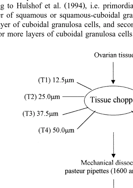
differed from each other according to the cut interval adjusted in the tissue chopper The
. Ž . Ž . Ž .
Mickle Laboratory Engineering, Gomshal, Surrey, UK : 12.5 T1 , 25.0 T2 , 37.5 T3 ,
Ž . Ž . Ž . Ž . Ž .
treatments. In all treatments the ovarian pieces were cut in the tissue chopper from the cortical to medullar side. The ovarian fragments obtained were then placed in 50 ml of
Ž . Ž .
PBS Phosphate Buffered Saline plus 5% goat serum Sigma , suspended 40 times with
Ž .
a large Pasteur pipette diameter—1600mm and 40 times with a smaller Pasteur pipette
Ždiameter—600mm . The suspensions were filtered successively through 500 and 100.
Ž .
mm nylon mesh filters Fig. 1 . Each treatment was repeated seven times.
To estimate the number of isolated preantral follicles per treatment, the suspension -100 mm was homogenized and then, two samples of 1 ml each were taken and
Ž .
examined separately at the inverted microscope Zeiss . The follicular diameters were measured with an ocular micrometer, considering the smaller diameter.
The isolated preantral follicles were classified by their morphological appearance
Ž .
according to Hulshof et al. 1994 , i.e. primordial follicles are oocytes surrounded by one layer of squamous or squamous-cuboidal granulosa cells; primary follicles have a single layer of cuboidal granulosa cells, and secondary follicles are oocytes surrounded by two or more layers of cuboidal granulosa cells.
2.3. QualitatiÕe analysis of isolated preantral follicles
To evaluate the quality of the isolated preantral follicles, variables such as integrity of the basement membrane, cellular density, presence or absence of picnotic bodies and integrity of the oocyte were observed. Follicular quality was evaluated only in the treatment that yielded the largest number of isolated preantral follicles.
2.3.1. Histology
Freshly isolated preantral follicles were briefly fixed in a solution containing 2% paraformaldehyde and 2.5% glutaraldehyde in 0.1 M cacodylate buffer, pH 7.2, for 30 min at room temperature. Subsequently, the follicles were embedded in drops of 200ml
Ž .
of 4% agar solution agarose, type VII, Sigma, St. Louis, MO, USA . The agar drops were fixed again in the same fixation solution described above for 3 h at room temperature. After the fixation procedure, they were dehydrated in a graded series of ethanol, clarified with xylol, and embedded in paraffin wax. Sections of 7mm were
Ž .
stained with periodic acid schiff PAS and haematoxylin.
2.3.2. Transmission electron microscopy
Some isolated follicles were embedded in agar drops and fixed as described above. After washing with sodium cacodylate buffer, postfixation in a solution containing 1% osmium tetroxide, 0.8% potassium ferricyanide and 5 mM calcium chloride in 0.1 M cacodylate buffer was performed. Subsequently, the samples were dehydrated in acetone
Ž .
and embedded in Spurr. Thin sections 70 nm were stained with uranyl acetate and lead citrate, and examined in a Jeol JEM 100 C transmission electron microscope.
2.4. Statistical Analysis
To compare the numbers of isolated preantral follicles among treatments, data were subjected to log transformation and ANOVA. Differences among individual means were
Ž .
tested using Duncan’s Multiple Range test SAS . Values were considered statistically significant when p-0.05.
3. Results
3.1. Isolation of oÕarian preantral follicles
Data regarding the effect of eight different intervals of serial sections in the tissue chopper on the number of isolated preantral follicles per ovary are included in Table 1. The mechanical treatment of goat ovaries provides a large number of isolated preantral follicles, irrespective of the interval used for serial sectioning. However, a large individual variation in the number of isolated follicles within the treatments was
Ž . Ž .
observed. The limits of this variation were 1550 T8 and 17220 T5 preantral follicles per ovary. The mean number of isolated preantral follicles per ovary increased from T1
Table 1
Ž .
Number of isolated ovarian preantral follicles mean"SEM at eight different sections interval
Ž . Ž .
Treatment section interval Number of follicles per ovary ns84 Mean"SEM Range
a,c
Ž .
T1 12.5mm 4937"1025 1625–7887
a,b,c
Ž .
T2 25.0mm 6441"1932 1637–13813
a,b,c
Ž .
T3 37.5mm 8052"1896 2375–15575
a,b,c
Ž .
T4 50.0mm 6971"1278 2725–10975
a,b
Ž .
T5 62.5mm 8589"1653 2875–17220
b
Ž .
T6 75.0mm 9664"1577 5650–16975
a,b,c
Ž .
T7 87.5mm 4957"684 2625–7175
c
Ž .
T8 100.0mm 3542"540 1550–4625
a,b,cŽ .
p-0.05 .
. Ž .
mm . However, only T6 75.0 mm showed a significantly higher number of isolated
Ž . Ž .
preantral follicles compared with T1 12.5mm and T8 100.0mm . Significantly more
Ž . Ž .
follicles were isolated in T5 62.5mm when compared with T8 100.0 mm .
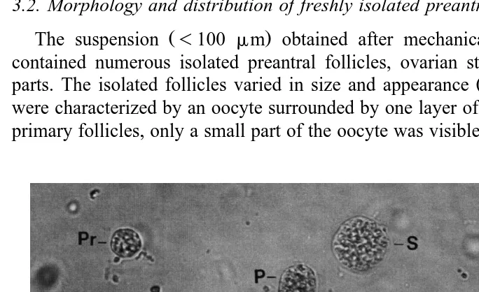
3.2. Morphology and distribution of freshly isolated preantral follicles
Ž .
The suspension -100 mm obtained after mechanical treatment of the ovaries contained numerous isolated preantral follicles, ovarian stroma cells and large tissue
Ž .
parts. The isolated follicles varied in size and appearance Fig. 2 . Primordial follicles were characterized by an oocyte surrounded by one layer of flattened granulosa cells. In primary follicles, only a small part of the oocyte was visible; the remainder was covered
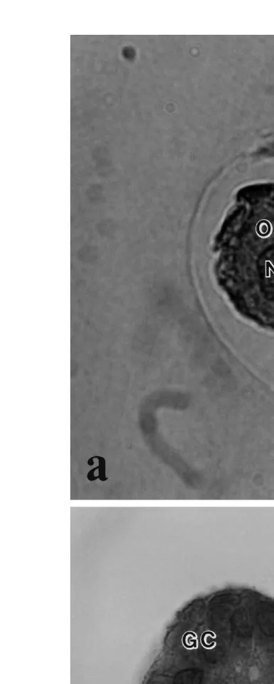
Ž . Ž .
Fig. 3. Histological section of a a morphologically normal follicle and b a degenerated follicle after mechanical isolation. O: Normal oocyte; Nu: Nucleus of a normal oocyte; GC: Granulosa cells; DO:
Ž .
by one layer of cuboidal granulosa cells. Secondary follicles had more than one layer of cuboidal granulosa cells. The oocyte was not always clearly visible because it was covered by granulosa cells.
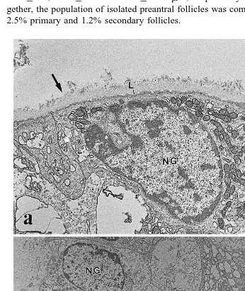
Ž .
Isolated primordial, primary and secondary follicles had a mean "SD diameter of 21.5"3.7, 34.7"6.1 and 65.3"22.3 mm, respectively. Taking the treatments to-gether, the population of isolated preantral follicles was composed of 96.3% primordial, 2.5% primary and 1.2% secondary follicles.
Ž .
Fig. 4. Electron micrographs of preantral follicles after mechanical isolation showing a the basement
Ž . Ž .
3.3. QualitatiÕe analysis of isolated preantral follicles
Ž .
In addition to the greater number of preantral follicles isolated, the T6 75.0 mm allowed the preservation of follicular integrity. Freshly isolated preantral follicles appeared healthy under the inverted microscope. They were spherical, with one or more organized layers of granulosa cells around the oocyte, surrounded by a basement membrane and with no antrum. No denuded oocytes were observed in this study.
Histological evaluation shows that after mechanical isolation 93% of preantral follicles were morphologically normal with a healthy oocyte surrounded by one or more
Ž .
well organized layers of granulosa cells without pycnotic nuclei Fig. 3a .
Histochem-Ž .
istry, using periodic acid schiff PAS , also revealed that the mechanically isolated follicles were surrounded by an intact basement membrane.
Ž .
The percentage of degenerated follicles was very low 7% and the degeneration was observed mainly in the oocyte. In degenerated follicles, the oocyte was contracted and
Ž .
strongly stained with PAS and its nuclear chromatin was also clumped Fig. 3b . The granulosa cells were apparently unaffected. The signs of atresia were not identified with the inverted microscope, but were detected only by histology.
Ultrastructural analysis of isolated follicles showed that the basement membrane is composed of a compact lamina in the inner part surrounded by a thick layer of collagen
Ž . Ž .
fibers Fig. 4a . The integrity of the oocyte and granulosa cells was confirmed Fig. 4b .
4. Discussion
The results of this study have shown that a large number of goat preantral follicles can be isolated using a simple mechanical procedure. Other mechanical procedures for the isolation of preantral follicles are described in the literature. Some authors isolated a
Ž
large number of ovarian preantral follicles from cows Figueiredo et al., 1993; Nuttinck
. Ž . Ž
et al., 1993; Hulshof et al., 1994 , ewes Amorim et al., 1998 , domestic cats Jewgenow
. Ž
and Pitra, 1993; Jewgenow and Goritz, 1995 and non-domestic felids Jewgenow and
¨
.Stolte, 1996; Jewgenow et al., 1997 . In addition, enzymatic procedures for the isolation
Ž
of follicles have been used in various mammalian species Nicosia et al., 1975; Eppig
.
and Downs, 1987; Daniel et al., 1989; Lazzari et al., 1992; Roy and Treacy, 1993 . Comparisons of our results with those of other authors are difficult to perform due to species differences, variation in follicle classification, and types of follicles included in the final count.
Ž .
Using the tissue chopper, Figueiredo et al. 1993 developed a simple mechanical method for isolating preantral follicles from bovine ovaries. This method consists in cutting the ovarian tissue in the tissue chopper adjusted to a 50.0 mm interval of serial
Ž .
sections. Using the same method, Rodrigues et al. 1998 isolated 1067 preantral follicles per goat ovary. The results of our research indicate that the method developed for cattle is not optimal for goats, since more follicles can be isolated from the goat ovary using a 75.0mm interval instead of a 50.0mm interval. The results of the present study demonstrated that the number of isolated preantral follicles is affected by the
Ž .
authors related that the interval of 87.5mm is the best to isolate ovine preantral follicles. In the present study, it seems that the use of cut intervals smaller than 75.0 mm may destroy preantral follicles, while the largest intervals may reduce the efficiency of mechanical dissociation of the ovarian fragments with Pasteur pipettes and thus prevent preantral follicle release from the ovarian tissue.
Great individual variation in the number of isolated follicles per ovary was observed in all intervals of serial sections tested in this study. These data are in accordance with
Ž
many other authors Jewgenow and Goritz, 1995; Jewgenow and Stolte, 1996; Amorim
¨
.et al., 1998; Rodrigues et al., 1998 . A variety of factors has been described which may
Ž . Ž
affect ovarian follicular populations, including age Peters, 1976 , breed Cahill et al.,
. Ž .
1979; Driancourt et al., 1985 , reproductive stage Erickson et al., 1976 , nutrition
ŽScaramuzzi et al., 1993 and genetic factors Erickson, 1966a,b; Cahill et al., 1979 .. Ž .
In the present study, no denuded oocytes were observed, probably because the basement membrane offers mechanical resistance and presumably protects the follicles
Ž .
from physical damage during isolation, as suggested by Figueiredo et al. 1995 . Histochemical and ultrastructural analysis demonstrated that mechanically isolated goat preantral follicles are surrounded by an intact basement membrane. Similar results were
Ž . Ž .
obtained by Figueiredo et al. 1994, 1995 and Amorim et al. 1998 using the tissue chopper to isolate bovine and ovine preantral follicles, respectively. In contrast, some studies have pointed out that proteolytic enzymes commonly used for isolating follicles
Ž
are detrimental to cell membranes Cavanaugh et al., 1963; Grob, 1964; Kono, 1969;
.
Post, 1971; Nicosia et al., 1975 . In vitro culture of enzymatically isolated preantral follicles, which are not surrounded by a basal lamina, shows a spreading of granulosa
Ž .
cells from the oocytes Maresh et al., 1990; Eppig, 1992 . In contrast, this latter alteration was not observed when mechanically isolated preantral follicles were cultured
Ž .
in vitro Figueiredo et al., 1994 . The culture of preantral follicles surrounded by a natural basement membrane has many advantages including preservation of follicular
Ž
morphology and maintenance of follicular adhesion to extracellular compounds
Fig-.
ueiredo et al., 1994 . Thus, the presence of a basement membrane around the isolated follicles is important for further studies of in vitro culture of goat preantral follicles.
The percentage of primordial follicles was greater than for primary and secondary follicles in all cut intervals tested. Similar proportions were reported in histological
Ž . Ž .
studies in ovine Driancourt et al., 1985 and bovine ovaries Erickson, 1966b .
Ž . Ž .
Rodrigues et al. 1998 and Amorim et al. unpublished results also showed similar results in the isolation of caprine and ovine preantral follicles, respectively. However,
Ž . Ž .
Hulshof et al. 1994 isolated a greater percentage of primary 57.2% than primordial
Ž .
follicles 12.4% from bovine fetal ovaries. These authors suggest that the lesser percentage of isolated primordial follicles may be due to these follicles being tightly embedded in the tunica albuginea, whereby a mild mechanical treatment is not sufficient to isolate primordial follicles.
The diameters of isolated primordial, primary and secondary follicles were similar to
Ž . Ž
the diameters reported for caprine Rodrigues et al., 1998 and ovine Amorim et al.,
.
unpublished results preantral follicles, but were smaller than those reported for preantral
Ž .
In conclusion, this work provides evidence that the number of isolated goat preantral follicles is affected by the interval of serial sections of ovarian tissue in the tissue chopper. The most desirable interval is 75.0mm because it yielded the largest number of intact preantral follicles from adult goat ovaries. In the future, the recovery and culture of a large number of preantral follicles will be essential to provide viable oocytes for in vitro maturation and fertilization techniques, for multiplying valuable or endangered animals.
Acknowledgements
This study was supported by the Laboratorio de Morfologia e Morfogenese and the
´
ˆ
Laboratorio de Microscopia Eletronica of University of Brasılia, Brazil and University´
ˆ
´
of Liege, Belgium. The authors thank Antonio Djalma Santos for technical assistance`
ˆ
and Zelia Ramos Madeira for the English correction of the manuscript.´
References
Amorim, C.A., Rodrigues, A.P.R., Lucci, C.M., Figueiredo, J.R., Gonc¸alves, P.B.D., 1998. Mechanical method for the isolation of preantral follicles from adult ovine ovaries. Arq. Fac. Vet. UFRGS, Porto
Ž .
Alegre 26 1 , 215, Suppl.
Cahill, L.P., Mariana, J.C., Mauleon, P., 1979. Total follicular populations in ewes of high and low ovulation´ rates. J. Reprod. Fertil. 55, 27–36.
Carroll, J., Whittingham, D.G., Wood, M.J., Telfer, E., Gosden, R.G., 1990. Extra-ovarian production of mature viable mouse oocytes from frozen primary follicles. J. Reprod. Fertil. 90, 321–327.
Cavanaugh, D.J., Berndt, W.O., Smith, T.E., 1963. Dissociation of heart cells by collagenase. Nature 200, 261–262.
Daniel, S.A.J., Armstrong, D.T., Gore-Langton, R.E., 1989. Growth and development of rat oocytes in vitro. Gamete Res. 24, 109–121.
Driancourt, M.A., Cahill, L.P., Bindon, B.M., 1985. Ovarian follicular populations and preovulatory enlarge-ment in booroola and control merino ewes. J. Reprod. Fertil. 73, 93–107.
Eppig, J.J., 1992. Growth and development of mammalian oocytes in vitro. Arch. Pathol. Lab. Med. 116, 379–382.
Eppig, J.J., Downs, S.M., 1987. The effect of hypoxanthine on mouse oocyte growth and development in vitro: maintenance of meiotic arrest and gonadotropin-induced oocyte maturation. Dev. Biol. 119, 313–321. Erickson, B.H., 1966a. Development and radio-response of the prenatal bovine ovary. J. Reprod. Fertil. 10,
97–105.
Erickson, B.H., 1966b. Development and senescence of the postnatal bovine ovary. J. Anim. Sci. 25, 800–805. Erickson, B.H., Reynolds, R.A., Murphree, R.L., 1976. Ovarian characteristics and reproductive performance
of the aged cow. Biol. Reprod. 15, 555–560.
Figueiredo, J.R., Hulshof, S.C.J., Van Den Hurk, R., Ectors, F.J., Fontes, R.S., Nusgens, B., Bevers, M.M., Beckers, J.F., 1993. Development of a combined new mechanical and enzymatic method for the isolation of intact preantral follicles from fetal, calf and adult bovine ovaries. Theriogenology 40, 789–799. Figueiredo, J.R., Hulshof, S.C.J., Van Den Hurk, R., Nusgens, B., Bevers, M.M., Ectors, F.J., Beckers, J.F.,
1994. Preservation of oocyte and granulosa cell morphology in bovine preantral follicles cultured in vitro. Theriogenology 41, 1333–1346.
Greenwald, G.S., Moor, R.M., 1989. Isolation and preliminary characterization of pig primordial follicles. J. Reprod. Fertil. 87, 561–571.
Grob, H.S., 1964. Enzymatic dissection of the mammalian ovary. Science 146, 73–74.
Hulshof, S.C.J., Figueiredo, J.R., Beckers, J.F., Bevers, M.M., Van Den Hurk, R., 1994. Isolation and characterization of preantral follicles from foetal bovine ovaries. The Veterinary Quarterly 16, 78–80. Jewgenow, K., Goritz, F., 1995. The recovery of preantral follicles from ovaries of domestic cats and their¨
characterization before and after culture. Anim. Reprod. Sci. 39, 285–297.
Jewgenow, K., Pitra, C., 1993. Hormone-controlled culture of secondary follicles of domestic cats. Therio-genology 39, 527–535.
Jewgenow, K., Stolte, M., 1996. Isolation of preantral follicles from nondomestic cats—viability and ultrastructural investigations. Anim. Reprod. Sci. 44, 183–193.
Jewgenow, K., Blottner, S., Lengwinat, T., Meyer, H.H.D., 1997. New methods for gamete rescue from gonads of non domestic felids. J. Reprod. Fertil. 51, 33–39, Suppl.
Kono, T., 1969. Destruction of insulin effector system of adipose tissue cells by proteolytic enzymes. J. Biol. Chem. 244, 1772–1778.
Lazzari, G., Galli, C., Moor, R.M., 1992. Centrifugal elutriation of porcine oocytes isolated from the ovaries of newborn piglets. Anal. Biochem. 200, 31–35.
Maresh, G.A., Timmons, T.M., Dunbar, B.S., 1990. Effects of extracellular matrix on the expression on specific ovarian proteins. Biol. Reprod. 43, 965–976.
Nicosia, S.V., Evangelista, I., Batta, S.K., 1975. Rabbit ovarian follicles. I. isolation technique and characteri-zation at different stages of development. Biol. Reprod. 13, 423–447.
Nuttinck, F., Mermillod, P., Massip, A., Dessy, F., 1993. Characterization of in vitro growth of bovine preantral ovarian follicles: a preliminary study. Theriogenology 39, 811–821.
Peters, H., 1976. The development and maturation of the ovary. Ann. Biol. Anim. Bioch. Biophys. 16, 271–278.
Post, G., 1971. Adsorption and activity of enzymes at the cell surface. tissue dissociation with proteolytic enzymes. Exp. Cell Res. 65, 359–367.
Rodrigues, A.P.R., Amorim, C.A., Lucci, C.M., Figueiredo, J.R., Gonc¸alves, P.B.D., Bem, A.R. 1998. Isolamento mecanico de folıculos ovarianos pre-antrais em cabras. Ciencia Rural, 28, in press.ˆ ´ ´ ˆ
Roy, S.K., Treacy, B.J., 1993. Isolation and long-term culture of human preantral follicles. Fertil. Steril. 59, 783–790.
Saumande, J., 1991. La folliculogenese chez les ruminants. Rec. Med. Vet. 167, 205–218.´ ` ´ ´