70
BACTERIAL CELLULOSE FROM RICE WASTE WATER AND ITS COMPOSITE WHICH ARE DEPOSITED NANOPARTICLE AS AN
ANTIMICROBIAL MATERIAL
Eli Rohaetia*, Endang W Laksonoa, and Anna Rakhmawatib
a
Chemistry Department, Faculty of Mathematics and Natural Science, Yogyakarta State University, Indonesia
b
Biology Education Department, Faculty of Mathematics and Natural Science, Yogyakarta State University, Indonesia
*email : [email protected]; [email protected]
DOI : 10.20961/alchemy.v12i1.946
Received 02 February 2016, Accepted 24 February 2016, Published 01 March 2016
ABSTRACT
Bacterial cellulose (C) and its composites were synthesized from rice waste water with addition of glycerol (G) and chitosan (Ch). Antibacterial activity of the C, the bacterial cellulose-chitosan composite (CCh), and the bacterial cellulose – glycerol - chitosan composite (CGCh) which were deposited silver nanoparticles against S. aureus, E. coli, and yeast C. albicans has been conducted. Silver nanoparticles was prepared by chemical reduction of a silver nitrate solution, a trisodium citrate as a reductor, and a PVA as a stabilizer. The UV-Vis spectroscopy is used to determine the formation of silver nanoparticles. The characterization was conducted on the bacterial celluloses and those composites including the functional groups by the FTIR, the mechanical properties by Tensile Tester, photos surfaces by SEM, and the test of the antibacterial activity against S. aureus, E. coli, and C. albicans by diffusion method. The silver nanoparticle characterization indicates that the silver nanoparticles are formed at a wavelength of 418.80 nm. The antibacterial test showed an inhibitory effect of the C, the CCh, and the CGCh which are deposited the silver nanoparticles against of S. aureus, E. coli, and C.albicans. The CGChs which are deposited silver nanoparticles has the highest antimicrobial activity against the Staphylococcus aureus ATCC 25923. The CGs which are deposited silver nanoparticles provide the highest antimicrobial activity against the E. coli ATCC 25922 and the yeast Candida albicans ATCC 10231.
Keywords: antimicrobial, bacterial cellulose, chitosan, glycerol, and silver nanoparticle.
INTRODUCTION
Utilization of household waste as a medium for the formation of bacterial cellulose
has not been widely studied. Thus doing research on the formation of bacterial cellulose
from rice waste water is valuable. Rice waste water can be made into cellulose through the
addition of sucrose, urea, and acetic acid (Hoenich, 2006). A cellulosic polymer is
reinforced by XRD diffractogram, IR spectrum, thermal properties, and surface image by
71
The acetobacter is the bacteria which is used to produce vinegar. When the vinegar
production process takes place, often membrane that resembles a gel form films on the
surface of the culture medium is found. This material is known as microbial cellulose.
(Ciechńska, 2004; Morones et al., 2005; and Baker et al., 2005).
The bacterial cellulose can be used to treat patients with kidney failure and as a
temporary skin substitute for treating burns. The cellulose can also be used as sewing
threads and implanted into the human body in surgery (Heru and Eli, 2010). Some
literature reveals that bacterial cellulose performed well enough to be used for wound
healing in biomedical purposes. The bacterial cellulose has high hydrophilicity properties
and can be used as an artificial blood vessel, non-allergenik, and can be sterilized without
affecting the characteristics of the material (Heru and Eli, 2010; Philips and Williams,
2000). However, cellulose is an excellent medium for the growth of microorganisms,
because the surface area is quite spacious and it is able to retain moisture. To overcome
these problems, it has been composited by using silver compounds. The antimicrobial
silver particles are influenced by particle size. The smaller the particle size, the greater the
antimicrobial effect is generated. (Fang et al., 2005; Lina et al., 2011; Kuusipalo et al.,
2005; and Anicuta et al., 2010). Silver nanoparticles are generally smaller than 100 nm and
containing silver as much 20 - 15000 atom (Brugnerotto et al., 2001).
Their superior properties of microbial cellulose and chitosan may be made of a
composite material that is experiencing the interaction between the molecules of chitosan
(unit glucosamine and N-acetylglucosamine) with the resulting cellulose chain. However,
cellulose and chitosan are media for the growth of microorganisms, because their surface
area are quite spacious and they are able to retain moisture. To overcome these problems,
many of the chemicals that have been used. Antimicrobial activity on cellulose fibers, such
as silver compounds have been widely used because it has a broad spectrum of
antibacterial activity showed low toxicity against mammalian cells. To improve the
antibacterial properties of the cellulose material, application of silver nanoparticles on
bacterial cellulose and bacterial cellulose - chitosan composite could be conducted. At the
nanoscale, particles of silver have a physical, chemical, and biological properties, as well
as antibacterial activity (Zhong and Xia, 2008). Several studies reveal the inhibitory
mechanism of silver nanoparticles on bacteria. The mechanism is assumed that the high
silver affinity for sulfur and phosphorus are the key factors of the antibacterial effect.
72
nanoparticles can react with sulfur-containing amino acids within or outside the membrane
that will have an impact on the survival of bacteria (Zhang et al., 2011)
Silver nanoparticles have a good antibacterial ability due to large surface area
which allows excellent contact with microorganisms. During the diffusion process, silver
nanoparticles approach the bacterial cell membrane and penetrate into the bacteria. Particle
monovalent silver ions (Ag+) cations capable of replacing hydrogen (H+) from the thiol
groups of proteins, decrease membrane permeability, and ultimately cause cell death.
Monovalent silver reaction with sulfhydryl compounds produce S-Ag clusters and are
more stable on the surface of bacterial cells (Brudnerotto et al., 2001). Morphological
changes that occurred in the E. coli after the treatment with silver nanoparticles could be
seen using TEM analysis. The bacteria were not given the external morphology of the
silver nanoparticles showed a normal form, but silver ions on E. coli for 2 hours showed
serious damage to the cell wall. The E.coli cells showed aberrant morphology, cell walls
have cracks and breaks (Zhang et al., 2011).
Based on the background, problems in this study include: the effect of the use of
rice waste water to the successful formation of bacterial cellulose by Acetobacter xylinum,
the effect of adding chitosan and glycerol as a plasticizer to biomaterial characteristics, the
successful preparation of silver nanoparticles, as well as the effect of the application silver
nanoparticles to antibacterial properties of bacterial celluloses and their composites. This
study aims to utilize rice waste water in the formation of bacterial cellulose by Acetobacter
xylinum, to study the effect of chitosan and glycerol as a plasticizer to the composite
biomaterial characteristics, to prepare and deposit of silver nanoparticles, and to study the
effect of the application of the silver nanoparticles toward the properties of the bacterial
celluloses and their composites.
METHODS
The tools which are used in this research, including SEM JEOL JSM T300, FT-IR
Shimadzu models prestige 21, Universal Testing Machine Zwick Z 0.5, Dumb Bell Ltd.
Japan Saitama Cutter SOL-100, MT-365 Mitotuyo dial Thickness Gage 2046F, XRD
instrument, UV-Vis instruments, Particle Size Analyzer, oven Memmert BE-500,
autoclaves, digital balance Mettler Toledo BV, Fine Coat Ion Sputter JGC models 1100,
pH sticks Merck®, wrapping paper, hot plate, thermometer, magnetic stirrer, and
glasswares. The materials which are used in this study include rice waste water, chitosan,
73
Acetobacter xylinum, Staphylococcus aureus, Escherichia coli, and the yeast of Candida
albicans which are obtained from the Agricultural Technology, Gadjah Mada University,
Yogyakarta, Indonesia.
Stage of the research is as follows: preparation of bacterial cellulose from rice
water media, preparation of composite bacterial cellulose - chitosan composite (CCh) by
immersion method of bacterial cellulose in a solution of 2 % chitosan, preparation of the
bacterial cellulose - glycerol composite (CG) with a concentration of 0.5 % glycerol
solution, preparation of bacterial cellulose - chitosan composite with the addition of
plasticizers such as glycerol solution 0.5 % (CGCh), preparation of silver nanoparticles by
reduction method using trisodium citrate, characterization of silver nanoparticles by UV
Visible Absorption spectrophotometry and Particle Size Analyzer, and deposition of silver
nanoparticles on the bacterial celluloses and theirs composite. The characterization of
physical and chemical properties of bacterial celluloses and theirs composite material
deposited silver nanoparticle including determination of functional groups by IR,
mechanical properties such as a strong break, elongation at break and Young's modulus by
tensile tester, surface observation by Scanning Electron Microscopy (SEM), and
antibacterial activity test by diffusion method.
Preparation the bacterial celluloses from rice waste water and theirs composites
One kilogram of rice was washed with distilled water as much as 1 liter (1 : 1), then
drained or filtered water with filter paper and placed in a container (bowl). The material is
referred to as the rice waste water. Twenty grams of sucrose, 1.0 gram of urea, and 1.0
gram glycerol were mixed in 200 mL of rice waste water. The rice waste water was poured
into Erlenmeyer that has been equipped with a magnetic stirrer, then stirred until dissolved.
When the pH of the mixture solution was ranged between 5 - 6, the mixture was acidified
by addition of glacial acetic acid to a pH range from 3 - 4. Subsequently the mixture was
cooled briefly and then poured in a warm state into a tray that has been sterilized and
cooled to room temperature. After chilling, the mixture was added 40 mL of Acetobacter
xylinum and was fermented for 7 - 14 days at room temperature. After 7 - 14 days, the
pellicle layer was taken and washed several times with tap water, then with distilled water,
then with hot water, then this pellicle layer was weighed. Then a solution of 2 % chitosan
with deacetylation degree of 73.78 % was poured onto the pellicle layer and dried in an
oven with a temperature between 37 - 40 °C. The bacterial cellulose and its composites
74 Preparation and application silver nanoparticle
Preparation of silver nanoparticles carried out by chemical reduction method. This
method uses a solution of AgNO3 as an oxidator and a solution of trisodium citrate as a
reducing agent. A total of 100 mL AgNO3 10-3 M and 0.5 grams of PVA in the three-neck
flask is then heated to a temperature of 90 °C while stirring using a magnetic stirrer. PVA
(polyvinyl alcohol) is used as a stabilizer of colloidal silver. The PVA is used to make
colloidal silver is not formed rapidly changing and unstable. The 10 % of trisodium citrate
solution is added dropwise. When the solution has reached a temperature of 90 °C,
dripping performed until the solution turns yellow. The solution which has turned yellow
was flowing nitrogen gas and the heating is stopped. The nitrogen gas is used to remove
oxygen flows resulting from the reduction process and to make cool the solution.
Expulsion of oxygen gas intended that colloidal silver is not agglomerated and oxidized
back. The measurement wavelength of maximum absorbance by using UV-Vis
spectrophotometry performed in the range of 190 - 500 nm. The application of silver
nanoparticles has been done by inserting the cellulose in a glass beaker containing silver
nanoparticles up entirely submerged. Cellulose membrane incorporated into the shaker at a
speed of 145 rpm for 60 minutes. Then the cellulose membrane was dried with a
temperature of 50 °C.
Characterization of functional group
This analysis used a set of tool FTIR - ATR and performed at the Leather
Technology Academy, Yogyakarta, Indonesia. A thin layer is clamped in place and then
put the device in the direction of the infrared beam. The result will be recorded onto paper
in the form of the intensity versus wave numbers.
Characterization of surface morphological
SEM image of bacterial cellulose and its composites was measured using SEM
instrument. This test was performed in Laboratory at the Center for Borobudur
Conservation Agency. Sample was cut in such a way, then was coated with ion coater
apparatus for approximately 5 minutes before vacuum process. Sample was introduced into
the electron gun. Then the sample was set with microstage to get the right focus and
accelerate voltage detector sets, 20 kilo volts.
Characterization of mechanical properties.
The characterization of mechanical properties was performed in Laboratory of
Biotechnology, Faculty of Agricultural Technology, UGM. The dried sample material was
75
specimen were put in dumbble cutting. Then the sample was measured with a micrometer
Mitutoyo thickness and both sides of the cutting result was attached to a Universal Testing
Machine. Power and panel were in the on position. Then the device is turned on and set to
the test speed = 10 mm/min and the specimens were observed to drop out, the test was
stopped when the specimen was broken. Data obtained in the form of percent elongation,
tensile strength, and F max.
Characterization of antibacterial activity
The antimicrobial activity against bacteria was undertaken with the following
stages. Sterilization of tools and media. Staphylococcus aureus isolates ATCC 25923 was
rejuvenated by microbial inoculation on medium NA and incubated for 24 hours at room
temperature. S. aureus ATCC 25923 was inoculated into NB medium in bottles and
incubated using the incubator temperature 37 oC. After 24 hours, optical density of S.
aureus ATCC 25923 in NB media was measured. If optical density (OD) of S. aureus
ATCC 25923 in NB media has shown 1, then 100 mL cultures of S. aureus inoculated into
solid media NA in petridish. Optical density 1 showed that the number of microbes in 1
mL of culture is as much as 1 x 10⁸. The microbial inoculation was done on solid media in
petridish using the spread plate method. 100 mL of liquid culture in NB media was moved
in petridish aseptically using a pipette tip. The bacterial cellulose and its composites were
placed on the culture of microbes in the petridish, then incubated at 37 °C. Inhibition zone
of the bacterial cellulose and its composites against microbes was observed and measured
using calipers for 24 hours. Clear zone around the bacterial cellulose and its composites
showed inhibition activity of the bacterial cellulose toward microbial growth. The
increasing diameter of the clear zone showed antibacterial activity.
DISCUSSION
The physical properties of the bacterial celluloses and theirs composites
The cellulose is produced as many as four types of the bacterial cellulose (C), the
bacterial cellulose - glycerol (CG), the bacterial cellulose - chitosan (CCh), the bacterial
cellulose – glycerol - chitosan (CGCh). The physical properties of the bacterial cellulose
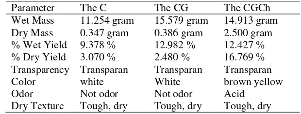
from rice waste water are summarized in Table 1.
Table 1 shows the physical properties of the bacterial celluloses and theirs
composites. The physical properties of the bacterial cellulose products without and with
the addition of glycerol (C and CG) have the same color, transparency, smell, and texture.
76
acidic odor. Odor which is generated also due to the solvent used in the preparation of
chitosan solution. The bacterial cellulose is capable of binding water (Zhong and Xia,
2008). Chitosan is able to enter into the pores of the microbial cellulose and the bacterial
cellulose surface coating so that the water that was in the air can not enter. Another
possibility, because hydrogen bond in chitosan interacts with the -OH group on the
cellulose so that water in the environment can not bind hydrogen, but not completely dried
cellulose chitosan glycerol because the water is still able to interact with the chitosan via
hydrogen bonds (Gadag and Shetty, 2006).
Table 1. Physical properties of the bacterial cellulose from rice waste water.
Parameter The C The CG The CGCh
Wet Mass 11.254 gram 15.579 gram 14.913 gram Dry Mass 0.347 gram 0.386 gram 2.500 gram % Wet Yield 9.378 % 12.982 % 12.427 % % Dry Yield 3.070 % 2.480 % 16.769 % Transparency Transparan Transparan Transparan
Color white White brown yellow
Odor Not odor Not odor Acid
Dry Texture Tough, dry Tough, dry Tough, dry
The physical properties of silver nanoparticle
The maximum wavelength of AgNO3 solution was at 216.20 nm region. The
spectrum measurement results of colloidal silver nanoparticles showed two largest peaks at
a wavelength of 423.40 nm and the maximum at 225.80 nm. The maximum peak at
wavelength of 225.80 nm in the spectrum of colloidal silver nanoparticles showed that
there is still a AgNO3 compound, it is possible because of the interaction between Ag with
H+ as a reaction product formed AgNO3 solution. A maximum peak at wavelength of
423.40 nm indicated the presence of silver nanoparticles (Lina et al., 2011 and Brugnerotto
et al., 2001). Based on Particle Size Analyzer results indicated that the reduction of silver
nitrate solution with a reducing agent of trisodium citrate has a median particle size of 74.8
nm and modus of 61.8 nm.
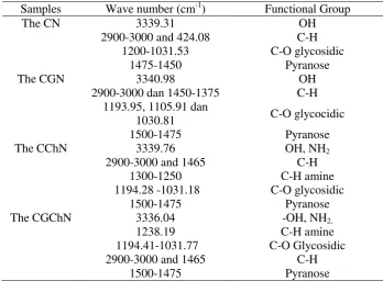
The functional group of the bacterial cellulose
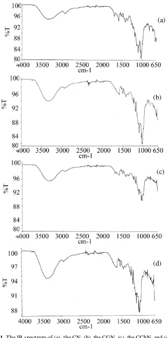
Analysis of the functional groups were performed using FTIR-ATR instrument.
Data analysis of functional groups in the form of IR spectra is shown in Figure 1. The IR
spectrum shows absorption of -OH groups of the CChN is lower than -OH group of the
CG. The IR spectrum of the CGCh shows typical absorption band for cellulosic functional
77
the functional groups of the CCh. Based on the IR spectrum interpretation, the functional
group of the bacterial cellulose and its composite are shown in Table 2.
Among the IR spectrum of the CGN, the CGN, the CChN, and the CGChN show
the similar functional group. The interpretation result of the IR spectrum can be shown in
Table 2.
Table 2. The functional group of the CN and the CGN.
Samples Wave number (cm-1) Functional Group
The CN 3339.31 OH
absorption of OH and NH2 groups. Absorption bands at wave number 1194.41 to 1031.77
cm-1 indicate the presence of CO glycosidic, the presence of three sharp absorption band in
the region and the absorption at 2900 - 3000 cm-1 indicate CH backed their uptake in
wave numbers of 1465 cm-1. Uptake of aromatic groups indicate their absorption in the
1500 - 1475 cm-1, the aromatic group is a group piran. The -OH absorption band on the
CGN showed its transmittance is smaller than the uptake -OH on the C. The smaller the
transmittance of the OH, the greater the absorbance of -OH, so the number of OH on the
CGN is more than- OH on the CN. The amount of hydrogen bonded can be seen from the
wide absorption band. If the absorption band is broad, it shows the -OH hydrogen bonding.
Based on the IR spectrum, it shows absorption band of -OH of the CGN is wider than the
CN. Thus, it indicates that the addition of glycerol will increase the number of hydrogen
bonds on the CN. The FTIR spectrum shows that the -OH absorption of the CGChN is
78
Figure 1. The IR spectrum of (a). the CN, (b). the CGN, (c). the CChN, and (d). The CGChN.
(a)
(b)
(c)
79
The addition of glycerol and chitosan on the bacterial cellulose increase the number
of -OH and amine groups in the cellulose. The addition of glycerol to the bacterial
cellulose - chitosan causes an increasing of hydrogen bonds in the bacterial cellulose, as
shown by the -OH absorption band in the bacterial cellulose -chitosan - glycerol (Zhong
and Xia, 2008). Results of functional groups by FTIR analysis showed aromatic ring and
vibration of amino group is characteristic of chitosan (Brugnerotto et al., 2001). This
characteristic absorptions alleged overlap with absorption aromatic ring, because the
cellulose and chitosan has an aromatic ring.
The Mechanical Properties of The Bacterial Cellulose
Based on the results of mechanical tests four samples of cellulose, the data
amounted strength at break, elongation at break and Young's modulus of microbial
cellulose, cellulose glycerol, cellulose selulsa chitosan and chitosan glycerol deposited
silver nanoparticles can be summarized in Table 3.
Table 3. The mechanical properties of the bacterial cellulose which is deposit silver nanoparticle.
Samples Strength at break (MPa) Elongation at break (%) Modulus Young (MPa)
The CN 54.878 20.3790 628.25
The CGN 87.560 9.7947 1268.30
The CChN 14.772 4.7842 660.55
The CGChN 7.9582 7.7829 249.18
Based on the Table 3, the CGN has the highest Young's modulus of 1268.3 MPa,
and the strength at break of 87.560 MPa. The CN has the highest elongation at break of
20.379 %. Thus, the CGN has the highest load bearing strength. This is likely due to the
initial structure of cellulose has a high crystallinity and addition of glycerol as internal
plasticizer can cause interacting between –OH of the cellulose and –OH of glycerol in
forming hydrogen bonds so that the CGN has stronger structure than the CN (Yunos and
Rahman, 2001). The addition of glycerol will generate interaction between -OH of
cellulose and –OH of glycerol through hydrogen bonding or dipole-dipole interactions
(Klaykruayat et al., 2010 and Kaushish, 2010). The more hydrogen bonds and high
crystallinity of the CGN cause it is able to withstand loads better than the CN. The
correlation between the increasing in hydrogen bonding and the tensile strength proved that
the increasing of hydrogen bonds will increase the tensile strength (Zhong and Xia, 2008;
Zhang et al., 2011; Liu et al., 2009; and Tien, 2010). Another possibility is the addition of
80
higher molecular mass shows the higher tensile strength than the lower molecular mass
(Brown, 2001). This may be a possibility when the glycerol was added, there was an
increase in the molecular mass of the cellulose so that a higher tensile strength of the CGN
than the CN.
The addition of chitosan turns down the strength at break of the CN. The reduction
of strength at break of the CChN could be caused the chitosan is a polymer with
amorphous structure, so the addition of chitosan to the CN will make the decreasing of
crystallinity of the CChN. The decreasing of the crystallinity of cellulose decreases the
load bearing strength of the CChN. A material is structurally strong because of high
crystallinity of a resistance to higher pressure, than the materials whose structure is
irregular and gives a lot of space around it (Liu et al., 2009). The addition to the
amorphous nature of the material that has high crystallinity will make crystalline
compound initially be semi-crystalline so many empty spaces that appeared causes of force
against the pressure to be reduced. The addition of the chitosan decreases elongation at
break significantly. Film corn starch occurs intermolecular bonds form hydrogen bonds
(Morones et al., 2005). This bond increases tensile strength but decreases elongation. This
is caused by hydrogen bonding in the formation of a crystal structure, that is rigid and
strong so that the elasticity of the material will decrease. The reason is based on glycerol
effect by disrupting intermolecular bonds in cellulose has a less rigid structure. Conversely,
if the intermolecular bonds in cellulose more and more due to the addition of chitosan, it
can be said that the elongation of the cellulose will decrease (Rechia et al., 2010).
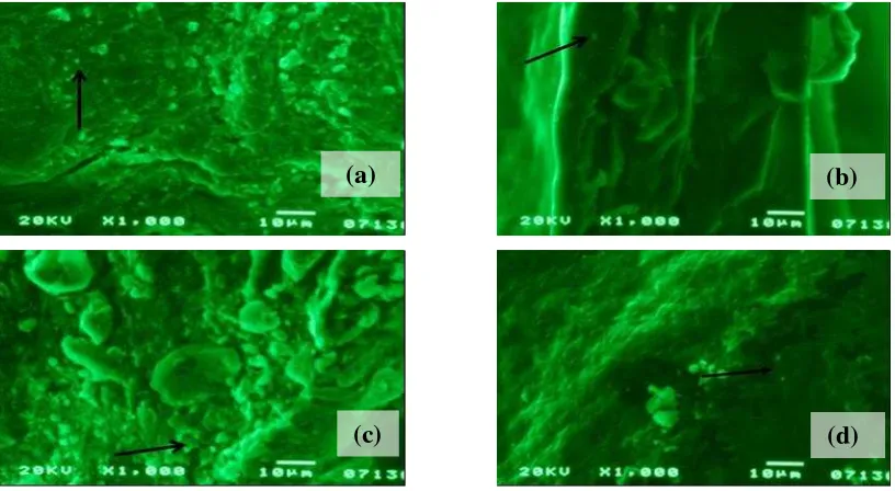
The SEM Photograph of The Bacterial Cellulose
Figure 2 shows the SEM photos of the bacterial celluloses and theirs composite
which are deposited nanoparticles silver. The photos are enough to prove the difference
between addition of chitosan and addition of glycerol on the cellulose. This proves that
chitosan is capable on coating the entire surface of the bacterial cellulose. The SEM results
indicate that silver nanoparticles also successfully deposited on the bacterial cellulose from
rice waste water. The suspected silver nanoparticles adsorbed on the bacterial cellulose.
Interactions between the bacterial cellulose and silver nanoparticles are chemical
adsorption which occurs through chemical bonds to form a covalent bond between -OH
groups on microbial cellulose with Ag in the silver nanoparticles. Figure 2 shows the SEM
photos of the bacterial celluloses and theirs composite which are deposited nanoparticles
81
Figure 2. The SEM photos of (a). the CN; (b). the CGN; (c). the CChN; (d). the CGChN.
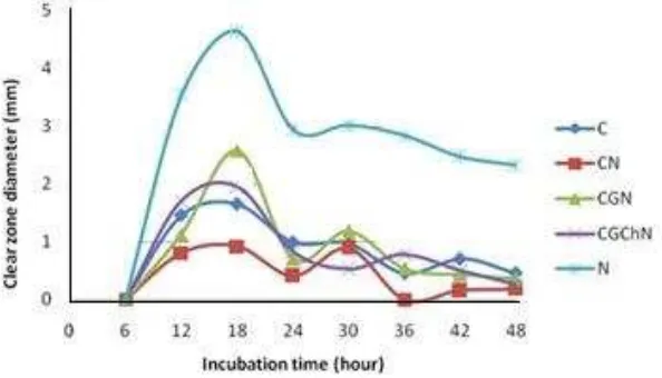
The antibacterial activity of the bacterial cellulose and its composite
Parameters of antimicrobial activity is apparent diameter of the clear zone around
the sample. A clear zone around the sample is formed because no microbes that can be
grown on the spot due to the antimicrobial activity of a compound in the sample. The
wider of the clear zone diameter indicates that the samples more effectively inhibit the
growth of microbes.
Figure 3. The Clear zone diameter of the bacterial cellulose and its composite against Staphylococcus aureus ATCC 25923.
Observation zone of inhibition was conducted for every 6 hours to know an
antimicrobial activity. Figure 3 shows that the samples against Staphylococcus aureus
ATCC 25923 have shown inhibition zone on the observation of the first 6 hours of
incubation, although still very narrow (except on pure cellulose samples). The CGN and
(a) (b)
82
the CGChN show inhibition zone diameter which began to decline after incubation for 42
hours. The CGChN has largest inhibition zone, while the C has the lowest inhibition zones.
ANOVA statistical test among sample types which were used against Staphylococcus
aureus ATCC 25923 shows that there are significant differences among the types of
samples. ANOVA statistical test among incubation time of Staphylococcus aureus ATCC
25923 shows a significance value of 0.744 (P > 0.05), meaning there is no significant
difference between the time of incubation of the samples against bacteria Staphylococcus
aureus ATCC 25923. Figure 4 shows that the testing of samples against yeast Candida
albicans ATCC 10231 has shown inhibition zone in the first 12 hours of observation. The
lowest antimicrobial on testing with Candida albicans is the CN, then the sample C and
CGChN, however the highest antifungal is indicated by the CGN sample The highest
inhibition is achieved at 18 hours incubation time.
Figure 4. The Clear zone diameter of the bacterial cellulose and its composite against Candida albicans ATCC 10231.
The samples have a inhibition of microbial activity against Candida albicans yeast
larger than a inhibition against Staphylococcus aureus. Differences in the effectiveness of
this inhibition may be due to the structure of the bacterial cell. The peptidoglycan of S.
aureus is a thick layer, serves as protection of antibacterial agents such as antibiotics,
toxins, chemicals, and degradative enzymes. Candida albicans cell wall is complex and
dynamic with a 100 - 400 nm thick. The outer layer of the cell wall of C. albicans consist
of mannoprotein. Mannoprotein represents 30 – 40 % of total cell wall polysaccharides and
determines the nature of the cell surface. C. albicans cell wall also contains sterols which
plays an important role as targets antimycotic and possibilities are the workings of
83
and sterols in the cell wall of Candida albicans causes high antimicrobial activity on the
samples against C. albicans.
Peptidoglycan in Staphylococcus aureus has teikoic acid, a polyalcohol which is
linked by phosphodiester bonds and usually has other sugars bonded with it. The teikoic
acids are negatively charged (having -COOH or -COO- group and phosphate group), thus
providing a negative charge on the surface of bacterial cells. Silver nanoparticles have a
positive charge (Ag+), making it easier to bind to the negatively charged surface of the
bacteria (Kim et al., 2007; Feng et al., 2000; and Jung et al., 2008). The -COO- group will
bind with Ag+. Then Ag+ will affect the proteins in the cell membrane. Therefore, the
silver nanoparticles react faster on the bacteria S. aureus compared with yeast C. albicans.
It marked only with a time of 6 hours has seen the diameter of inhibition zone in S. aureus
bacteria.
Candida albicans cell wall has a lot of mannoprotein which has positively charged amine
groups. The positive charge on the surface of C. albicans is a little slow to react to the silver
nanoparticles which were also charged positive. This is due to the tendency of the same charge for
this slow reaction. The reaction is marked by inhibition zone in yeast C. albicans after 12 hours of
incubation. The silver nanoparticles will affect the sulfhydryl groups on the protein surface
(mannoprotein). The number of mannoprotein which is influenced by silver nanoparticles, then the
resulting optimal inhibition.
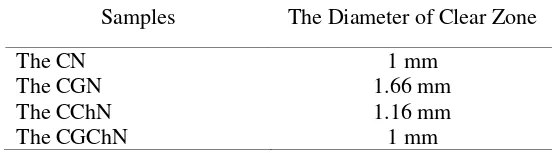
The antibacterial activity of the cellulose samples which are deposited silver nanoparticles
against E.coli ATCC 25922 are shown in Table 4. The antibacterial activity test of all samples
shows positive results. The largest clear zone is given by the CGN, then the CChN, the latter the
CN and the CGChN. Based on these results, the CGN is the most effective against the E. coli
ATCC 25922 compared to the three other types of cellulose.
Table 4. The antibacterial activity of the bacterial cellulose and its composite against E.coli ATCC25922.
Samples The Diameter of Clear Zone
The CN 1 mm
The CGN 1.66 mm
The CChN 1.16 mm
The CGChN 1 mm
The clear zone diameter of the CChN is greater than the CN, this is caused by the regular
structure of the CChN. Therefore, it is difficult for the silver nanoparticles to interact
electrostatically with functional groups of the CChN because of steric hindrance. The composition
of the cellulose with chitosan will change cellulose structure. Chitosan is a polymer amorphous so
84
decline with the addition of chitosan. Composite structures are more amorphous and the silver
nanoparticles easier attack to functional groups by electrostatic interaction. The CGChN samples
showed a positive antibacterial against E. coli ATCC 25922 and has a inhibition diameter of 1
mm. The CGChN has physical properties similar to the CChN but the CGChN contains plasticizer
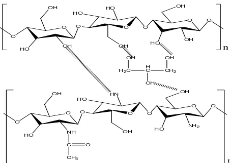
glycerol. The CGN molecular structure is shown in Figure 5. Based on these interactions seem that
the CGN has a structure steric bulk and a lot of obstacles causing the silver nanoparticles is
difficult to interact electrostatically with functional groups such as -OH and NH2. (Tien, 2010;
Smith et al., 1982; Helander et al., 2003; and Desi, 2006). If the silver nanoparticles is difficult to
interact with the functional groups, the antibacterial activity will be low due to at least silver
nanoparticles deposited on the CGChN. However, based on Figure 3 and 4, silver nanoparticles
showed the highest antibacterial activity, this is in accordance with the literature that silver
nanoparticles act as an antibacterial (Rai and Bai, 2010; Agus and Sri, 2010).
O O
Figure 5. Interaction among glycerol, chitosan, and cellulose.
CONCLUSION
Based on the results of the research and the discussion, it can be concluded that the
rice waste water can be used as a medium for bacterial growth in the formation of the
bacterial cellulose. The bacterial cellulose and the bacterial cellulose - glycerol composite,
which are deposited silver nanoparticles, have functional groups -OH, CH, CO glycosidic
and pyranose. The bacterial cellulose – chitosan (CCh) and the bacterial cellulose –
glycerol - chitosan (CGCh) have functional groups -OH, NH2, CN amide, CO glycosidic,
CH, and pyranose. The bacterial cellulose - glycerol, which is deposited silver
nanoparticles (CGN), has a strength at break of 1268.3 MPa. The CGN has the highest
Young's modulus. The CNs have an elongation at break of 20.379 %. The SEM photos of
the CN shows that the silver nanoparticles has been deposited on the surface of bacterial
85
and the CGChN against the growth of S. aureus, E. coli, and C.albicans. The CGChNs
provide the highest antimicrobial activity against Staphylococcus aureus ATCC 25923.
The CGNs provide the highest antimicrobial activity against E. coli ATCC 25922 and the
yeast Candida albicans ATCC 10231.
ACKNOWLEDGEMENT
Acknowledgements submitted to the Ministry of Research and Technology of the
Republic of Indonesia, which has provided financial support through the Research
Incentive SINas 2014.
REFERENCES
Agus, H., and Sri, B. H, 2010, Aplikasi Nanopartikel Perak pada Serat Katun sebagai Produk jadi Tekstil Antimikroba, Jurnal Kimia Indonesia, vol. 5, no. 1, pp. 1- 6.
Anicuta, S. G., Dobre, L., Stroesc,a M., and Jipa, I., 2010, Fourier Transform Infrared (FTIR) Spectroscopy for Characterization of Antimicrobial Films Containing Chitosan, Analele UniversităŃii din Oradea Fascicula: Ecotoxicologie, Zootehnie şi Tehnologii de Industrie Alimentară, pp. 1234-1240.
Baker, C. V., Pradhan, L. P., Pochan, D. J., and Shah, S. I., 2005, Synthesis and Antibacterial Properties of Silver Nanoparticles, Journal Nanoscience Nanotechnology, vol. 5, pp. 244-249.
Brown, M. E., 2001, Introduction of Thermal Analysis : Techniques and Applications 2nd Edition (Dordrecht : Kluwer Academic Publishers).
Brugnerotto, J., Lizardi, J., Goycoolea, F. M., Arguella-Monal, W., Desbrieres, J., and Rinaudo, M, 2001, An Infrared Investigation in Relation with Chitin and Chitosan Characterization, Polymer, vol. 42, no. 8, pp. 3569-3580.
Ciechańska, D., 2004, Multifunctional Bacterial Cellulose/Chitosan Composite Materials for Medical Application, Fiber & Textiles in Eastern Europe, vol. 12, pp. 69-72.
Desi, A. Y., 2006, Hubungan antara Aktivitas Antibakteri Kitosan dan Ciri Permukaan Dinding Sel Bakteri, Thesis, IPB.
Fang, J. C., Zhong, R., and Mu, R., 2005, The Study of Deposited Silver Particulate Films by Simple Method for Efficient SERS, Chemical Physics Letters, vol. 401, no. 1, pp. 271-275.
Feng, Q. L., Wu, J., Chen, G. Q., Cui, F. Z., Kim, T. N., and Kim, J. O., 2000, A Mechanistic Study of The Antibacterial Effect of Silver Ions on Escherichia coli and Staphylococcus aureus, Journal of Biomedical Material Research, vol. 52, no. 4, pp. 662-668.
86
Helander, I. M., Nurniaho, E. L., Ahvenainen, R., Rhoades, J., and Roller, S., 2001, Chitosan Disrupts The Barrier Properties of The Outer Membrane of Gram-negative Bacteria, Journal of Food Microbiology, vol. 7, no. 1, pp. 235-244.
Heru, P. and Eli, R., 2010, Pembuatan Bioplastik dari Limbah Rumah Tangga sebagai Bahan Edible Film Ramah Lingkungan, Laporan Penelitian, Yogyakarta : UNY
Hoenich, N., 2006., Cellulose for Medical Applications: Past, Present, and Future, BioResources, vol. 1, no. 2, pp. 270-280.
Jung, K. W., Koo, H. C., Kim, K. W., Shin, S., Kim, S. H., and Park, Y. H., 2008, Antibacterial Activity and Mechanism of Action of the Silver Ion in Staphylococcus aureus and Escherichia coli, Journal American Society for Microbiology, vol. 74, no. 7, pp. 2171-2178.
Kaushish, J. P., 2010, Manufacturing Process 2nd Edition, New Delhi : PHI Learning Private Limited.
Kim, Jung Sung, Eunye Kuk, Kyeong Nam Yu, Jong-Ho Kim, Sung Jin Park, Hu Jang Lee, So Hyun Kim, Young Kyung Park, Cheol-Yong Hwang, Yong-Kwon Kim, Yoon-Sik Lee, Dae Hong Jeong, andMyung-Haing Chao, 2007, Antimicrobial effects of silver nanoparticles, Nanotechnology, Biology and Medicine, vol. 3, no. 1, pp. 95-101.
Klaykruayat, B., Siralertmukul, K., and Srikulkit, K., 2010, Chemical Modification of Chitosan with Cationic Hyperbranched Dendritic Polyamidoamine and Its Antimicrobial Activity on Cotton Fabric, Carbohydrate Polymers, vol. 80, no. 1, pp. 97–207.
Kuusipalo, J., Kaunisto, M., Laine, A., and Kellomaki, M., 2005, Chitosan as a Coating Additive in Paper and Paperboard, TAPPI Journal, vol. 4, no. 8, pp. 17-21.
Lina, F., Yue, Z., Jin, Z., and Guang, Y., 2011, Bacterial Cellulose for Skin Repair Materials, Biomedical Engineering – Frontiers and Challenges (Croatia : InTech).
Liu, X., Gao, G., Dong, L., Ye, G., and Gu, Y., 2009, Correlation between Hydrogen-Bonding Interaction and Mechanical Properties of Polyimide Fibers, Polymer Advanced Technology, vol. 20, pp. 362-366. Antibacerials, Science Against Microbacterial Pathogens : Comunicating Current Research and Technological Advance, pp. 197-209.
Rechia, L. M., Morona, J. B., Zepon, K. M., Soldi, V., and Kanis, L. A., 2010, Mechanical properties and total hydroxycinnamic derivative release of starch/glycerol/Melissa officinalis extract films, Brazilian Journal of Pharmacy Science, vol. 46, no. 3, pp. 491-497.
87
Tien, B., 2010, Modifying Cellulose to Create Protective Material for Firefighters, (http://cosmos.ucdavis.edu/archives/2010/cluster8/TIEN_Benjamin.pdf)
Yunos, M. B. Z., and Rahman, W. A., 2011, Effect of Glycerol on Performance Rice Straw/Starch Based Polymer, Journal of Applied Science, vol. 11(13), pp. 2456-2459.
Zhang, H., Deng, L., Yang, M., Min, S., Yang, L., and Zhu, L., 2011, Enhancing Effect of Glycerol on the Tensile Properties of Bombyx mori Cocoon Sericin Films, International Journal of Molecul Science, vol. 12(5), pp. 3170-3181.