SYNTHESIS OF POLYETHYLENE OXIDE (PEO)–CHITOSAN HYDROGEL PREPARED BY
GAMMA RADIATION TECHNIQUE
Erizal
1.*and Thamrin Wikanta
21
Centre for the Applications Isotope and Radiation Technology, National Nuclear Agency, Jl. Lebak Bulus Raya no.49, Jakarta 12440
2
Centre Research for Product Processing and Biotechnology Marine & Fisheries, Jl. K.S. Tubun VI, Jakarta 10260
Received August 19, 2010; Accepted April 26, 2011
ABSTRACT
Crosslinked PEO hydrogel containing chitosan for wound dressing has been prepared by gamma radiation technique. Chitosan solutions with different concentration (0.5-2% w/v) have been blended with 5% aqueous solution of PEO and irradiated at the doses 20-40 kGy by gamma rays. The copolymers were characterized by Fourier transform infra red spectroscopy and their physico-chemical properties of hydrogels were evaluated in terms of gel fraction, swelling ratio, elongation at break and antimicrobial activities. It was found that under maximum condition of incorporation 1% chitosan with irradiation dose of 20 kGy, PEO-chitosan hydrogels with high gel fraction (85%), swelling ratio (10 g/g) and elongation at break (145%) were obtained. The examination of the microbe penetration shows that the prepared hydrogels can be considered as a good barrier against microbes. Thus, PEO-chitosan hydrogel showed satisfactory properties for use as a wound dressing.
Keywords:Gamma radiation, polyethylene oxide, chitosan, hydrogel, wound dressing
INTRODUCTION
Wound healing is an active area of research on accounts of its importance in the treatment of burns, prevention of post surgical adhesions and cosmetics surgery [1]. The purpose served by dressing includes protecting wounds, promoting healing and providing, retaining or removing moisture. Wound repairing is a complex process involving an integrate response by many different cell types and growth factors to achieve rapid restoration of skin integrity and protective function after injury [2]. In recent years, there have been tremendation in advances in the design and composition of bandages and dressings and there are numerous wound care materials. Some wound dressing types are biosynthetic dressings [3], hydrocolloid dressing [4], composite dressing [5], gauze [6], transparent film and hydrogels [7-8]. These dressings are differ in their architecture and compositions.
Hydrogel dressings are based upon a variety of different polymers. Some polymers are crosslinked to impact degree of structural stability to that the final product which often taken the form of the sheet used or application to relatively shallow surface of wound [9]. Hydrogel wound dressing has humectants properties such that the wound is kept hydrated to prevent any scabbing or drying out so that the wound is allowed to heal from inside out. The absorption of secretion causes an expansion of the crosslink in the polymer chains
making room for the inclusion of foreign bodies such as bacteria detritus and odor molecules that are irreversibly taken up along the liquid [10]. The hydrogel dressing’s removal is almost painless because hydrogel does not adhere to the wound surface. Hydrogel stays permanently moist and can be easily removed after prolong application without pain and risk of wound irritation. Due to the above reasons the hydrogel wound dressing are highly accepted by the patient [11]. Wound dressing materials should be chemically inert and free from leachable impurities. Most important criteria to select materials for wound dressing are their biocompatibility.
Instrumentation
Volumetric glasses were used for preparing reagents, polyethylene oxide and chitosan solution. The FT-IR spectra of crosslinking of hydrogels were recorded on Shimadzu spectrophotometer using solid KBr pellets after completely drying the sample at 120 °C for 1 h.
Procedure
Preparation of PEO-chitosan hydrogels
A 5% aqueous solution of PEO was prepared in distilled water. Different quantities of chitosan (0.5–2%) in 1% acetic acid solution were dissolved in the above solution at room temperature to get the final mixture, respectively. The mixture was poured into polyethylene plastic bags ( 20 cm x 10 cm ), sealed and then irradiated by gamma rays from 60Co source (dose rate: 10 kGy/h) at room temperature. The samples were irradiated at doses from 10 kGy to 40 kGy. The hydrogels obtained were washed directly for gel fraction determination whereas for swelling studies, the hydrogels were washed several times with distilled water to remove the unreacted polymer and dried in the vacuum oven at 60 °C, until constant weight.
Gel fraction
The hydrogel samples (2 cm x 2 cm x 0,5 cm) in distilled water were taken into shaker incubator at room temperature to remove soluble fraction for 24 h. The gel was dried to constant weight under vacuum to determine the insoluble fraction in the samples gravimetrically and was calculated as
Gel Fraction (%) =Wg/Wox 100, (1)
whereWgis the weight of dry gel after extraction andWo
is the initial weight of the gel.
Swelling studies
The degree of swelling was determined by gravimetric method. The gel samples (dried to constant
measurement. Both end of the pieces were firmly clamped in the jaws of a testing machine. One jaw was fixed and the other was movable. The movable jaw moved at the rate of 30 mm/min. at room temperature. The resultant data was showed at the recorder. This procedure was repeated for three times for each result. The elongation at break was calculated from the following equation
Elongation at break = L1/Lox 100% (3)
where Lois the original length and L1is the final length.
Penetration of bacteria
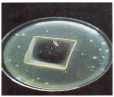
A 102 aseptic Bacillus pumilus suspension and PCA (Plate Count Agar) solutions were prepared in 200 mL distilled water, then 0.1 mL of bacteria suspension were taken into petridish glass containing PCA solution, homogenized until solidification. The hydrogels (5 cm x 7 cm x 0.2 cm) were put in the middle of the media and incubated at room temperature. The bacteria’s penetration through the hydrogels was monitored after one week.
RESULT AND DISCUSSION
FT-IR spectra
FT-IR spectrum of pure PEO, chitosan and PEO-chitosan hydrogels are shown in Fig. 1. For pure PEO polymer (curve 1) the characteristic bands at 2600–3000 cm-1, 2000–2400 cm-1, and 900–1300 cm-1 correspond to hydroxyl group, C-H stretching, and C-O-C group, respectively. For chitosan (curve 2) the characteristic bands at 3450 cm-1, 2920 cm-1, 1660 cm-1, 1590 cm-1, 1380 cm-1, and 1150–1040 cm-1 correspond to hydroxyl group, CH2 stretching vibration
of pyranose ring, C=O in amide group,NH2 in amino
group, CH3 in amide group and C-O-C in glycosidic
Fig 1. FT-IR spectrum of [1] PEO [2] Chitosan and [3] PEO-chitosan hydrogel
Fig 2.Effect of chitosan concentration on the gel fraction of PEO-chitosan hydrogels at different radiation doses with 5% PEO
Fig 3. Effect chitosan concentration on the swelling ratio of PEO-chitosan hydrogels (formed at 20 kGy) in different time
Fig 4.Effect of chitosan concentration on elongation at break of PEO-chitosan hydrogels (formed at 20 kGy)
Gel fraction
Fig. 2 shows the effects of irradiation dose and the variation of chitosan amount in hydrogels on gel fraction of PEO-chitosan hydrogels. Chitosan is a natural polysaccharide, which degrades on irradiation by breakdown of the main chains [14]. However in the system studied, PEO is the main component that is known polymerizes and crosslink in aqueous medium [15]. When the mixture containing PEO and chitosan is irradiated an interpenetrating polymer network (IPN) is formed with the chemical crosslinking of PEO and physical crosslinking of chitosan [16].
The gel fraction of the hydrogel increases up to the maximum condition with increasing of the 1% chitosan concentration after which the gel fraction decreases. Thus, there is only slight decrease in the gel fraction with
increasing of irradiation dose in this study. This result indicated that the hydrogel with high gel fraction of 85% can be produced in the presence of 1% chitosan at the dose of 20 kGy. Soren et al. [12] and Yang et al. [17] reported that the hydrogels with the gel fraction nearly 80% are suitable to be applied as wound dressings.
Effect of chitosan concentration on swelling ratio
might say that the hydrogels can not absorb all the wound effusion. According to Yang et al. [17] if the hydrogel with swelling ratio 1/2 times lower than its ideal value it was needed to be changed every 2 or 3 days for the applications as exudates wound dressings.
Effect of chitosan concentration on elongation at break
Elongation at break is an important physical factor of hydrogels representing its flexibility when it is used for wound dressings. Until now, there are no available references related with the standard values of elongation at break for wound dressing. Generally, the more flexible of hydrogels, the more easily to follow the skin surface contour. The measurement of elongation at break of hydrogels conducted in this experiment in order to get a supporting data for specification of products [12,17].
Fig. 4 illustrates the dependence of elongation at break of PEO-chitosan hydrogels on the chitosan concentrations. The original elongation at break of hydrogel is 145% at chitosan concentration 0.5%. With the increase of chitosan concentration, the elongation at break of hydrogel keeps constant at chitosan concentration 1% and then starts to decrease and keeps to the end. Thus it is reasonable to say that the chitosan concentration at the range of 0.5–1% is the maximum condition for elongation at break of hydrogel can be reached. The decrease of elongation at break over than chitosan concentration 1% may result from the uncrosslinks of chitosan which distributed unhomogenously in the hydrogel matrix producing harder and less extensible hydrogels. These results were consistent with the increase chitosan concentration, the gel fraction of hydrogel decrease as obtained above.
Microbe penetration test
Effect of Bacillus pumilus bacterial growth on hydrogel in the PCA agar media is presented in Fig. 5. It
inhibition or bacterial penetration inhibition into the hydrogel and practically no bacteria was growth in the hydrogel. So, the crosslinked PEO-chitosan hydrogel prepared in this manner could be considered as a good barrier against the microbe. This is a very important property of hydrogel for wound dressing to protect wound from infections.
CONCLUSION
Based on the research, PEO-chitosan hydrogel can be synthesized by gamma irradiation techniques with specific physical properties: gel fraction (85%) at chitosan concentration 1%, swelling ratio 10 g/g, an elongation at break (145%), and an antibacterial or bacterial growth inhibition activities. This method has advantages such as: convenient preparation, possibility of hydrogel formation with a variety of hydrophilic polymers, and not necessary to add any crosslinker. This hydrogel is suitable for wound dressings.
ACKNOWLEDGEMENT
We are gratefully acknowledged to colleagues in irradiation facilities PATIR-BATAN which has been providing the samples irradiation and to Prof. Ir. Sugiharto and Dra. Krisna MLR, M.Sc. for their valuable comments and discussions.
REFERENCES
1. Risbud, M., Hardikar, A., and Bhonde, R., 2000,J. BioSci.,25, 1, 25–31.
2. Park, J.E., and Barbul, A., 2004,Am. J. Surg.,187, 511–516.
3. Davydova, G.A., Selezneva, I.I., Savinttseva, and Gavrilyuk, B., 2008,Bull. Exp. Biol. Med., 145, 52– 58.
5. Korycky, R., 2009,Struct. Multidiscip. Optim., 39, 3, 283–296.
6. Li, S-T., Cao, B., Deng, W-L., and Li, Z., 2009,Chin. J. Integr. Med.,15, 4, 279–283.
7. Chandroth, K.S., and Tholath, E.A., 2010, Colloid Polym. Sci., 288, 3, 297–306.
8. Erizal, 2008,Indo. J. Chem., 8, 2, 271–278. 9. Thomas, S., 1996,Wounds, 8, 5, 145–149.
10. Pai, K., Banthia, A.K., and Mayumdar, D.K., 2006,J. Biol. Res.,9, 23–29.
11. Krasner, D., and Kane, D., 1997, Chronic Wound Care: A Clinical Sourcebook for Healthcare Professionals, Health Management Publications, 2nd ed., 336–343.
12. Sorens, D.A, and Malik, S., 2005, USA Patent 6967261.
13. Michael, P.T., and Stephen, E.H., 1997, An Introduction to Polysaccharide Biotechnology, Taylor & France, 147–149.
14. Maolin, Z., Hongfei, H., Yoshii, F., and Makuuchi, K., 2000,Radiat. Phys. Chem.,57, 3-6, 459–464. 15. Rosiak, J.M., Ulanski, P., Pajewski, A., Yoshii, F.,
and Makuuchi, K., 1995,Radiat. Phys. Chem., 46, 2, 161–168.
16. Jing, R., Yancun, Z., Jiuqiang, L., and Hongfei, H., 2001,Radiat. Phys. Chem., 62, 2-3, 277–281. 17. Yang, Z., Rao, Z., Ling, Y.S., and Zhu, Y.N., 2008,
![Fig 1. FT-IR spectrum of [1] PEO [2] Chitosan and [3]PEO-chitosan hydrogel](https://thumb-ap.123doks.com/thumbv2/123dok/872120.820838/3.595.56.295.109.304/fig-ft-spectrum-peo-chitosan-peo-chitosan-hydrogel.webp)