Cytotoxic Triterpenoids from the Stembark of
Aglaia argentea
(Meliaceae)
Ace Tatang Hidayat
1,2, Kindi Farabi
1, Desi Harneti
1, Nurlelasari
1, Rani Maharani
1,2,
Ida Nurfarida
2, Unang Supratman
1,2,*, and Yoshihito Shiono
31Department of Chemistry, Faculty of Mathematics and Natural Sciences, Universitas Padjadjaran, Jatinangor 45363, Indonesia
2Central Laboratory of Universitas Padjadjaran, Jatinangor 45363, Indonesia
3Department of Food, Life, and Environmental Science, Faculty of Agriculture, Yamagata University, Tsuruoka, Yamagata 997-8555, Japan
Received May 5, 2017; Accepted November 18, 2017
ABSTRACT
Four dammarane-type triterpenoid compounds, dammar-24-en-3α-ol (1), 3-epi-cabraleahydroxy lactone (2), (E)-25-hydroperoxydammar-23-en-3β,20-diol (3), and dammar-24-en-3β, 20-diol (4), were isolated from the methanolic extract of the stembark ofAglaia argentea. Compounds,1-4, were isolated for first time from this plant. The structure of isolated compounds were elucidated by spectroscopic methods including one and two-dimensional NMR as well as mass spectrometric analysis. Compounds 1-4, along with a known synthetic analog, 20-hydroxy-dammar-24-en-3-on (5), were evaluated their cytotoxic activity against P-388 murine leukemia cells in vitro. The IC50 values of compounds, 1-5 were 9.09 ± 0.10, 68.53 ± 0.08, 5.89 ± 0.08, 22.40 ± 0.11, and 11.53 ± 0.08 µg/mL, respectively. Among the dammarane-type triterpenoids, compounds1,3,4and5having opened side chain showed the stronger activity, where’s compound2with a cyclic side chain showed weak or no activity. In addition, compound 3showed the strongest activity, indicate that hydroperoxy group at side chain increase cytotoxic activity.
Keywords:Aglaia argentea; cytotoxic activity; dammarane-type triterpenoids; P-388 murine leukemia
ABSTRAK
Empat senyawa triterpenoid tipe damaran, damar-24-en-3α-ol (1), 3-epi-kabraleahidroksi lakton (2), (E)-25-hidroksi-damar-23-en-3β,20-diol (3), dan damar-24-en-3β,20-diol (4), telah disolasi dari ekstrak metanol dari kulit batang Aglaia argentea. Senyawa 1-4, diisolasi untuk pertama kali dari tumbuhan ini. Senyawa 1-4, bersama dengan senyawa analog sintetik yang telah dikenal, dammar-24-en,20-ol-3-on (5), dievaluasi aktivitas sitotoksiknya terhadap sel murin leukemia P-388 secara in vitro. Nilai IC50senyawa1-5 berturut-turut adalah 9,09 ± 0,10, 68,53 ± 0,08, 5,89 ± 0,08, 22,40 ± 0,11, dan 11,53 ± 0,08 µg/mL. Diantara senyawa triterpenoid tipe damaran, senyawa1,3, 4, dan 5 yang memiliki rantai samping terbuka menunjukkan aktivitas lebih kuat, sementara senyawa 2 dengan rantai samping siklik menunjukkan aktivitas lemah atau tidak aktif. Sebagai tambahan, senyawa 3 menunjukkan aktivitas sitotoksik paling kuat, mengindikasikan bahwa gugus hidroperoksi pada rantai samping meningkatkan aktivitas sitotoksik.
Kata Kunci: Aglaia argentea; aktivitas sitotoksik; triterpenoid tipe damaran; sel murin leukemia P-388
INTRODUCTION
Dammarane-type triterpenoids have been gaining worldwide attention for a long time because of their potent bioactivities [1]. Dammarane-type triterpenoids are distributed in genera of Panax (Araliaceae), Gynostemma (Cucurbitaceae), Aralia (Araliaceae), Aglaia (Meliaceae), Bacopa (Scrophulariaceae), Copernicia (Arecaceae), Celastrus (Celastraceae), Forsythia (Oleaceae), Myrica (Myrica), Rhus (Anacardiaceae), Polyscias (Araliaceae) and Sapindus (Sapindus) [2-6]. The basic molecular skeleton of
dammarane-type triterpenoids comprises tetracyclic moiety and side chain moiety [1]. The bioactivities and chemical properties of dammarane-type triterpenoids have been investigated, and several pharmacological effects have been disclosed, including anti-fatigue, antihyperglycemic, antiobesity, anticancer, anti-HIV, antioxidant, antiaging, immunostimulatory, antiatherosclerotic and antihypertensive effects [7-9]. The genus Aglaia is the largest genus of the family of Meliaceae comprises more than 100 species distributed mainly in India, Indonesia, Malaysia and parts of the Western Pacific region [10]. The genus of
Aglaia traditionally used to moisturizing the lungs, reducing fever and for treating the contused wound, coughs and skin diseases [11]. In Indonesia, flowers of A. odoratatraditionally used as an insect repellent [12].
Some species ofAglaiahave been phytochemically investigated previously with unique biological activity, such as antifungal sesquiterpenoids [13-14], diterpenoids [15-16], insecticidal and cytotoxic rocaglate derivatives [17-18], lignans [19-20], cytotoxic and antiviral dammarane-type triterpenoids [21,24] and antitumor cycloartane-type triterpenoids [4-5,8].
In our continuous search for cytotoxic constituents against P-388 murine leukemia cells from Indonesian Aglaia plants, we isolated and described two new cytotoxic triterpenoids, aglinone, and aglinin E, from the bark of A. smithii [23], a new stigmastane steroid, 3,4-epoxy-(22R,25)-tetrahydrofuran-stigmast-5-en, two new bisamide compounds, eximiamide A and B, from the bark ofA. eximia[24-25] and one lignan [20].
In the further screening for cytotoxic compounds from Indonesia Aglaia plants, we found that the n-hexane and ethyl acetate extract of A. argentea exhibited a cytotoxic activity against P-388 murine leukemia cells with IC50 values of 26.72 and 15.49
µg/mL, respectively. We report herein the isolation, structural elucidation of triterpenoid compounds (1-4) and a synthetic analogue,5, together with their cytotoxic activity.
EXPERIMENTAL SECTION
Materials
The bark of A. argentea was collected in Bogor Botanical Garden, Bogor, West Java Province, Indonesia in June 2015. The plant was identified by the staff of the Bogoriense Herbarium, Bogor, Indonesia and a voucher specimen (No. Bo-1288718) was deposited at the herbarium.
Instrumentation
Melting points were measured on an electrothermal melting point apparatus and are uncorrected. The IR spectra were recorded on a Perkin-Elmer Spectrum-100 FT-IR in KBr. Mass spectra were obtained with a Synapt G2 mass spectrometer instrument. NMR data were recorded on a JEOL ECZ-600 spectrometer at 600 MHz for 1H and 150 MHz for 13C and TMS as an internal
standard. Column chromatography was conducted on silica gel 60 (Kanto Chemical Co., Inc., Japan). TLC plates were precoated with silica gel GF254(Merck, 0.25
mm) and detection was achieved by spraying with 10% H2SO4in EtOH, followed by heating.
Procedure
Extraction and isolation
The dried bark (2.5 kg) of A. argentea was extracted with methanol exhaustively (12 L) at room temperature for 5 days. After removal of the solvent under vacuum, the viscous concentrated of MeOH extract (133.5 g) was first suspended in H2O and then
partitioned with n-hexane, EtOAc, and n-butanol, successively. The n-hexane soluble fraction (26.3 g) was fractionated by vacuum liquid chromatography on silica gel 60 using a gradient n-hexane and EtOAc to give nine fractions (A–I). Fraction C (2.68 g) was chromatographed on a column of silica gel, eluted with a gradient of n-hexane–EtOAc (10:0–1:1), to give nine subfractions (C01–C09). Subfraction C05 was chromatographed on a column of silica gel, eluted with CH2Cl2:CHCl3 (9.75:0.25), to give four subfractions
(C05A-C05D). Subfraction C05B was separated on preparative TLC, eluted with n-hexane–EtOAc (9:1), to give 1 (5.3 mg). Fraction D (1.65 g) was chromatographed on a column of silica gel, eluted with a gradient ofn-hexane–EtOAc (10:1–1:10), to give six subfractions (D01–D06). Subfraction D04 was recrystallized in EtOAc, to give2(27.6 mg). Subfraction D05 was chromatographed on a column of silica gel, eluted with a gradient of n-hexane–EtOAc (10:1–1:10) to afford five subfractions (D05A-D05E). Subfraction D05D was chromatographed on a column of silica gel, eluted with a gradient of CHCl3–EtOAc (10:1–1:10) to
give 3 (4.9 mg). The EtOAc soluble fraction (12.1 g) was fractionated by column chromatography on silica gel using a gradient n-hexane-EtOAc to give eight fractions (J–Q). Fraction K (927.6 mg) was chromatographed on a column of silica gel, eluted with n-hexane–EtOAc (10:1–1:1), to give four subfractions (K01–K04). Subfraction K02 was separated on preparative TLC silica gel GF254, eluted with
n-hexane:EtOAc (7:3), to give4(40.3 mg).
Compound 4 (12.0 mg) was dissolved in anhydrous pyridine (1 mL) in a vial (4 mL), and CrO3
(20.0 mg) was then added. After standing at room temperature overnight, the reaction mixture was separated through a small silica gel (1 g) column (0.5 x 4.2 cm), eluted withn-hexane:Me2CO (4:1, 20 mL). The
elution was evaporated to dryness under reduced pressure at 45 °C, to give the oxidation product of 5, 20-hydroxy-dammar-24-en-3-on (Rf 0.55; 4.2 mg). Dammar-24-en-3α-ol (1). White needle-like crystals, m.p. 152-153 °C; IR (KBr)max3345, 2937, 2870, 1464,
1379, 1056 cm-1; 1H-NMR (CDCl3, 600 MHz) and13
C-NMR (CDCl3, 125 MHz), see Table 1.
3-epi-Cabraleahydroxy lactone (2).White amorphous powder, m.p. (decomposed); IR (KBr)max3477, 2942,
1715, 1471, 1387, 1075 cm-1;1H-NMR (CDCl3, 600 MHz),
Fig 1.Structures of compounds1-5
13C-NMR (CDCl3, 125 MHz), see Table 1; HR-TOFMS
(positive ion mode) m/z 417.3105 [M+H]+, (calcd.
C27H44O3,m/z416.3290).
(E)-25-Hydroperoxydammar-23-en-3β,20-diol (3). Colorless oil; IR (KBr) max 3436, 2945, 1651, 1456,
1074, 847 cm-1; 1H-NMR (CDCl3, 600 MHz), 13C-NMR
(CDCl3, 125 MHz), see Table 1; HR-TOFMS (positive ion
mode) m/z 477.3951 [M+H]+, (calcd. C30H52O4, m/z
476.3866).
Dammar-24-en-3β,20-diol (4). White amorphous powder, m.p. (decomposed); IR (KBr) max3369, 2939,
1639, 1458, 1109 cm-1;1H-NMR (CDCl3, 600 MHz),13
C-NMR (CDCl3, 125 MHz), see Table 2; HR-TOFMS
(positive ion mode) m/z 445.0527 [M+H]+, (calcd.
C30H52O2,m/z444.3967).
20-Hydroxy-dammar-24-en-3-on (5).White amorphous powder, m.p. (decomposed);1H-NMR (CDCl3, 600 MHz)
and13C-NMR (CDCl3, 125 MHz), see Table 2.
Determination of Cytotoxic Activity
The P388 murine leukemia cells were seeded into 96-well plates at an initial cell density of approximately 3 x 10⁴ cells cm-3. After 24 h of incubation for cell
attachment and growth, varying concentrations of samples were added. The compounds added were first dissolved in DMSO at the required concentration. Subsequent six desirable concentrations were prepared using PBS (phosphoric buffer solution, pH = 7.30–7.65). Control wells received only DMSO. The assay was terminated after a 48 h incubation period by adding MTT reagent [3-(4,5-dimethylthiazol-2-yl)-2,5-diphenyl tetra zolium bromide; also named as thiazol blue] and the
incubation was continued for another 4 h, in which the MTT-stop solution containing SDS (sodium dodecyl sulphate) was added and another 24 h incubation was conducted. Optical density was read by using a microplate reader at 550 nm. IC50 values were taken
from the plotted graph of percentage live cells compared to control (%), receiving only PBS and DMSO, versus the tested concentration of compounds (µg/mL). The IC50 value is the concentration required
for 50% growth inhibition. Each assay and analysis was run in triplicate and averaged [26-28].
RESULT AND DISCUSSION
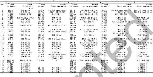
Compound 1 was obtained as white needle-like crystal. The molecular formula of compound 1 was C30H52O based on the analysis of NMR and thus
required five degrees of unsaturation, originating from one pair of C sp2 and the remaining tetracyclic triterpenoids. The IR spectra showed absorption peaks at 3345 cm-1 (OH), 2937 and 2870 cm-1 (C-H sp3), 1464 cm-1 (C=C), 1379 cm-1 (gem-dimethyl groups),
and 1056 cm-1 (C-O). The 1H-NMR (CDCl
3 600 MHz)
spectrum showed the presence of seven tertiary methyl
groups, resonating at δH 0.95 (H-18), 0.85 (H-19),
1.62 (H-26), 1.56 (H-27), 0.96 (H-28), 0.79 (H-29), and
0.88 (H-30) and one secondary methyl at δH 1.10 (d,
J = 6.5 Hz, H-21). There was one olefinic methine
group, resonating at δH5.09 (1H, t,J= 7 Hz, H-24) and
one oxymethine resonating at δH 3.64 (1H, d, J = 2.5
Hz, H-3) which indicates that the hydroxy group was attached in C-3. The proton pairing was also confirmed
HO HO
O O
1 2
3 OH
OOH
HO HO
OH
4 1
3 5
7 9
12
15 17 18 19
21
30 20
28
23
25
29
26 27
Fig 2.Selected HMBC and1H-1H COSY correlations for1-4
with the 1H-1H COSY spectrum (Fig. 2). The 13C-NMR
(CDCl3 150 MHz) and DEPT 135° spectra showed the
presence of eight methyl groups, exhibiting the characteristics of triterpenoid compounds [21-22], one
olefinic methine at δC 125.3 (C-24), one olefinic
quaternary carbon at δC 130.5 (C-25), and an
oxymethine group at δC 75.0 (C-3). The HMBC
cross-peaks (Fig. 2) from CH3-28 (δH 0.96), CH3-29 (δH 0.79),
and the methylene protons at H-2 (δH 1.47) to the
oxymethine carbon at C-3 (δC 75.0) indicated the
presence of a hydroxy group at C-3. The correlation which was arising from CH3-26 (δH1.62) and CH3-27 (δH
1.56) to C-25 (δC 130.5) and C-24 (δC 124.3) indicated
that position of a double bond at C-24/C-25. The
conformation of C-3 was assigned as α based on
coupling constant of H-3 (J = 2.6 Hz) and by comparing to those of reference [29]. These functionalities accounted for one of five total degrees of unsaturation, and the remaining four degrees of unsaturation were consistent with the triterpenoid skeleton. A comparison of the NMR data of 1 with dammar-24-en-3α-ol [30] revealed that the structures of the two compounds were very similar; consequently, compound 1 was identified
as dammar-24-en-3α-ol.
Compound 2 was obtained as a white amorphous powder. Its molecular composition C27H44O3, was
established from the HR-ESI-TOFMS spectrum (m/z 417.3105, [M+H]+) together with NMR data (Table 1).
The IR spectra showed absorption peaks at 3477 cm-1
(OH), 2942 cm-1(C-Hsp3), 1715 cm-1(C=O), 1471 and
1379 cm-1 (gem-dimethyl groups), and 1075 cm-1
(C-O). The 1H-NMR (CDCl
3 600 MHz) spectrum
showed the presence of six tertiary methyl groups,
resonating at δH 0.92 (CH3-18), 0.82 (CH3-19),
1.33 (CH3-21), 0.91 (CH3-28), 0.81 (CH3-29), and
0.87 (CH3-30) and one oxymethine group, resonating at
δH 3.37 (1H, s, H-3) which was indicated the presence
of dammarane-type triterpenoid skeleton [30-31]. The proton pairing was also confirmed with the1H-1H COSY
spectrum (Fig. 2). The 13C-NMR (CDCl
3 150 MHz)
spectra showed 27 carbons and classified by DEPT 135° experiment as six methyl groups, exhibiting the characteristics of trisnor-triterpenoid compounds [31],
one carbonyl lactone at δC176.9 (C-24), an oxymethine
group at δC75.0 (C-3), and an oxygenated quaternary
carbon at δC90.3 (C-20). The HMBC cross-peaks (Fig.
2) from CH3-28 (δH 0.91), CH3-29 (δH 0.81), and the
methylene protons at H-2 (δH 1.40) to the oxymethine
carbon at C-3 (δC 76.3) indicated the presence of a
hydroxy group at C-3. The correlation which was
arising from H-22 (δH 1.47) and H-23 (δH 2.52) to C-24
(δC 176.9) and C-20 (δC90.3) indicated that position of
lactone in C-20/C-24. The conformation of C-3 was
assigned as α based on coupling constant of H-3
(J= 0) [22,31]. These functionalities accounted for one of six total degrees of unsaturation, and the remaining
Table 1.NMR Data (600 MHz for1H and 150 MHz for13C, in CDCl3) for1-5
five degrees of unsaturation were consistent with the triterpenoid skeleton with lactone ring at the side chain. A comparison of the NMR data of 2 with cabraleahydroxy lactone [22] revealed that the structures of the two compounds were very similar, consequently, compound 2 was identified as a 3-epi-cabraleahydroxy lactone.
Compound 3 was obtained as a colorless oil. Its molecular composition C30H52O4, was established from
the HR-ESI-TOFMS spectrum (m/z 477.3951, [M+H]+)
together with NMR data (Table 1). The IR spectra showed absorption peaks at 3436 cm-1(OH), 2945 cm-1
(C-H sp3), 1651 cm-1 (C=C), 1456 cm-1 (gem-dimethyl
groups), 1076 cm-1(C-O), and 847 cm-1(O-O). The 1
H-NMR (CDCl3 600 MHz) spectrum showed the presence
of eight tertiary methyl groups, resonating at δH 0.94
(CH3-18), 0.83 (CH3-19), 1.11 (CH3-21), 1.34 (CH3-26),
1.33 (CH3-27), 0.96 (CH3-28), 0.76 (CH3-29), and 0.85
(CH3-30), one oxymethine group, resonating at δH 3.19
(1H, dd,J = 4.8, 11.4 Hz, H-3), and two methine sp2 at
δH5.76 (1H, dd,J= 7.8, 16.2 Hz) and 5.60 (1H, dd, J=
4.8, 16.2 Hz, H-24), which was indicated the presence of dammarane-type triterpenoid skeleton. The proton pairing was also confirmed with the 1H-1H COSY
spectrum (Fig. 2). The 13C-NMR (CDCl3 150 MHz)
spectra showed 30 carbons and classified by DEPT 135° experiment as eight methyl groups, an oxymethine group
at δC79.1 (C-3), two oxygenated quaternary carbons at
δC 75.2 (C-20) and 82.2 (C-24), and two methinesp2at
δC 127.4 (C-23) and 137.4 (C-24). One oxygenated
quaternary carbon at δC 82.2 (C-24) was more
deshielded, indicate that hydroperoxy group attaches at
C-24 [26]. The HMBC cross-peaks (Fig. 2) from H-28
(δH 0.96), H-29 (δH0.76), and the methylene protons at
H-2 (δH 1.44) to the oxymethine carbon at C-3 (δC79.1)
indicated the presence of a hydroxy group at C-3. The
correlation which was arising from H-23 (δH 5.76) and
H-24 (δH 5.60) to C-22 (δC 43.4) and C-25 (δC 82.2)
suggest the position of a double bond at C-23/C-24.
The conformation of C-3 was assigned as β based on
coupling constant of H-3 (J = 4.8, 11.4 Hz) [22]. These functionalities accounted for one of five total degrees of unsaturation, and the remaining four degrees of unsaturation were consistent with the triterpenoid skeleton. A comparison of the NMR data of 3with
(E)-25-hydroperoxydammar-23-en-3β,20-diol [22] revealed
that the structures of the two compounds were very similar; consequently, compound 3 was identified as 3(E)-25-hydroperoxydammar-23-en-3β,20-diol.
Compound 4 was obtained as a colorless oil. Its molecular composition C30H52O2, was established from
the HR-ESI-TOFMS spectrum (m/z 445.0527, [M+H]+)
together with NMR data (Table 1). The IR spectra showed absorption peaks at 3369 cm-1(OH), 2939 cm-1
(C-H sp3), 1639 cm-1 (C=C), 1458 cm-1 (gem-dimethyl
groups), and 1109 cm-1 (C-O). The 1H-NMR (CDCl 3
600 MHz) spectrum showed the presence of eight
tertiary methyl groups, resonating at δH0.93 (CH3-18),
0.82 (CH3-19), 1.13 (CH3-21), 1.66 (CH3-26),
1.59 (CH3-27), 0.91 (CH3-28), 0.81 (CH3-29), and
0.86 (CH3-30), one oxymethine group, resonating at
δH3.37 (1H, t,J= 4.5 Hz, H-3), and one methinesp2at
δH 5.10 (1H, t, J = 5.4 Hz, H-24) which was indicated
the presence of dammarane-type triterpenoid skeleton.
Table 2.Cytotoxic activity of compounds1–5against P-388 murine leukemia cells Compounds IC50(µg/mL)
Dammar-24-en-3α-ol (1) 9.09 ± 0.10
3-epi-Cabraleahydroxy lactone (2) 68.53 ± 0.08 (E)-25-Hydroperoxydammar-23-en-3β,20-diol (3) 5.89 ± 0.08
Dammar-24-en-3β,20-diol (4) 22.40 ± 0.11
20-Hydroxy-dammar-24-en-3-on (5) 11.53 ± 0.08
The proton pairing was also confirmed with the 1H-1H
COSY spectrum (Fig. 2). The 13C-NMR (CDCl 3
150 MHz) spectra showed 30 carbons and classified by DEPT 135° experiment as eight methyl groups, an
oxymethine group at δC 76.4 (C-3), one oxygenated
quaternary carbon at δC75.5 (C-20), one methinesp2 at
δC 124.8 (C-24) and one quartenary sp2 carbon at δC
131.7 (C-25). The HMBC cross-peaks (Fig. 2) from CH3
-28 (δH 0.91), CH3-29 (δH 0.81), and the methylene
protons at H-2 (δH1.43) to the oxymethine carbon at C-3
(δC 76.4) indicated the presence of a hydroxy group at
C-3. The correlation which was arising from CH3-21
(δH1.13) and CH3-22 (δH 1.44) to C-20 (δC75.5) confirm
that another hydroxy group at C-20. The position of a double bond at C-24/C-25 evidenced by the correlation between CH3-26 (δH 1.66), CH3-27 (δH 1.59), and H-23
(δH 2.02) to C-24 (δC 124.8) and C-25 (δC 131.7). The
conformation of C-3 was assigned as β based on
coupling constant of H-3 (J = 4.5 Hz) [33-34]. These functionalities accounted for one of five total degrees of unsaturation, and the remaining four degrees of unsaturation were consistent with the triterpenoid skeleton. A comparison of the NMR data of 4 with
dammar-24-en-3β,20-diol [30,35] revealed that the
structures of the two compounds were very similar; consequently, compound 4 was identified as
dammar-24-en-3β,20-diol.
Compound 5 was obtained as a colorless oil. Its molecular composition C30H50O2, was established from
the NMR data (Table 1). The1H-NMR (CDCl3600 MHz)
spectrum showed similar with 4. The difference was no
signal for oxymethin at δH 3.37 (1H, t,J = 4.5 Hz, H-3),
replace by carbonyl ketone. Indicate that oxidation product of4has formed.
The cytotoxic activity of the isolated compounds 1 -4and semi-synthetic compound5was evaluated against the P-388 murine leukemia cells according to a method described [23-24,28]. Artonin E (IC50 0.75 µg/mL) was
used as positive control [27]. The results are shown in Table 2. Among all dammarane-type triterpenoid compounds, (E )-25-hydroperoxydammar-23-en-3β,20-diol (3), having a hydroperoxy group and straight side chain, showed the strongest activity among the dammarane-type triterpenoids tested, whereas 3-epi-cabraleahydroxy lactone (2) showed weak activity, indicated the releasing of three carbons and lactonization in side chain, significantly decreasing the cytotoxic activity. These results suggested that a
hydroperoxy group in the side chain may be some important structural features for cytotoxic activity in dammarane-type triterpenoids.
CONCLUSION
Four dammarane-type triterpenoid compounds,
dammar-24-en-3α-ol (1), 3-epi-cabraleahydroxy lactone (2), (E)-25-hydroperoxydammar-23-en-3β,20-diol (3),
and dammar-24-en-3β,20-diol (4), were isolated from the methanolic extract of the stembark of Aglaia argentea. Compounds, 1-4, along with a known synthetic analog, 20-hydroxy-dammar-24-en-3-on (5), were evaluated their cytotoxic activity against P-388 murine leukemia cellsin vitro. Among the dammarane-type triterpenoids, compounds 1, 3, 4 and 5 having opened side chain showed the stronger activity, where’s compound 2 with cyclic side chain showed weak or no activity. In addition, compound 3 showed the strongest activity, indicate that hydroperoxy group at side chain increase cytotoxic activity.
ACKNOWLEDGEMENT
This investigation was financially supported by Directorate General of Higher Education, Ministry of Science, Technology and Higher Education, Indonesia (Postgraduate Grant, 2015-2016 by US). We thank Mrs. Suzany Dwi Elita at Department of Chemistry, Faculty of Mathemathics and Natural Sciences, Institute Technology Bandung, Indonesia for cytotoxicity bioassay.
REFERENCES
[1] Cao, J., Zhang, X., Qu, F., Guo, Z., and Zhao, Y., 2015, Dammarane triterpenoids for pharmaceutical use: A patent review (2005-2014), Expert Opin. Ther. Pat., 25 (7), 805–817.
[2] Kim, D.H., 2012, Chemical diversity of Panax ginseng, Panax quinquifolium, and Panax notoginseng,J. Ginseng Res., 36 (1), 1–15. [3] Yang, W.Z., Hu, Y., Wu, W.Y., Ye, M., and Guo,
D., 2014, Saponins in the genus Panax L. (Araliaceae): A systematic review of their chemical diversity,Phytochemistry, 106, 7–24.
[4] Connolly, J.D., and Hill, R.A., 2003, Triterpenoids, Nat. Prod. Rep., 20 (6),
640–659.
[5] Connolly, J.D., and Hill, R.A., 2008, Triterpenoids, Nat. Prod. Rep., 25 (4), 794–830.
[6] Kim, J.H., and Han, Y.N., 2011, Dammarane-type saponins from Gynostemma pentaphyllum, Phytochemistry, 72 (11-12), 1453–1459.
[7] Jia, L., Zhao, Y., and Liang, X.J., 2009, Current evaluation of the millennium phytomedicine— ginseng (II): Collected chemical entities, modern pharmacology, and clinical applications emanated from traditional Chinese medicine, Curr. Med. Chem., 16 (22), 2924–2942.
[8] Kuo, R.Y., Qian, K., Morris-Natschke, S.L., and Lee, K.H., 2009, Plant-derived triterpenoids and analogues as antitumor and anti-HIV agents, Nat. Prod. Rep., 26 (10), 1321–344.
[9] Man, S., Gao, W., Zhang, Y., Huang, L., and Liu, C., 2010, Chemical study and medical application of saponins as anti-cancer agents,Fitoterapia, 81 (7), 703–714.
[10] Pannell, C.M., 1992, A Taxonnomic Monograph of the Genus Aglaia Lour. (Meliaceae), Kew Bulletin Additional Series 16, London, H.M.S.O.
[11] Proksch, P., Edrada, R., Ebel, R.A., Bohnenstengel, F.I., and Nugroho, B.W., 2001, Chemistry and biological activity of rocaglamide derivatives and related compounds in Aglaia species (Meliaceae), Curr. Org. Chem., 5 (9), 923–938.
[12] Joycharat, N., Thammavong, S., Voravuthikunchai, S.P., Plodpai, P., Mitsuwan, W., Limsuwan, S., and Subhadhirasakul, S., 2010, Chemical constituents and antimicrobial properties of the essential oil and ethanol extract from the stem of Aglaia odorata Lour,Nat. Prod. Res., 28 (23), 2169–2172.
[13] Liu, S., Liu, S.B., Zuo, W.J., Guo, Z.K., Mei, W.L., and Dai, H.F., 2014, New sesquiterpenoids from Aglaia odoratavar.microphyllinaand their cytotoxic activity,Fitoterapia, 92, 93–99.
[14] Cai, X.H., Wang, Y.Y., Zhao, P.J., Li, Y., and Luo, X.D., 2010, Dolabellane diterpenoids from Aglaia odorata,Phytochemistry, 71 (8-9), 1020–1024. [15] Yodsaoue, O., Sonprasit, J., Karalai, C.,
Ponglimanont, C., Tewtrakul, S., and Chantrapromma, S., 2012, Diterpenoids and triterpenoids with potential anti-inflammatory activity from the leaves of Aglaia odorata, Phytochemistry, 76, 83–91.
[16] Ishibashi, F., Satasook, C., Isman, M.B., and Towers, G.H.N., 1993, Insecticidal 1H-cyclopentatetrahydro[b]benzofurans from Aglaia odorata,Phytochemistry, 32 (2), 307–310.
[17] Wu, T.S., Liou, M.J., Kuoh, C.S., Teng, C.M., Nagao, T., and Lee, A.H., 1997, Cytotoxic and
antiplatelet aggregation principles from Aglaia elliptifolia,J. Nat. Prod., 60 (6), 606–608.
[18] Nugroho, B.W., Edrada, R.A., Wray, V., Witte L., Bringmann, G., Gehling, M., and Proksch, P., 1999, An insecticidal rocaglamide derivatives and related compounds from Aglaia odorata (Meliaceae),Phytochemistry,51 (3), 367–376. [19] Wang, B.G., Ebel, R., Wang, C.Y., Edrada, R.A.,
Wray, V., and Proksch, P., 2004, Aglacins I−K,
three highly methoxylated lignans from Aglaia cordata,J. Nat. Prod.,67 (4), 682–684.
[20] Sianturi, J., Harneti, D., Darwati, Mayanti, T., Supratman, U., and Awang, K., 2016, A New
(–)- 5′,6-dimethoxyisolariciresinol-(3″,4″-dimethoxy)-3α-O-β-D-glucopyranoside from the bark of Aglaia eximia (Meliaceae), J. Nat. Prod. Res., 30 (19), 2204–2208.
[21] Roux, D., Martin, M., Adeline, M., Sevenet, T., Hadi, A.H., and Pais, M., 1998, Foveolins A and B, dammarane triterpenes from Aglaia foveolata, Phytochemistry, 49 (6), 1745–1748.
[22] Mohamad, K., Sévenet, T., Dumontet, V., Paı̈s, M., Tri, M.V., Hadi, H., Awang, K., and Martin, M.T., 1999, Dammarane triterpenes and pregnane steroids from Aglaia lawii and A. tomentosa, Phytochemistry, 51 (8), 1031–1037.
[23] Harneti, D., Tjokronegoro, R., Safari, A., Supratman, U., Loong, X.M., Mukhtar, M.R., Mohamad, K., Awang, K., and Hayashi, H., 2012, Cytotoxic triterpenoids from the bark of Aglaia smithii (Meliaceae), Phytochem. Lett., 5 (3), 496– 499.
[24] Harneti, D., Supriadin, A., Ulfah, M., Safari, A., Supratman, U., Awang, K., and Hayashi, H., 2014, Cytotoxic constituents from the bark of Aglaia eximia(Meliaceae),Phytochem. Lett., 8, 28–31. [25] Sianturi, J., Purnamasari, M., Darwati., Harneti, D.,
Mayanti, T., Supratman, U., Awang, K., and Hayashi, H., 2015, New bisamide compounds from the bark ofAglaia eximia(Meliaceae),Phytochem. Lett., 13, 297–301.
[26] Alley, M.C., Scudiero, D.A., Monks, A., Hursey, M.L., Czerwinski, M.J., Fine, D.L., Abbott, B.J., Mayo, J.G., Shoemaker, R.H., and Boyd, M.R., 1988, Feasibility of drug screening with panels of human tumor cell lines using a microculture tetrazolium assay,Cancer Res., 48 (3), 589–601. [27] Hakim, E.H., Achmad, S.A., Juliawaty, L.D.,
Makmur, L., Syah, Y.M., Aimi, A., Kitajima, M., Takayama, H., and Ghisalberti, E.L., 2007, Prenylated flavonoids and related compounds of the Indonesian Artocarpus (Moraceae), J. Nat. Med., 61 (2), 229–236.
[28] Sahidin, Hakim, E.H., Juliawaty, L.D., Syah, Y.M., Din, L.B., Ghisalberti, E.L., Latip, J., Said, I.M.,
and Achmad, S.A., 2005, Cytotoxic Properties of Oligostilbenoids from the Tree Barks of Hopea dryobalanoides, Z. Naturforsch., C: Biosci., 60 (9-10), 723–727.
[29] Xie, B.J., Yang, S.P, Chen, H.D., and Yue, J.M., 2007, Agladupols A–E, triterpenoids from Aglaia duperreana,J. Nat. Prod., 70 (9), 1532–1535. [30] Zhang, F., Wang, J.S., Gu, Y.C., and Kong, L.Y.,
2010, Triterpenoids fromAglaia abbreviataand their cytotoxic activities, J. Nat. Prod., 73 (12), 2042– 2046.
[31] Esimone, C.O., Eck, G., Nworu, C.S., Hoffmann, D., Überla, K., and Proksch, P., 2010, Dammarenolic acid, a secodammarane triterpenoid fromAglaiasp. shows potent anti-retroviral activity in vitro, Phytomedicine,17 (7), 540–547.
[32] Awang, K., Loong, X.M., Leong, K.H., Supratman, U., Litaudon, M., Mukhtar, M.R., and Mohamad, K.,
2012, Triterpenes and steroids from the leaves of Aglaia exima (Meliaceae), Fitoterapia, 83 (8), 1391–1395.
[33] Breitmaier, E., 2002,Structure Elucidation by NMR in Organic Chemistry: A Practical Guide, John Wiley and Sons, London, 1–249.
[34] Cysne, J.B., Braz-Filho, R., Assunção, M.V., Uchoa, D.E., Silveira, E.R., and Pessoa, O.D., 2006, 1H and 13C NMR spectral assignments of
four dammarane triterpenoids from carnauba wax, Magn. Reson. Chem., 44 (6), 641–643.
[35] Phongmaykin, J., Kumamoto, T., Ishikawa, T., Suttisri, R., and Saifah, E., 2008, A new sesquiterpene and other terpenoid constituents of Chisocheton penduliflorus, Arch. Pharmacal Res., 31 (1), 21–27.