Brain Structure and Function
Bruce S. McEwen
Studies of the hippocampus as a target of stress and stress hormones have revealed a considerable degree of struc-tural plasticity in the adult brain. Repeated stress causes shortening and debranching of dendrites in the CA3 region of the hippocampus and suppresses neurogenesis of dentate gyrus granule neurons. Both forms of structural remodeling of the hippocampus appear to be reversible and are mediated by glucocorticoid hormones working in concert with excitatory amino acids (EAA) and N-methyl-D-aspartate (NMDA) receptors, along with transmitters such as serotonin and the GABA-benzodiazepine system. Glucocorticoids, EAA, and NMDA receptors are also involved in neuronal damage and death that is caused in pyramidal neurons by seizures and by ischemia. A similar mechanism may be involved in hippocampal damage caused by severe and prolonged psychosocial stress. Studies using magnetic resonance imaging have shown that there is a selective atrophy of the human hippocampus in a number of psychiatric disorders, as well as during aging in some individuals, accompanied by deficits in declarative, spatial, and contextual memory performance. It is therefore important to appreciate how hippocampal dysfunction may play a role in the symptoms of the psychiatric illness and, from a therapeutic standpoint, to distinguish between a permanent loss of cells and a reversible remodeling to develop treatment strategies to prevent or reverse deficits. Remodeling of the hippocam-pus may be only the tip of the iceberg; other brain regions may also be affected. Biol Psychiatry 2000;48: 721–731 © 2000 Society of Biological Psychiatry
Key Words: Hippocampus, dendritic remodeling,
neuro-genesis, depression, posttraumatic stress disorder, glu-cocorticoids, hypothalamic–pituitary–adrenal axis
Introduction
T
he adult brain is more plastic than previously believed. Remodeling of synaptic contacts and dendrites in the hypothalamus with the onset of lactation (Michaloudi et al1997; Stern and Armstrong 1998) and growth and branching of dendrites of cerebrocortical neurons in an enriched envi-ronment and after training (Greenough and Bailey 1988; Withers and Greenough 1989) are two examples of such plasticity. Recent studies on the hippocampal formation of the brain provides further examples of adult brain plasticity, which is regulated by hormones in adult life and during brain development. The hippocampus is involved in memory and in episodic, declarative, contextual, and spatial learning, as well as being a component in the control of autonomic and vegetative functions such as corticotropin secretion (Eichen-baum et al 1992; Jacobson and Sapolsky 1991; Phillips and LeDoux 1992). The hippocampus is also vulnerable to damage by stroke and head trauma and susceptible to damage during aging and repeated stress (Sapolsky 1992), and hip-pocampal atrophy has been reported in a number of psychi-atric disorders, as will be discussed below.
Hippocampal neurons express receptors for circulating adrenal steroids (McEwen et al 1968), and work in many laboratories has shown that the hippocampus has two types of adrenal steroid receptors, Type I (mineralocorti-coid) and Type II (glucocorti(mineralocorti-coid), that mediate a variety of effects on neuronal excitability, neurochemistry, and structural plasticity (DeKloet et al 1998). Many of these hormone effects do not occur alone but rather in the context of ongoing neuronal activity. In particular, exci-tatory amino acids and NMDA receptors, as well as serotonin, play an important role in the functional and structural changes produced in the hippocampal formation by steroid hormones. This article reviews the adaptive plasticity in the hippocampus produced by circulating adrenocortical hormones acting in many cases in concert with excitatory amino acid neurotransmitters, and it also considers some of the ways in which adaptive plasticity gives way to permanent damage. The implications for hippocampal function and its role in the pathophysiology of psychiatric illnesses is discussed.
An Overview of Hormonally Regulated
Plasticity in the Hippocampus
There are three types of plasticity in the hippocampal formation in which adrenal steroids play a role. First,
From Harold and Margaret Milliken Hatch Laboratory of Neuroendocrinology, The Rockefeller University, New York, New York.
Address reprints requests to Bruce S. McEwen, Ph.D., Rockefeller University, Laboratory of Neuroendocrinology, Box 165, 1230 York Avenue, New York NY 10021.
Received March 17, 2000; revised June 14; accepted June 19, 2000.
adrenal steroids participate along with excitatory amino acids in regulating neurogenesis of dentate gyrus granule neuron (Gould et al 2000) in which acute stressful expe-riences can suppress the ongoing neurogenesis (for re-views, see Gould et al 2000; McEwen 1999). We believe that these effects may be involved in fear-related learning and memory because of the anatomic and functional connections between the dentate gyrus and the amygdala (Ikegaya et al 1997), a brain area important in memory of aversive and fear-producing experiences (LeDoux 1995). Second, adrenal steroids participate along with excita-tory amino acids in a reversible stress-induced remodeling of dendrites in the CA3 region of hippocampus of male rats and tree shrews, a process that affects only the apical dendrites and results in cognitive impairment in the learning of spatial and short-term memory tasks (McEwen 1999; McEwen and Sapolsky 1995).
Third, adrenal steroids reversibly and biphasically mod-ulate excitability of hippocampal neurons and influence the magnitude of long-term potentiation, as well as pro-ducing long-term depression (DeKloet et al 1998; Kerr et al 1994; McEwen and Sapolsky 1995; Pavlides et al 1996). These effects on neuronal responses may be involved in biphasic effects of adrenal secretion on excitability and cognitive function and memory during the diurnal rhythm and after stress (Diamond et al 1996; McEwen and Sapolsky 1995). In particular, acute nonpainful novelty stress inhibits primed-burst potentiation and memory (Di-amond et al 1994, 1996).
Reversible Remodeling of Dendrites
Investigating the process of dendritic remodeling in the hippocampus of rats and tree shrews (formerly called atrophy; see next paragraph for explanation) provides one potential explanation of the hippocampal shrinkage that is seen in human subjects using magnetic resonance imaging (MRI; see below). Furthermore, the neurochemistry and neuroendocrinology of this process offers possibilities for pharmacologic intervention and either blocking or revers-ing hippocampal atrophy. In animal models usrevers-ing rats and tree shrews, dendritic length and branching are assessed by morphometry after silver staining of neurons with the single section Golgi technique. Furthermore, electron microscopy has revealed that stress and glucocorticoids alter morphology of presynaptic mossy fiber terminals in the stratum lucidum region of CA3 (for a review, see McEwen 1999; Figure 1).
The first study in a rat model showed that 21 days of corticosterone treatment or 21 days of 6-hour-per-day restraint stress caused remodeling of apical dendrites of CA3 pyramidal neurons (for a review, see McEwen and Sapolsky 1995). Subsequently, chronic restraint stress for
21 days in rats caused apical dendrites of CA3 pyramidal neurons to decrease in length and branching, and psycho-social stress over 28 days was found to cause the same type of dendritic remodeling (which we previously called “atrophy”) in the tree shrew. In the rat, recent evidence indicates that dendritic remodeling is reversible within 7 to 10 days after the termination of 21 days of daily restraint stress (Conrad et al 1999). Hence, we have dropped the term “atrophy” and refer to this process as “dendritic remodeling.” Nonetheless, shrinkage of the human hip-pocampus in depressive illness and other disorders will be referred to as “atrophy.”
Pharmacology and Neurochemistry of Dendritic Remodeling
Stress- and corticosterone-induced remodeling were pre-vented by the antiepileptic drug, phenytoin (Dilantin), thus implicating the release and actions of excitatory amino acids because phenytoin blocks glutamate release and antagonizes sodium channels and possibly also T-type calcium channels that are activated during glutamate-induced excitation. This result is consistent with evidence that stress induces release of glutamate in hippocampus and other brain regions, and NMDA receptor blockade is also effective in preventing stress-induced dendritic re-modeling (for reviews, see McEwen 1999; McEwen and Sapolsky 1995).
Glutamate is not the only transmitter involved in den-dritic remodeling. Other participating neurotransmitters include GABA and serotonin, and the evidence thus far for their involvement may be summarized as follows (for a review, see McEwen 1999; McEwen and Sapolsky 1995). First, inhibitory interneurons have a significant role in controlling hippocampal neuronal excitability (Freund and Buzsaki 1996), and the involvement of the GABA-benzo-diazepine receptor system is implicated by the ability of a benzodiazepine, adinazolam, to block dendritic remodel-ing (Magarinos et al 1999). Second, serotonin is released by stressors; and tianeptine, an atypical tricyclic antide-pressant that enhances serotonin reuptake and thus reduces extracellular 5HT levels, prevents both stress- and corti-costerone-induced dendritic remodeling of CA3 pyramidal neurons. In contrast, the inhibitors of serotonin reuptake, fluoxetine and fluvoxamine, failed to block remodeling (Magarinos et al 1999). Other antidepressant treatments have not yet been tried.
psychosocial stress in rats, both dominants and subordi-nates show dendritic remodeling as well as downregula-tion of 5HT transporter expression in the CA3 region (McKittrick et al 1996).
Because both phenytoin and tianeptine block corticoste-rone- and stress-induced remodeling of CA3 pyramidal neurons (McEwen and Sapolsky 1995), serotonin released by stress or by corticosterone may interact pre- or postsyn-aptically with glutamate released by stress or by cortico-sterone, and the final common path may involve
interac-tive effects between serotonin and glutamate receptors on the dendrites of CA3 neurons innervated by mossy fibers from the dentate gyrus. There is evidence for interactions between serotonin and NMDA receptors, indicating that serotonin potentiates NMDA receptor binding, as well as activity of NMDA receptors, and may do so via 5-HT2
receptors (Mennini et al 1991; Rahmann and Neumann 1993; Figure 1).
Following upon the widespread activation of NMDA receptors, the increased levels of intracellular calcium may
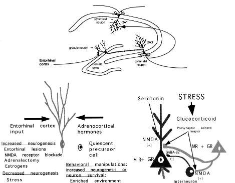
Figure 1. Schematic diagram of the role of neurotransmitters and glucocorticoids in regulating neurogenesis and dendritic remodeling in the dentate gyrus–CA3 system of the hippocampal formation. Granule neurons are replaced in adult life, and neurogenesis and apoptotic neuronal death are regulated by stress as well as by seizurelike activity. Granule neurons send mossy fibers to both the CA3 pyramidal neurons and to interneurons in the hilus, which in turn send inhibitory projections to the CA3 pyramidal neurons. The balance between the excitatory input and the inhibitory tone from the interneurons is presumed to be important to the excitability of CA3 neurons. Evidence summarized in the text indicates that excitatory amino acid release during repeated stress, aided by circulating glucocorticoids, leads to a reversible remodeling of apical dendrites over 3 to 4 weeks in rats and tree shrews. Serotonin also participates, possibly by aiding the excitatory amino acid activity at the N-methyl-D-aspartate (NMDA) receptor, and reduced
make the dendritic cytoskeleton become depolymerized or undergo proteolysis (McEwen and Sapolsky 1995) thus accounting for the dendritic remodeling. This, along with evidence that glucocorticoids enhance calcium currents in the hippocampus (Joels and Vreugenhil 1998; Kerr et al 1992), suggest that calcium channel blockers might also be effective in regulating dendritic remodeling. This possibil-ity has not yet been tested.
Stress is also reported to alter the expression of the neurotrophins, brain-derived neurotrophic factor (BDNF) and NT-3, in the hippocampus (Smith et al 1995; Ueyama et al 1997). In our hands, however, conditions that cause dendritic remodeling, such as repeated restraint stress or psychosocial stress, do not appear to change neurotrophin expression in hippocampus (Kuroda and McEwen 1998), indicating that neurotrophins are probably not directly involved in the mechanism of dendritic remodeling. This does not exclude the possibility that neurotrophin deple-tion or suppression might be involved in permanent neuronal loss resulting from more severe and prolonged stress (Uno et al 1989).
Along the same line, a number of antidepressant treat-ments, including selective serotonin reuptake inhibitors (SSRIs), increase the levels of BDNF messenger RNA (mRNA) in hippocampus of unstressed rats (Nibuya et al 1995), but these treatments have not been tried in conjunc-tion with repeated stress. We noted above that SSRIs fail to prevent dendritic remodeling; yet tianeptine, which does prevent dendritic remodeling, did not increase BDNF mRNA in hippocampus of unstressed or stressed rats (Kuroda et al 1998). Therefore, the relationship with the ability of a treatment or drug to increase BDNF mRNA levels in hippocampus and the ability of that same treat-ment to alter stress-induced changes in BDNF mRNA levels or to affect dendritic remodeling remain important and unresolved topics in sorting out the relevance of these findings to the treatment of depressive illness.
Role of Glucocorticoids in Dendritic Remodeling
What is the role of glucocorticoids in dendritic remodeling in the hippocampus? Glucocorticoid treatment causes dendritic remodeling, and stress-induced remodeling is blocked by treatment with an adrenal steroid synthesis blocker, cyanoketone (McEwen and Sapolsky 1995), in-dicating a role for endogenous glucocorticoids in stress-induced dendritic remodeling. Nonetheless, treatment with agents such as Dilantin and tianeptine do not exert their effects in preventing dendritic remodeling by altering glucocorticoid secretion during stress because these same agents prevented glucocorticoid-induced dendritic remod-eling (McEwen et al 1995). Thus, glucocorticoids can drive the remodeling of dendrites via a mechanism that
involves activation of excitatory amino acid and serotonin release (Figure 1).
Corticosterone dissolved in the drinking water at a concentration of 400mg/mL is able to do this (Magarinos et al 1998), and this allowed us to demonstrate in a “therapeutic” model that tianeptine treatment could re-verse, over several weeks, the dendritic remodeling caused by corticosterone treatment even while the treatment was continued along with the tianeptine (Magarinos et al 1999). Yet the studies with oral corticosterone revealed another, unresolved paradox, namely, that when cortico-sterone treatment was combined with repeated stress for 21d, no remodeling of CA3 dendrites was observed (Magarinos et al 1998). Clearly, stress and oral corticoste-rone are doing somewhat different things, and the two pathways block each other proximally to the reorganiza-tion of dendrites.
Because of the diversity of neurotransmitter systems involved in stress- and glucocorticoid-induced dendritic remodeling, as noted above, it is not so surprising that there are multiple ways in which glucocorticoids affect the excitatory amino acid system (for a review, see McEwen 1999). In general, stress and glucocorticoids seem to tip the balance from inhibition toward excitation. First, there are glucocorticoid effects on the expression of mRNA levels for specific subunits of GABAa receptors in CA3 and the dentate gyrus; both low and high levels of CORT have different effects on GABAa receptor subunit mRNA levels and receptor binding. Second, adrenal steroids modulate expression of NMDA receptors in hippocampus, with chronic glucocorticoid exposure leading to increased expression of NMDA receptor binding and elevation of both NR2A and NR2B subunit mRNA levels. Third, adrenal steroids regulate extracellular levels of glutamate, in that adrenalectomy markedly reduces the level of extracellular glutamate following restraint stress. One possible mechanism for this effect is that mossy fiber terminals in the stratum lucidum contain presynaptic kainate receptors that positively regulate glutamate re-lease, and kainate receptors in the CA3 stratum lucidum are decreased in density by adrenalectomy and restored to normal by corticosterone replacement (for reviews, see McEwen 1999; McEwen and Sapolsky 1995).
Reorganization and Depletion of Synaptic Vesicles in Mossy Fiber Terminals
showed a marked depletion and rearrangement of vesicles, with more densely packed clusters localized in the vicinity of active synaptic zones. Moreover, compared with control subjects, rats undergoing 21 days of repeated restraint stress increased the area of the mossy fiber terminal occupied by mitochondrial profiles, which implies a greater, localized energy generating capacity. A single stress session did not produce these changes either imme-diately after or on the day following the restraint session (Magarinos et al 1997). In MFT from stressed rats, the redistribution of vesicles and their localization near the active synaptic zones, together with more mitochondria, suggests that more vesicles may be available for glutamate release, although this possibility remains to be tested directly by electrophysiology and microdialysis. Further-more, the synaptic vesicle reorganization in MFT may be useful in future studies to provide insights into possible molecular mechanisms of the effects of stress and stress mediators on glutamate release, involving expression and phosphorylation of synaptic vesicle docking proteins such as synapsin I (Magarinos et al 1997).
Does Dendritic Remodeling Play a Protective Role in CA3 Apical Dendrites?
The mossy fiber terminals reside on the proximal regions of the CA3 apical dendrites that are remodeled by chronic stress, and chronic stress does not reduce their numbers (Magarinos et al 1997). If, indeed, chronic stress makes these giant synapses more efficient in releasing glutamate, then what is the significance of the remodeling of the apical dendrites? To answer this question, it is important to understand some key features of the CA3 region. In terms of circuitry, there are a series of recurrent feedback loops within the CA3 region that reactivate the mossy fiber system and sustain CA3 excitation, as in the so-called SPW or “sharp waves” (Buzsaki 1986). The SPW waves are postulated to be part of a circuit subserving memory of sequences of events (Lisman 1999). As reviewed in McEwen (1999), CA3 pyramidal neurons send axon col-laterals to nearby CA3 neurons; moreover, CA3 neurons have a multiplicity of calcium channel types that contrib-ute to the activation of calcium currents by low voltage changes, and pyramidal neurons in subregion CA3c that lies closest to the hilus send excitatory axons back to the hilar region and affect the dentate gyrus itself. Thus the CA3 region has an intrinsic instability that can be driven by stimulation via the perforant pathway, and the CA3 apical dendritic remodeling might be a protective adapta-tion to limit the increased excitatory input via the recurrent feedback loops. Moreover, the synchronized firing of the SPW waves during chronic stress or seizures may drive the reorganization of vesicles within the mossy fiber
terminals. Indeed, collateral activation of CA3 neurons by other CA3 neurons would help explain the blockade of dendritic remodeling by treatment with antagonists to NMDA receptors because the stratum lucidum of the CA3 region does not express NMDA receptors (for a review, see McEwen 1999).
As reviewed in McEwen (1999), chronic stress may produce a graded response that is less drastic than that produced when seizures are elicited either by kainate treatment or perforant pathway stimulation. CA3 pyrami-dal neurons display a high vulnerability to kainic acid administration, an effect that requires the integrity of the mossy fiber pathway. The CA3 hippocampal subregion is also damaged by seizures evoked by stimulation of the perforant path, and this involves the activation of the dentate gyrus (DG)–MFT–CA3 pathway. In epilepsy, an interesting parallel exists with synaptic vesicle clustering found in the chronic stress model (see above). That is, gerbils that are genetically prone to epilepsy show MFT synaptic vesicle clustering that could be blocked by the disruption of the perforant pathway (Farias et al 1992).
Stress activation of the hippocampus also shows a strong trend to increase seizure susceptibility. In a recent electrophysiologic study of the effects of stress on long-term potentiation in the hippocampus (C. Pavlides et al, unpublished data), high-frequency stimulation (HFS) of the commissural/associational and MF inputs to CA3 produced epileptic afterdischarges in 56% of acutely stressed animals 48 hours after the stress, and 29% of the chronically stressed animals 48h after the last stress, but in only 9% of the non-stressed control animals. No epileptic afterdischarges were seen in the medial perforant path to DG input. The rats showing seizures were removed from the analysis of long-term potentiation (LTP) that is de-scribed below. The increased incidence of seizures is consistent with the possibility of stress-induced mossy fiber sprouting, since in epilepsy there is sprouting of mossy fibers that generate a recurrent excitatory circuit involving aberrant granule cell-granule cell synapses; see McEwen (1999) for references. Moreover, long-term po-tentiation itself appears to be capable of inducing mossy fiber sprouting (Noguchi et al 1990).
Stress-Induced Changes in Hippocampal Electrophysiology
LTP in the laconosum–molecular layer of CA3 after stimulation of the commissural–associational pathway. This inhibition was significant compared with that in animals receiving a single stress session and to control animals that were briefly handled but not subjected to the restraint stress. Chronic, but not acute, stress produced an inhibition of LTP in the dentate gyrus granule cell layer with stimulation of the medial perforant pathway. Mossy fiber LTP was not affected by repeated stress or by acute stress. In a second experiment, animals were subjected to a similar stress paradigm, and a current source density analysis was performed that revealed significant chronic stress-induced shifts in the current sources and sinks in the apical dendrites and pyramidal cell layers of the CA3 field, but not in the DG. These findings are consistent with the morphologic findings for effects of stress on dendrites of CA3 neurons. Furthermore, they suggest that chronic stress produces changes in the input– output relationship in the hippocampal trisynaptic circuit, which could affect information flow through this structure (C. Pavlides et al, unpublished data).
Gender Differences in Dendritic Remodeling
All of the experiments described above were carried out on male rats. We previously reported that female rats subjected to repeated restraint stress failed to show the remodeling of apical dendrites of CA3 pyramidal neurons (Galea et al 1997). The mechanism for this gender difference is unclear. It may reflect a protection by ovarian hormones or a developmentally programmed difference in hippocampal neuroanatomy. Gender differences in hip-pocampal neuroanatomy are known to exist (Juraska 1991). There also are reported gender differences in vulnerability of the hippocampus to damage in rats under-going cold swim stress (Mizoguchi et al 1992) and in vervet monkeys undergoing psychosocial stress (Uno et al 1989). As discussed above, however, the relationship of dendritic remodeling to permanent hippocampal damage is too complex at this stage to make a simple link of these findings to each other.
Neurogenesis in the Dentate Gyrus
Neurogenesis in the dentate gyrus of adult rodents was reported several decades ago (for a review, see Gould et al 2000) but never fully appreciated until recently, and the reactivation of this topic occurred in an unusual manner (McEwen 1999). First, bilateral adrenalectomy of an adult rat was shown to increase granule neuron death by apoptosis (Gould et al 1990). Subsequently, neurogenesis was also found to increase following adrenalectomy in adults rats, as well as in the developing dentate gyrus. In
adult rats, very low levels of adrenal steroids, sufficient to occupy Type I adrenal steroid receptors, completely blocks dentate gyrus neuronal loss; but in newborn rats, Type II receptor agonists protect against neuronal apopto-sis (for reviews, see Gould et al 2000; McEwen 1999; Figure 1). This is consistent with the fact that dentate neuronal loss in the developing rat occurs at much higher circulating steroid levels than in the adult, and it represents another example of the different ways that the two adrenal steroid receptor types are involved in hippocampal func-tion (Lupien and McEwen 1997).
In adult rats, newly born neurons arise in the hilus, very close to the granule cell layer, and then migrate into the granule cell layer, presumably along a vimentin-staining radial glial network that is also enhanced by adrenalec-tomy (Gould et al 2000). Most neuroblasts labeled with [3H] thymidine lack both Type I and Type II adrenal steroid receptor, indicating steroidal regulation occurs via messengers from an unidentified steroid-sensitive cell (Gould et al 2000). It has been reported that neurogenesis declines in the aging rodent (Kempermann et al 1998) and rhesus monkey (Fallah et al 1998) dentate gyrus. Recent studies of aging rats showed that adrenalectomy could reverse the decline in dentate gyrus neurogenesis (Cam-eron and McKay 1999), suggesting that they are the result of age-related increases in hypothalamic–pituitary–adrenal (HPA) activity and glucocorticoid levels that have been reported (Landfield et al 1994; Sapolsky et al 1986). Besides glucocorticoids, excitatory amino acids acting through NMDA receptors inhibit ongoing dentate gyrus neurogenesis, whereas other neurochemical and hormonal agents are known that stimulate neurogenesis, including serotonin and estrogens (Gould et al 2000).
The question of whether dentate gyrus neurogenesis is a widespread phenomenon among mammals was addressed by studies showing that neurogenesis occurs in the mar-moset, a New World primate, as well as in an Old World primate species, the rhesus monkey and in the adult human dentate gyrus (Gould et al 2000). Thus changes in size of the human hippocampus, described below, may include changes in neuron number in the dentate gyrus.
(Galea et al 1996), although acute restraint stress does not inhibit neurogenesis (K. Pham et al, unpublished data). Acute psychosocial stress in the adult tree shrew, involv-ing largely visual cues, inhibits neurogenesis (Gould et al 1997). Inhibition of neurogenesis is also seen in the dentate gyrus of the marmoset after acute psychosocial stress (Gould et al 1998).
Chronic psychosocial stress in the tree shrew results in a more substantial inhibition of neurogenesis than after a single acute stressful encounter (Gould et al 2000); more-over, the dentate gyrus is 30% smaller in the chronically stressed tree shrew, although the granule neuron number only shows a trend for reduction (E. Gould et al, unpub-lished data). This finding suggests that there may be other changes such as remodeling of dendritic branching to account for the decrease in dentate gyrus volume. On the other hand, repeated restraint stress does not reduce dentate gyrus neuron number or alter the survival of granule neurons formed before the beginning of chronic stress (K. Pham et al, unpublished data). This indicates that not all stressors will alter dentate gyrus neuron number, even though they may cause remodeling of dendrites in CA3 neurons.
Changes in dentate gyrus volume appear to have con-sequences for cognitive functions subserved by the hip-pocampus. In the enriched environment studies (Kemper-mann et al 1997), increased dentate gyrus volume was accompanied by better performance on spatial learning tasks. Chronic stress, on the other hand, impairs spatial learning and memory in the tree shrew (Ohl and Fuchs 1999). This may be due to the decreased dentate gyrus volume as well as to remodeling of dendrites of CA3 pyramidal neurons and dentate granule neurons (see above).
Chronic stress has another interesting effect on the dentate gyrus. In the restraint stress model in the rat, 21 days of repeated stress increased expression of cells with polysialic acid neural cell adhesion molecule (PSA-NCAM) in the inner granule cell layer of the dentate gyrus while at the same time decreasing the rate of neurogenesis found in the dentate; a single acute stress did not produce either effect (K. Pham et al, unpublished data). The number of cells expressing PSA-NCAM was 10-fold larger than the number of newly BrdU labeled neurons, indicating that chronic stress has an effect on the subsequent properties of DG granule neurons. Glucocorticoids may be involved in regulating the process that adds polysialic acid to NCAM, and because PSA-NCAM is associated with the movement of cells and their processes, these findings are consistent with the overall increased plasticity produced by repeated stress in the DG-CA3 region of the hippocampus (K. Pham et al, unpublished data).
Stress, Glucocorticoids, and Cognition
Stress and glucocorticoids have specific effects on cogni-tive function in humans and in animal models. Adrenal steroids and stressful experiences produce short-term and reversible deficits in episodic and spatial memory in animal models and in humans (de Quervain et al 2000; Lupien and McEwen 1997), whereas repeated stress also impairs cognitive function in animal models and repeated glucocorticoid elevation or treatment in humans is accom-panied by cognitive dysfunction (McEwen and Sapolsky 1995). There also are declines in cognitive function in aging humans that are correlated with progressive eleva-tions in HPA activity over 3 to 4 years (see below).
Acute effects of stress or glucocorticoid administration are evident within a time span ranging from a few hours to a day and are generally reversible and quite selective to the task or particular situation (Lupien and McEwen 1997).Adrenal steroid effects are implicated in both selec-tive attention, as well as in memory consolidation (Lupien and McEwen 1997) and retrieval, and such actions are consistent with the effects of adrenal steroids on the modulation of long-term potentiation and primed-burst potentiation (see above). Nonetheless, some acute actions of stress may involve other mechanisms than glucocorti-coids, including endogenous opioid neuropeptides in the case of painful stressors such as shock (for a summary, see McEwen et al 1995). With regard to nonpainful stressors, exposure of rats to a novel environment resulted in a rapid and reversible impairment of plasticity in vivo in the CA1 region, and this effect may involve the actions of glucocor-ticoids (Diamond et al 1996).
Repeated stress that produces dendritic remodeling in the CA3 region impairs hippocampal-dependent learning. Rats that received 21 days of restraint stress were impaired in performance on an eight-arm radial maze when they were trained starting one day after the end of stress but not when trained 18 days later, whereas a subsequent study showed that 21 days of repeated restraint stress impaired the short-term (4-hour) retention of a spatial recognition memory in a hippocampus-dependent Y-maze task; again, stress impairment was prevented by tianeptine treatment during the stress regimen (McEwen 1999). We now know that dendritic remodeling is reversible within 7 to 10 days after the end of stress (Conrad et al 1999). The impairment was in the same direction, but not as great as, impairment found in aging rats. Moreover, stress effects were pre-vented by prior treatment of rats with phenytoin or with tianeptine under the same conditions in which both drugs prevented the stress-induced remodeling of CA3c pyrami-dal neurons (for a review, see McEwen and Sapolsky 1995).
such as spatial and episodic memory, occur in human subjects and are correlated with increases in HPA activity over 3 to 4 years (for a review, see McEwen et al 1999). Recent evidence has revealed that the most severely impaired individuals have a significantly smaller hip-pocampal volume compared with the least impaired indi-viduals (Lupien and McEwen 1998).
An important aspect of stressful experiences is the developmental influence of early stress and of neonatal handling on the life course of aging and age-related cognitive impairment. As discussed elsewhere (Meaney et al 1988, 1994), such early experiences can either increase or decrease the rate of brain aging through a mechanism in which the activity of the HPA axis appears to be involved. The early experiences are believed to set the level of responsiveness of the HPA axis and autonomic nervous system in such a way that these systems either overreact in animals subject to early unpredictable stress or underreact in animals exposed to the neonatal handling procedure.
Long-term stress also accelerates a number of biological markers of aging in rats, including increasing the excit-ability of CA1 pyramidal neurons via a calcium-dependent mechanism and causing an apparent loss of hippocampal pyramidal neurons (Kerr et al 1991). An important factor may be the enhancement by glucocorticoids of calcium currents in hippocampus (Kerr et al 1992), in view of the key role of calcium ions in destructive as well as plastic processes in hippocampal neurons (Mattson 1988, 1992). Another aspect making the aging hippocampus more vulnerable may be the persistence of excitatory amino acid release after the termination of a stressful experience (Lowy et al 1995).
Atrophy of the Hippocampus and Other
Brain Structures in Psychiatric Disorders
The human brain shows signs of atrophy as a result of elevated glucocorticoids and severe, traumatic stress (e.g., holocaust survivors; Sapolsky 1992). Advances in brain imaging techniques have allowed for a regional analysis of the atrophy of various brain structures. Recent evidence indicates that the human hippocampus is particularly sensitive in this respect and tends to show greater changes than do other brain areas, especially in Cushing’s syn-drome, recurrent depressive illness, posttraumatic stress disorder (PTSD), schizophrenia, and aging before overt dementia (Bogerts et al 1993; Bremner et al 1995; Fuku-zako et al 1996; Gurvits et al 1996; Lupien et al 1998; Sheline et al 1996, 1999; Starkman et al 1992).
The diversity of conditions in which atrophy occurs raises two questions, namely, whether there is a common mechanism and whether the atrophy is permanent or reversible. Based on what we have summarized above, the
atrophy might be due to one of at least four different processes: 1) a reduced volume of Ammon’s horn or dentate gyrus due to reduced dendritic branching, 2) a reduction in dentate gyrus neuron number due to a suppression of neurogenesis, 3) a decreased rate of neuron survival, 4) permanent neuron loss. In addition, it is noteworthy that atrophy of other brain regions has been reported in depressive illness (e.g., prefrontal cortex; Drevets et al 1997) and amygdala (Sheline et al 1998). Moreover, new evidence suggests that glial cell depletion may contribute to atrophy of brain regions such as the prefrontal cortex and amygdala (Drevets et al 1998; Ongur et al 1998; Rajkowska et al 1999; Sheline et al 1998), and the contribution of glial cell changes must now be consid-ered for the hippocampus.
Because of the high density of intracellular receptors for adrenal steroids in hippocampus, it is tempting to attribute the occurrence of hippocampal atrophy solely to the actions of glucocorticoids. As summarized above, the hippocampus shows influences of adrenal steroids on plasticity, as well as on the loss and damage to hippocam-pal neurons in conditions such as ischemia and aging (Landfield and Eldridge 1994; McEwen and Sapolsky 1995; Sapolsky 1992; Sapolsky et al 1986); however, adrenal steroids produce their effects on plasticity (see earlier discussion) and on damage in ischemia and aging (see above references) by acting in concert with neuro-modulators and neurotransmitters, in particular the endog-enous excitatory amino acids. Nonetheless, the role of glucocorticoids should not be ignored. Glucocorticoids are elevated in Cushing’s syndrome and may also be some-what elevated in depressive illness, but this is probably not the case for PTSD, at least at the time the PTSD subjects are studied, except as there are elevations in glucocorti-coids associated with the diurnal rhythm and stressful experiences that take place on a daily basis.
a more reactive stress hormone profile will expose them-selves to more cortisol and experience more stress-ele-vated neural activity than will other people who can more easily habituate to psychosocial challenges.
In this regard, events related to the course of illness in recurrent depressive illness may involve distinct pathways of selective and repeated elevations of glucocorticoid hormones in relation to the individual experiences and reactivities. We are largely ignorant of the history of the depressed individual as far as endocrine function and neurochemical activity, as well as responses to stressful life experiences. In both disorders, a long-term pattern of increased neurochemical, autonomic and HPA reactivity to experiences may underlie a progression of neuronal structural changes involving atrophy that might lead to permanent damage, including neuronal loss. Regarding the neurochemical aspects, there is need to measure the activity of excitatory amino acids in the brain during recurrent depressive illness because neural activity is likely to be a major factor in the long-term atrophy of key brain regions such as hippocampus, amygdala, and prefrontal cortex.
Regarding reversibility or irreversibility of these struc-tural changes, treatment with drugs such as phenytoin or tianeptine, both of which block stress-induced dendritic remodeling in the CA3 or rats and tree shrews, is a potential means of testing both the mechanism and at the same time demonstrating the reversibility of human hip-pocampal atrophy. There is already some indication that hippocampal atrophy in Cushing’s syndrome is at least partially reversible (Starkman et al 1999). On the other hand, there may be irreversible loss of hippocampal neurons, and some of the evidence in the MRI of recurrent depressive illness is consistent with this possibility (She-line et al 1996). In so far as atrophy of the hippocampus and accompanying cognitive impairment are signs of reversible neuronal remodeling, they may be treatable with agents that block the neuronal remodeling in animal models. On the other hand, where atrophy involves neu-ronal loss, treatment strategies should focus on the earlier traumatic or recurrent events, and it may be possible to devise strategies to reduce or prevent neuronal damage.
Research in the author’s laboratory on the topic of this article is supported by National Institutes of Health Grant Nos. MH 41256 and Center Grant No. MH58911, as well as by funding from the Health Foundation (New York) and Servier (France).
Aspects of this work were presented at the conference “Depression in the Twenty-First Century: New Insights into Drug Development and Neurobiology,” February 21–22, 2000, Dana Point, California. The conference was sponsored by the Society of Biological Psychiatry through an unrestricted educational grant provided jointly by Pharmacia & Upjohn and Janssen Pharmaceutica.
References
Bogerts B, Lieberman JA, Ashtair M, Bilder RM, De Greef G, Lerner G, et al (1993): Hippocampus-amygdala volumes and psychopathology in chronic schizophrenia. Biol Psychiatry 33:236 –246.
Bremner DJ, Randall P, Scott TM, Bronen RA, Seibyl JP, Southwick SM, et al (1995): MRI-based measurement of hippocampal volume in patients with combat-related post-traumatic stress disorder. Am J Psychiatry 152:973–981. Buzsaki G (1986): Hippocampal sharp waves: Their origin and
significance. Brain Res 398:242–252.
Cameron HA, McKay DG (1999): Restoring production of hippocampal neurons in old age. Nat Neurosci 2:894 – 897. Conrad CD, Magarinos AM, LeDoux JE, McEwen BS (1999):
Repeated restraint stress facilitates fear conditioning indepen-dently of causing hippocampal CA3 dendritic atrophy. Behav
Neurosci 113:902–913.
DeKloet ER, Vreugdenhil E, Oitzl MS, Joels M (1998): Brain corticosteroid receptor balance in health and disease. Endocr
Rev 19:269 –301.
de Quervain DJ, Roozendaal B, Nitsch RM, McGaugh JL, Hock C (2000): Acute cortisone administration impairs retrieval of long-term declarative memory in humans. Nat Neurosci 3:313–314. Diamond DM, Fleshner M, Ingersoll N, Rose GM (1996): Psychological stress impairs spatial working memory: Rele-vance to electrophysiological studies of hippocampal func-tion. Behav Neurosci 110:661– 672.
Diamond DM, Fleshner M, Rose GM (1994): Psychological stress repeatedly blocks hippocampal primed burst potentia-tion in behaving rats. Behav Brain Res 62:1–9.
Diamond DM, Fleshner M, Rose GM (1996): Psychological stress impairs spatial working memory: Relevance to electro-physiological studies of hippocampal function. Behav
Neuro-sci 110:661– 672.
Drevets WC, Ongur D, Price JL (1998): Neuroimaging abnor-malities in the subgenual prefrontal cortex: Implications for the pathophysiology of familial mood disorders. Mol
Psychi-atry 3:220 –226.
Drevets WC, Price JL, Simpson JR Jr, Todd RD, Reich T, Vannier M, Raichle ME (1997): Subgenual prefrontal cortex abnormalities in mood disorders. Nature 386:824 – 827. Eichenbaum H, Otto T (1992): The hippocampus—what does it
do? Behav Neural Biol 57:2–36.
Fallah M, Fuchs E, Tanapat P, Reeves AJ, Gould E (1998): Hippocampal neurogenesis in old world primates declines with aging. Soc Neurosci Abstr 24:1993.
Farias PA, Low SQ, Peterson GM, Ribak CE (1992): Morpho-logical evidence for altered synaptic organization and struc-ture in the hippocampal formation of seizure-sensitive gerbils.
Hippocampus 2:229 –246.
Freund TF, Buzsaki G (1996): Interneurons of the hippocampus.
Hippocampus 6:345– 470.
Fukuzako H, Fukuzako T, Hashiguchi T, Hokazono Y, Takeuchi K, Hirakawa K, et al (1996): Reduction in hippocampal formation volume is caused mainly by its shortening in chronic schizo-phrenia: Assessment by MRI. Biol Psychiatry 39:938 –945. Galea LAM, McEwen BS, Tanapat P, Deak T, Spencer RL,
CA3 pyramidal neurons in response to chronic restraint stress.
Neuroscience 81:689 – 697.
Galea LAM, Tanapat P, Gould E (1996): Exposure to predator odor suppresses cell proliferation in the dentate gyrus of adult rats via a cholinergic mechanism. Soc Neurosci Abstr 22:1196. Gould E, Beylin A, Tanapat P, Reeves A, Shors TJ (1999):
Learning enhances adult neurogenesis in the hippocampal formation. Nat Neurosci 2:260 –265.
Gould E, McEwen BS, Tanapat P, Galea LAM, Fuchs E (1997): Neurogenesis in the dentate gyrus of the adult tree shrew is regulated by psychosocial stress and NMDA receptor activa-tion. J Neurosci 17:2492–2498.
Gould E, Tanapat P, McEwen BS, Flugge G, Fuchs E (1998): Proliferation of granule cell precursors in the dentate gyrus of adult monkeys is diminished by stress. Proc Natl Acad Sci
U S A 95:3168 –3171.
Gould E, Tanapat P, Rydel T, Hastings N (2000): Regulation of hippocampal neurogenesis in adulthood. Biol Psychiatry 48: 715–720.
Gould E, Woolley C, McEwen BS (1990): Short-term glucocor-ticoid manipulations affect neuronal morphology and survival in the adult dentate gyrus. Neuroscience 37:367–375. Greenough WT, Bailey CH (1988): The anatomy of a memory:
convergence of results across a diversity of tests. Trends
Neurosci 11:142–147.
Gurvits TV, Shenton ME, Hokama H, Ohta H, Lasko NB, Gilbertson MW, et al (1996): Magnetic resonance imaging study of hippocampal volume in chronic, combat-related posttraumatic stress disorder. Biol Psychiatry 40:1091–1099. Ikegaya Y, Saito H, Abe K (1997): The basomedial and baso-lateral amygdaloid nuclei contribute to the induction of long-term potentiation in the dentate gyrus in vivo. Eur
J Neurosci 8:1833–1839.
Jacobson L, Sapolsky R (1991): The role of the hippocampus in feedback regulation of the hypothalamic-pituitary-adrenocor-tical axis. Endocr Rev 12:118 –134.
Joels M, Vreugdenhil E (1998): Corticosteroids in the brain. Mol
Neurobiol 17:87–198.
Juraska JM (1991): Sex differences in “cognitive” regions of the rat brain. Psychoneuroendocrinology 16:105–119.
Kempermann G, Kuhn HG, Gage FH (1997): More hippocampal neurons in adult mice living in an enriched environment.
Nature 586:493– 495.
Kempermann G, Kuhn HG, Gage FH (1998): Experience-induced neurogenesis in the senescent dentate gyrus. J
Neu-rosci 18:3206 –3212.
Kerr DS, Campbell LW, Thibault O, Landfield PW (1992): Hippocampal glucocorticoid receptor activation enhances voltage-dependent Ca21 conductances: Relevance to brain aging. Proc Natl Acad Sci U S A 89:8527– 8531.
Kerr DS, Huggett AM, Abraham WC (1994): Modulation of hippocampal long-term potentiation and long-term depression by corticosteroid receptor activation. Psychobiology 22:123– 133.
Kerr S, Campbell L, Applegate M, Brodish A, Landfield P (1991): Chronic stress-induced acceleration of electrophysi-ologic and morphometric biomarkers of hippocampal aging.
J Neurosci 11:1316 –1324.
Kirschbaum C, Prussner JC, Stone AA, Federenko I, Gaab J,
Lintz D, et al (1995): Persistent high cortisol responses to repeated psychological stress in a subpopulation of healthy men. Psychosom Med 57:468 – 474.
Kuroda Y, McEwen BS (1998): Effect of chronic restraint stress and tianeptine on growth factors, GAP-43 and MAP2 mRNA expression in the rat hippocampus. Mol Brain Res 59:35–39. Landfield PW, Eldridge JC (1994): Evolving aspects of the glucocorticoid hypothesis of brain aging: Hormonal modula-tion of neuronal calcium homeostasis. Neurobiol Aging 15: 579 –588.
LeDoux JE (1995): In search of an emotional system in the brain: Leaping from fear to emotion and consciousness. In: Gazza-niga M, editor. The Cognitive Neurosciences. Cambridge, MA: MIT Press, 1049 –1061.
Lisman JE (1999): Relating hippocampal circuitry to function: Recall of memory sequences by reciprocal dentate-CA3 interactions. Neuron 22:233–242.
Lowy MT, Wittenberg L, Yamamoto BK (1995): Effect of acute stress on hippocampal glutamate levels and spectrin proteol-ysis in young and aged rats. J Neurochem 65:268 –274. Lupien SJ, DeLeon MJ, De Santi S, Convit A, Tarshish C, Nair
NPV, et al (1998): Cortisol levels during human aging predict hippocampal atrophy and memory deficits. Nat Neurosci 1:69 – 73.
Lupien SJ, McEwen BS (1997): The acute effects of corticoste-roids on cognition: Integration of animal and human model studies. Brain Res Rev 24:1–27.
Magarinos AM, Deslandes A, McEwen BS (1999): Effects of antidepressants and benzodiazepine treatments on the den-dritic structure of CA3 pyramidal neurons after chronic stress.
Eur J Pharmacol 371:113–122.
Magarinos AM, Orchinik M, McEwen BS. (1998): Morpholog-ical changes in the hippocampal CA3 region induced by non-invasive glucocorticoid administration: a paradox. Brain
Res 809:314 –318.
Magarinos AM, Verdugo Garcia JM, McEwen BS (1997): Chronic restraint stress alters synaptic terminal structure in hippocampus. Proc Natl Acad Sci U S A 94:14002–14008. Mattson MP (1988): Neurotransmitters in the regulation of
neuronal cytoarchitecture. Brain Res Rev 13:179 –212. Mattson MP (1992): Calcium as sculptor and destroyer of neural
circuitry. Exp Gerontol 27:29 – 49.
McEwen BS (1999): Stress and hippocampal plasticity. Annu
Rev Neurosci 22:105–122.
McEwen BS, Albeck D, Cameron H, Chao HM, Gould E, Hastings N, et al (1995): Stress and the brain: A paradoxical role for adrenal steroids. In: Litwack GD, editor. Vitamins
and Hormones. New York: Academic Press, 371– 402.
McEwen BS, de Leon MJ, Lupien S, Meaney MJ (1999): Corticosteroids, the aging brain and cognition. Trends
Endo-crinol Metab 10:92–96.
McEwen BS, Sapolsky RM (1995): Stress and cognitive func-tion. Curr Opin Neurobiol 5:205–216.
McEwen BS, Weiss J, Schwartz L (1968): Selective retention of corticosterone by limbic structures in rat brain. Nature 220: 911–912.
de-creases binding to 5HT transporter sites and reduces dendritic arbors in CA3 of hippocampus. Soc Neurosci Abstr 22:2060. Meaney M, Aitken D, Berkel H, Bhatnager S, Sapolsky R (1988): Effect of neonatal handling of age-related impair-ments associated with the hippocampus. Science 239:766 – 768.
Meaney MJ, Tannenbaum B, Francis D, Bhatnagar S, Shanks N, Viau V, et al (1994): Early environmental programming hypothalamic-pituitary-adrenal responses to stress. Semin
Neurosci 6:247–259.
Mennini T, Miari A (1991): Modulation of 3H glutamate binding by serotonin in rat hippocampus: An autoradiographic study.
Life Sci 49:283–292.
Michaloudi HC, Majdoubi ME, Poulain DA, Papadopoulos GC, Theodosis DT (1997): The noradrenergic innervation of identified hypothalamic magnocellular somata and its contri-bution to lactation-induced synaptic plasticity. J
Neuroendo-crinol 9:17–23.
Mizoguchi K, Kunishita T, Chui DH, Tabira T (1992): Stress induces neuronal death in the hippocampus of castrated rats.
Neurosci Lett 138:157–160.
Nibuya M, Morinobu S, Duman RS (1995): Regulation of BDNF and trkB mRNA in rat brain by chronic electroconvulsive seizure and antidepressant drug treatments. J Neurosci 15: 7539 –7547.
Noguchi S, Higashi K, Kawamura M (1990): A possible role of the b-subunit of (Na, K):-ATPase in facilitating correct assembly of thea-subunit into the membrane. J Biol Chem 265:5991–5995.
Ohl F, Fuchs E (1999): Differential effects of chronic stress on memory processes in the tree shrew. Cogn Brain Res 7:379–387. Ongur D, Drevets WC, Price JL (1998): Glial loss in the
subgenual prefrontal cortex in familial mood disorders. Soc
Neurosci Abstr 24:990.
Pavlides C, Ogawa S, Kimura A, McEwen BS (1996): Role of adrenal steroid mineralocorticoid and glucocorticoid recep-tors in long-term potentiation in the CA1 field of hippocam-pal slices. Brain Res 738:229 –235.
Phillips RG, LeDoux JE (1992): Differential contribution of amygdala and hippocampus to cued and contextual fear conditioning. Behav Neurosci 106:274 –285.
Rahmann S, Neumann RS (1993): Activation of 5-HT2 receptors facilitates depolarization of neocortical neurons by N-methyl-D-aspartate. Eur J Pharmacol 231:347–354.
Rajkowska G, Miguel-Hidalgo JJ, Wei J, Dilley G, Pittman SD, Meltzer HY, et al (1999): Morphometric evidence for
neuro-nal and glial prefrontal cell pathology in major depression.
Biol Psychiatry 45:1085–1098.
Sapolsky R (1992): Stress, the Aging Brain and the Mechanisms
of Neuron Death. Cambridge, MA: MIT Press.
Sapolsky R, Krey L, McEwen BS (1986): The neuroendocrinol-ogy of stress and aging: The glucocorticoid cascade hypoth-esis. Endocr Rev 7:284 –301.
Sheline YI, Gado MH, Price JL (1998): Amygdala core nuclei volumes are decreased in recurrent major depression.
Neuro-report 9:2023–2028.
Sheline YI, Sanghavi M, Mintun MA, Gado MH (1999): De-pression duration but not age predicts hippocampal volume loss in medically healthy women with recurrent major depres-sion. J Neurosci 19:5034 –5043.
Sheline YI, Wang PW, Gado MH, Csernansky JC, Vannier MW (1996): Hippocampal atrophy in recurrent major depression.
Proc Natl Acad Sci U S A 93:3908 –3913.
Sherry DF, Jacobs LF, Gaulin SJ (1992): Spatial memory and adaptive specialization of the hippocampus. Trends Neurosci 15:298 –303.
Smith MA, Makino S, Kvetnansky R, Post RM (1995): Stress and glucocorticoids affect the expression of brain-derived neurotrophic factor and neurotrophin-3 mRNAs in the hip-pocampus. J Neurosci 15:1768 –1777.
Starkman M, Gebarski S, Berent S, Schteingart D (1992): Hippocampal formation volume, memory dysfunction, and cortisol levels in patients with Cushing’s syndrome. Biol
Psychiatry 32:756 –765.
Starkman MN, Giordani B, Gebrski SS, Berent S, Schork MA, Schteingart DE (1999): Decrease in cortisol reverses human hippocampal atrophy following treatment of Cushing’s dis-ease. Biol Psychiatry 46:1595–1602.
Stern JE, Armstrong WE (1998): Reorganization of the den-dritic trees of oxytocin and vasopressin neurons of the rat supraoptic nucleus during lactation. J Neurosci 18:841– 853.
Ueyama T, Kawai Y, Nemoto K, Sekimoto M, Tone S, Senba E (1997): Immobilization stress reduced the expression of neurotrophins and their receptors in the rat brain. Neurosci
Res 28:103–110.
Uno H, Ross T, Else J, Suleman M, Sapolsky R (1989): Hippocampal damage associated with prolonged and fatal stress in primates. J Neurosci 9:1705–1711.
Withers GS, Greenough WT (1989): Reach training selectively alters dendritic branching in subpopulations of layer II-III pyramids in rat motor-somatosensory forelimb cortex.