www.elsevier.com/locate/ibmb
Autoinduction of nuclear hormone receptors during metamorphosis
and its significance
Jamshed R. Tata
*National Institute for Medical Research, The Ridgeway, Mill Hill, London NW7 1AA, UK
Received 31 October 1999; received in revised form 31 December 1999; accepted 25 January 2000
Abstract
Metamorphosis is a most dramatic example of hormonally regulated genetic reprogramming during postembryonic development. The initiation and sustenance of the process are under the control of ecdysteroids in invertebrates and thyroid hormone, 3,39 ,5-triiodothyronine, in oviparous vertebrates. Their actions are inhibited or potentiated by other endogenous or exogenous hormones — juvenile hormone in invertebrates and prolactin and glucocorticoids in vertebrates. The nuclear receptors for ecdysteroids and thyroid hormone are the most closely related members of the steroid/retinoid/thyroid hormone receptor supergene family. In many pre-metamorphic amphibia and insects, the onset of natural metamorphosis and the administration of the exogenous hormones to the early larvae are characterized by a substantial and rapid autoinduction of the respective nuclear receptors. This review will largely deal with the phenomenon of receptor autoinduction during amphibian metamorphosis, although many of its features resemble those in insect metamorphosis.
In the frogXenopus, thyroid hormone receptor autoinduction has been shown to be brought about by the direct interaction between the receptor protein and the thyroid-responsive elements in the promoter of its own gene. Three lines of evidence point towards the involvement of receptor autoinduction in the process of initiation of amphibian metamorphosis: (1) a close association between the extent of inhibition or potentiation by prolactin and glucocorticoid, respectively, and metamorphic response in whole tadpoles and in organ and cell cultures; (2) thyroid hormone fails to upregulate the expression of its own receptor in obligatorily neotenic amphibia but does so in facultatively neotenic amphibia; and (3) dominant-negative receptors known to block hormonal response prevent the autoinduction of wild-typeXenopus receptors in vivo and in cell lines.
Autoinduction is not restricted to insect and amphibian metamorphic hormones but is also a characteristic of other nuclear receptors (e.g., retinoid, sex steroids, vitamin D3receptors) where the ligand is involved in a postembryonic developmental function.
A wider significance of such receptor autoregulation is that the process may also be important for mammalian postembryonic development. 2000 Published by Elsevier Science Ltd. All rights reserved.
Keywords:Amphibian metamorphosis; Thyroid hormone; Prolactin; Thyroid hormone receptors; Receptor autoinduction
1. Introduction
Thyroid hormone (TH) is synthesized and secreted in virtually every cold- and warm-blooded vertebrate examined (Gorbman and Bern, 1962). During evolution it has been put to different uses in different organisms as a hormonal signalling molecule, thus generating a remarkable multiplicity of physiological actions. With the discovery that thyroid hormone receptor (TR) is a member of the nuclear steroid/thyroid hormone
super-* Tel.:+44-181-959-3666; fax:+44-181-913-8583. E-mail address:[email protected] (J.R. Tata).
0965-1748/00/$ - see front matter2000 Published by Elsevier Science Ltd. All rights reserved. PII: S 0 9 6 5 - 1 7 4 8 ( 0 0 ) 0 0 0 3 5 - 7
2. Amphibian metamorphosis
Following the discovery by Gudernatsch (1912) that frog larvae fed on extracts of mammalian thyroid glands underwent precocious metamorphosis spontaneously, detailed analysis of the functional role of the developing larval thyroid gland, and the availability of pure l
-thy-roxine, confirmed that the process of metamorphosis was obligatorily dependent on thyroid hormone.
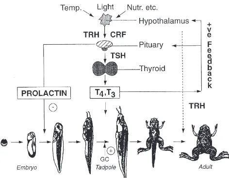
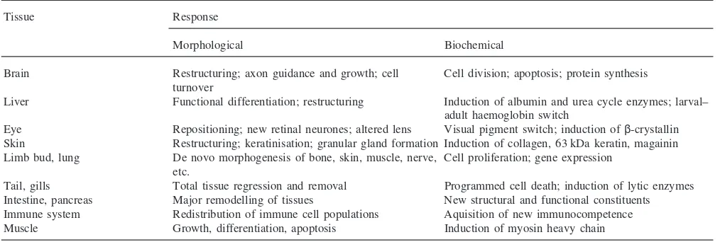
This obligatory requirement of thyroid hormone for amphibian metamorphosis offers an ideal model to explore its developmental actions in vertebrates, in the same way as for ecdysteroids in invertebrates. This simi-larity can be discerned from Fig. 1, which depicts how environmental cues and neuroendocrine cascades initiate metamorphosis in amphibia (Gilbert et al., 1996; Tata, 1997). Thyroid hormone provokes diverse and multiple morphological, physiological and biochemical responses in virtually every tissue of the amphibian tadpole, some of which are listed in Table 1 for different tissues of the pre-metamorphic tadpole. These range from de novo morphogenesis, repatterning, functional reprogramming, and partial and total regression. Before extending this article to its receptors, it is useful at this point to briefly consider the special characteristics of the role of TH in amphibian metamorphosis.
The thyroidectomized anuran larva will continue to grow without undergoing metamorphosis until adminis-tration of l-thyroxine (T4) or 3,39,5-triiodo-l-thyronine
(T3) at any stage thereafter causes resumption of the
Fig. 1. Schematic representation of the hormonal regulation of amphibian metamorphosis. In response to environmental cues, the dor-mant thyroid gland of the tadpole is activated to produce the thyroid hormones T4and T3by the hypothalamic and pituitary hormones TRF,
CRF and TSH. Thyroid hormone (TH) is obligatorily required to initiate and maintain the metamorphosis, its action being potentiated by glucocorticoid hormone and retarded by prolactin. Nutr., nutritional factors; TRH, thyrotrophin-releasing hormone; CRF, cortcotrophin-releasing factor; TSH, thyoid-stimulating hormone; T4, l-thyroxine;
T3, triiodo-l-thyronine; GC, glucocorticoid hormone.
arrested differentiation and leads to metamorphosis. This not only establishes the obligatory requirement of TH for metamorphosis but also indicates that the genetic programme for this postembryonic development is stable and not determined temporally. The competence for metamorphosis is established well before the tadpole’s thyroid gland has developed functionally (Tata, 1968), which means that TR must be expressed constitutively early in development. Transplantation and organ culture studies with early tadpole tissues established that the hormone does not determine the developmental pro-gramme but only initiates it, and that it can induce dia-metrically opposite developmental changes in different tissues (Tata et al., 1991; Ishizuya-Oka and Shimoz-awa, 1991).
Although the culture studies confirmed that TH acts directly on its target tissues, it is also known that other hormones, factors released by neighbouring tissues or environmentally transmitted signals can modify the response of a given tissue to TH. Among important endocrine factors are glucocorticoid hormone (GC), corticotrophin-releasing hormone (CRF) and prolactin (PRL). As seen in Fig. 1, exogenous GC and CRF are known to potentiate, while PRL blocks, both natural and TH-induced metamorphosis in whole tadpoles and organ cultures (Kikuyama et al., 1993; Tata, 1997). The modu-lation of TH action by glucocorticoids and PRL has pro-vided a useful tool in analysing the action of TH, especially in organ cultures of tadpole tissues.
Some of the responses of the amphibian larval tissues to TH, seen in Table 1, are the manifestation of a new genetic programme activated by the hormone, whereas others represent the acceleration or slowing down of the expression of a programme that had already been initiated before the action of the hormone (Tata, 1997). It is well known that stimulation of transcription is one of the earliest biochemical responses ofXenopustadpole tissues to exogenous T3, and that blocking the
stimu-lation of transcription will prevent most of the down-stream biochemical and physiological responses to TH (Tata 1966, 1998). What is of particular interest also are the direct-response genes, namely those whose transcrip-tion is initiated by TH in the presence of inhibitors of protein synthesis. Brown, Shi and colleagues have been able to classify genes inXenopustadpoles which are up-or downregulated by T3 and those that are
direct-response genes (Shi, 1996). Most direct-direct-response genes are upregulated during metamorphosis, even in tissues programmed for total tissue regression.
Table 1
Diversity of morphological and biochemical responses to thyroid hormone during amphibian metamorphosis
Tissue Response
Morphological Biochemical
Brain Restructuring; axon guidance and growth; cell Cell division; apoptosis; protein synthesis turnover
Liver Functional differentiation; restructuring Induction of albumin and urea cycle enzymes; larval– adult haemoglobin switch
Eye Repositioning; new retinal neurones; altered lens Visual pigment switch; induction ofβ-crystallin Skin Restructuring; keratinisation; granular gland formation Induction of collagen, 63 kDa keratin, magainin Limb bud, lung De novo morphogenesis of bone, skin, muscle, nerve, Cell proliferation; gene expression
etc.
Tail, gills Total tissue regression and removal Programmed cell death; induction of lytic enzymes Intestine, pancreas Major remodelling of tissues New structural and functional constituents Immune system Redistribution of immune cell populations Aquisition of new immunocompetence Muscle Growth, differentiation, apoptosis Induction of myosin heavy chain
apoptosis being a common feature of programmed cell death during development.
3. Thyroid hormone receptors
The interaction between thyroid hormone and its receptors in the cell nucleus is the crucial step that initiates the molecular and biochemical chain of events leading to the physiological response of the target cell to the hormone.
TRs are members of an evolutionarily highly con-served supergene family of steroid/thyroid hormone/retinoid nuclear receptors that function as ligand-inducible transcription factors (Mangelsdorf and Evans, 1995; Chatterjee et al., 1997; Laudet et al., 1992; Baniahmad et al., 1997). In all vertebrates they are enco-ded by two genes, termed TRα and TRβ, from which are generated multiple isoforms according to the tissue and species. The modular structure of nuclear receptors comprising the N-terminus, DNA-binding and ligand-binding domains is now well known. TRs belong to the subgroup that includes nuclear receptors for retinoic and 9-cis-retinoic acids (RARs and RXRs, respectively), vit-amin D3 (VDR) and peroxisome proliferators (PPAR).
Unliganded TRs are constitutively located in the nucleus as components of chromatin; by combining with TRs, T3activates the transcription of its target genes by
inter-acting with thyroid-response elements (TREs) in their promoters, the most common motif being AGGTCAnnnnAGGTCA and known as direct repeat plus 4 (DR+4).
The mechanism by which TR regulates transcription is not fully understood, but three important features of the receptor are relevant (Chatterjee et al., 1997). First, unlike other nuclear receptors, unliganded TR acts as a strong repressor, the repression being relieved by the ligand. Second, although TR monomer and homodimer
can interact with TRE, the physiologically active form is the heterodimer formed with RXR, a property also shared by other members of its subgroup of nuclear receptors. Third, two groups of proteins that have recently been identified as repressors (CoR) or co-activators (CoAc) are thought to be essential for the transactivation function of the receptor. The role of hor-mone binding to the ligand-binding domain of TR would be to cause the dissociation of CoR from its inactive complex with the receptor while at the same time facilit-ating the recruitment of CoAc to form a transactivational complex. Also recently, much interest has been gener-ated by the finding that co-repressors and co-activators have histone acetylase or deacetylase activities (Pazin and Kadonaga, 1997; Wong et al., 1998). It will not be surprising that more components participating in such complexes and structurally organized as chromatin are discovered in the near future.
4. Autoregulation of TRs during metamorphosis
In Xenopustadpoles normal metamorphosis does not begin until the larval thyroid gland becomes functional, which can be about six weeks after fertilization. The onset and rapid acceleration of metamorphosis correlates well with the build-up of circulating T3 (Tata, 1997).
concentrations of all three constituents decline very shar-ply. This correlation raised the question as to whether the hormone itself regulates the expression of its own receptor genes.
Biochemical, in situ hybridization and immunocyto-chemical analyses of TR mRNAs and proteins have clearly shown that exogenous TH can precociously upre-gulate TR gene expression in all tissues of the pre-meta-morphic tadpole, irrespective of whether or not they undergo de novo morphogenesis, total regression or restructuring (Tata, 1997; Shi, 1996). The upregulation of TR genes, which can also be reproduced in Xenopus
cell lines with similar kinetics to those seen in whole tadpoles, is among the most rapid responses to TH in amphibia, is more marked for theβthan theαgene, and is the result of direct activation of their transcription (Tata et al., 1993; Kanamori and Brown, 1992; Machuca and Tata, 1992; Tata, 1996). The advantage of studying receptor autoinduction in tissue culture is not only greater precision in establishing its kinetics, but it also allows one to investigate the mechanism of the process by DNA transfection.
The possibility that TR can interact with its own gene promoter to produce the autoinduction was strengthened by the finding that the XenopusTR promoter comprises two or more functional DR+4-type TRE sequences (Ranjan et al., 1994; Machuca et al., 1995). These stud-ies also demonstrated that TR–RXR heterodimers, but not TR monomers or homodimers, specifically interacted with TREs in the promoter of the Xenopus TRβ gene, and that the heterodimer could regulate the transcription of TR. However, since RXRs are also heterodimeric partners of other nuclear receptors (Chambon, 1995; Mangelsdorf and Evans, 1995), it is not surprising that the expression of RXR does not closely parallel that of TR genes during the autoinduction process in Xenopus
tissues (Iwamuro and Tata, 1995).
Further evidence that TRs could act directly on the promoters of their own genes has come from studies on dominant-negative (d-n) TRs in whole Xenopustadpole tissues and cell lines (Ulisse et al., 1996). A large num-ber of d-n TRβ1 mutant receptors in man, associated with the syndrome of generalized thyroid hormone resistance, have been shown to bind TREs, but not TH, and inhibit transactivation by wild-type TRs in a domi-nant-negative manner (Refetoff et al., 1993). It is there-fore significant that human mutant TRβs and a synthetic mutantXenopusTRβwere able to inhibit autoinduction of wild-type TRβwhen transfected into XTC-2 cells, in a manner whereby the strength of the dominant-negative effect of the mutant TRs correlated well with the dose of T3, heterodimerization with RXR and the binding of
the heterodimers with various TREs (Ulisse et al., 1996). More interestingly, transfection of tadpole tail muscle in vivo showed that d-n mutant TRs prevented wild-type TRβautoinduction in pre-metamorphicXenopustadpole
tissues. These results now lead to the important question of how relevant is the autoinduction of TR to the process of metamorphosis itself.
5. Significance of autoinduction of TRs during amphibian metamorphosis
There is good indirect evidence from two separate observations of an intimate relationship between TR gene expression and the regulation of amphibian meta-morphosis by thyroid hormone.
There is good correlation between the inhibition and potentiation of metamorphosis by PRL and GC, respect-ively, and the inhibition or enhancement of autoinduc-tion of TR and RXR genes in several tadpole tissues during natural or T3-induced metamorphosis (Baker and
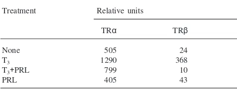
Tata, 1992; Tata, 1997). This correlation is particularly marked for the antagonism between TH and PRL, as can be discerned from Table 2.
PRL blocks the upregulation of both TRα and β mRNAs induced by T3, but does not affect the
constitut-ive expression of the receptor genes in whole pre-meta-morphic tadpoles and in organ cultures. The conclusion that TR autoinduction is necessary for the activation of downstream TR target genes is further supported by the inhibition by PRL of the activation by T3 of the
down-stream genes encoding albumin, 63 kDa keratin and stro-melysin-3 genes. Interestingly, the synthetic glucocort-icoid dexamethasone, which potentiates T3-induced
metamorphosis, elevates the levels of both TR and RXR mRNAs but PRL suppresses only that of TR when all three hormones are added to organ cultures of pre-meta-morphic Xenopus tadpole tails. The mechanism of the anti-metamorphic action of PRL in amphibia remains unknown.
Another indirect indication that the autoinduction of TR genes is closely linked to amphibian metamorphosis comes from comparative studies of TR in neotenic amphibia, i.e., those that do not undergo metamorphosis
Table 2
Relative accumulation of TRαandβmRNAs in pre-metamorphic Xen-opustadpoles treated with T3and prolactin (PRL)a
Treatment Relative units
a Batches of 20 stage 50 Xenopus tadpoles were treated with
2×1029M T
3with or without 0.1 iu PRL/ml for 4 days before relative
Table 3
Association between TR autoinduction and response to thyroid hor-mones (T3, T4) of spontaneously metamorphosing and facultatively or
obligatorily neotenic amphibia
Species Metamorphosis Endogenous TR genes T3,T4
Expresseda Autoinducedb
Xenopus Spontaneous Yes Yes Yes Ambystoma Facultatively No Yes Yes
neotenic
Necturus Obligatorily Yes Yes No neotenic
aOnly TRαdetectable inNecturus. b By exogenous TH.
spontaneously (Tata et al., 1993; Yaoita and Brown, 1990; Safi et al., 1997). Facultatively neotenic amphibia such as the mexican axolotl or the tiger salamander (Ambystoma), which do not go through metamorphosis normally, will do so if exogenous TH is given, while obligatory neotenic amphibia such asNecturusand Pro-teus do not respond to TH. As can be seen in Table 3, low levels of TR mRNAs can be detected inAmbystoma
tissues which can be upregulated by the administration of T3, in parallel with a partial metamorphic response
(loss of tail fin, growth of limbs, excretion of nitrogen as urea), as inXenopus. In contrast, only TRαtranscripts could be detected in tissues of Necturus in which T3
failed to upregulate the expression of TRαorβmRNA.
6. Auto-upregulation of other nuclear receptors
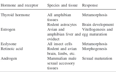
The phenomenon of autoinduction is not restricted to thyroid hormone receptors but has been observed for other nuclear receptors of growth and developmental hormones and signals (Tata et al., 1993; Tata, 1996). Some examples of auto-upregulation of nuclear recep-tors are presented in Table 4. Of some relevance to TRs
Table 4
Autoinduction of nuclear receptors by some hormones while exerting their growth and developmental actions
Hormone and receptor Species and tissue Response
Thyroid hormone All amphibian Metamorphosis tissues
Rodent astrocytes Brain development Estrogen Avian and Vitellogenesis and
amphibian liver and egg maturation oviduct
Ecdysone All insect cells Metamorphosis Retinoic acid Rodent and avian Morphogenesis
brain, limbs, etc.
Androgen Mammalian male Sexual maturation sexual accessory
tissues
are the three isoforms of receptors for retinoic acid (RARs) which is well known to serve an important func-tion in mammalian early embryogenesis and postembry-onic development (Chambon, 1995). These three recep-tor genes are all induced by retinoic acid and, interestingly, they also contain retinoic-response elements in their promoters through which the liganded receptor regulates its own gene (Tata, 1998). Of greater relevance, however, to this conference on juvenile hor-mone (JH) is the autoinduction of ecdysteroid receptors (EcRs) in insects. Furthermore, among all the members of the steroid/thyroid hormone/retinoic acid nuclear receptor supergene family, TRs and EcRs are the most closely related members (Mangelsdorf and Evans, 1995). Thummel’s and Riddiford’s groups have established accurately the temporal coordination of EcR expression with that of other regulatory genes before, during and after different stages of natural metamorphosis and fol-lowing pulses of exogenous ecdysone inDrosophilaand
Manduca, respectively (Thummel, 1996; Hiruma et al., 1999). It is therefore most significant that juvenile hor-mone has recently been shown to prevent the autoinduc-tion of EcR-A and EcR-B1 mRNAs in larval epidermis of Manduca (Hiruma et al., 1999). It is not known whether the mechanism of this inhibitory effect of JH in invertebrates is comparable to that of prolactin in amphibia. What can, however, be concluded from the examples cited in Table 4 is that nuclear receptor autoin-duction is a general phenomenon during postembry-onic development.
7. Significance of autoinduction of nuclear receptors
There is now good evidence to suggest that receptor upregulation can be closely linked to the biological activity of its ligand. The model presented in Fig. 2,
ely based on the autoinduction of TR during amphibian metamorphosis, is generally applicable to to all nuclear receptors in the context only of a developmental action of their respective ligands (Tata 1996, 1998). If upregul-ation of a given receptor is a neccessary first step leading to the sequential activation of its target genes whose pro-ducts specify the final biological action, the model pre-dicts a possible dual threshold of receptor numbers underlying the process of receptor autoinduction, on the one hand, and the activation of the target genes on the other. Thus, the genes encoding the relevant nuclear receptors would be constitutively expressed to produce very low levels of functional receptor in the target tissues from the very early stages of development. The unli-ganded receptor would be inactive or, as in the case of TR, may even act as a strong transcriptional repressor of genes with TREs in their promoters. Upon functional maturation of the appropriate endocrine gland and secretion of the hormone, or administration of exogenous hormone, the low levels of the liganded receptor would be sufficient to activate the genes encoding its own receptor but not for the receptor’s target genes. For the latter, it is proposed that, as the concentration of the hor-mone reaches higher levels, concentrations of the liganded receptor, well above the lower threshold levels required to upregulate its own receptor, would now be able to activate different sets of genes in different tissues, each depending on distinct developmental pro-grammes. An intriguing question relates to the factors involved in maintaining low levels of a given receptor prior to the onset of a developmental change and high levels during the change or inductive process. One such factor could be the involvement of other hormones or signalling molecules, as exemplified by prolactin and glucocorticoids in TH-regulated metamorphosis.
Hormonal cross-regulation of nuclear receptors is therefore an impotant additional consideration.
Acknowledgements
I am particularly grateful to all my laboratory col-leagues, too numerous to be mentioned individually, whose work has inspired much of this article.
References
Baker, B.S., Tata, J.R., 1992. Prolactin prevents the autoinduction of thyroid hormone receptor mRNAs during amphibian metamor-phosis. Dev. Biol. 149, 463–467.
Baniahmad, A., Eggert, M., Renkawitz, R., 1997. In: Papavassilou, A.G. (Ed.), Transcription Factors in Eukaryotes. Springer, Heidel-berg, pp. 95–123.
Chambon, P., 1995. The molecular and genetic dissection of the reti-noid signaling pathway. Rec. Progr. Horm. Res. 50, 317–332. Chatterjee, V.K.K., Clifton-Bligh, R.J., Matthews, C., 1997. The
ster-oid hormone superfamily of receptors. In: Rumsby, G., Farrow, S.M. (Eds.), Molecular Endocrinology. Genetic Analysis of Hor-mones and Their Receptors. Bios, Oxford, pp. 73–101.
Eliceiri, B., Brown, D.D., 1994. Quantitation of endogenous thyroid hormone receptorsαand βduring embryogenesis and metamor-phosis inXenopus laevis. J. Biol. Chem. 269, 24459–24465. Fairclough, L., Tata, J.R., 1997. An immunocytochemical analysis of
thyroid hormone and β proteins during natural and thyroid hor-mone-induced metamorphosis inXenopus. Dev. Growth Differen. 39, 273–283.
Gilbert, L.I., Tata, J.R., Atkinson, B.G. (Eds.), 1996. Metamorphosis. Postembryonic Reprogramming of Gene Expression in Amphibian and Insect Cells. Academic Press, San Diego, CA.
Gorbman, A., Bern, H.A., 1962. A Textbook of Comparative Endocrin-ology. John Wiley, New York.
Gudernatsch, J.F., 1912. Feeding experiments on tadpoles. Arch. Ent-wicklungsmech. Organ. 35, 457–483.
Hiruma, K., Shinoda, T., Malone, F., Riddiford, L.M., 1999. Juvenile hormone modulates 20-hydroxyecdysone-inducible ecdysone receptor and ultrasppiracle gene expression in the tobacco hornwormManduca sexta. Dev. Genes Evol. 209, 18–30. Ishizuya-Oka, A., Shimozawa, A., 1991. Induction of metamorphosis
by thyroid hormone in anuran small intestine cutured organotyp-icallyin vitro. In Vitro Cell. Dev. Biol. 274, 853–857.
Iwamuro, S., Tata, J.R., 1995. Contrasting patterns of expression of thyroid hormone and retinoid X receptor genes during hormonal manipulation ofXenopustadpole tail regression in culture. Molec. Cell Endocrinol. 113, 235–243.
Kanamori, A., Brown, D.D., 1992. The regulation of thyroid hormone βgenes by thyroid hormone inXenopus laevis. J. Biol. Chem. 267, 739–745.
Kikuyama, S., Kawamura, K., Tanaka, S., Yamamoto, K., 1993. Aspects of amphibian metamorphosis. Hormonal control. Int. Rev. Cytol. 145, 105–148.
Laudet, V., Ha¨nni, C., Coll, J., Catzeflis, F., Ste´helin, D., 1992. Evol-ution of the nuclear receptor gene superfamily. EMBO J. 11, 1003–1013.
Machuca, I., Tata, J.R., 1992. Autoinduction of thyroid hormone recep-tor during metamorphosis is reproduced inXenopusXTC-2 cells. Mol. Cell. Endocrinol. 87, 105–113.
Machuca, I., Esslemont, G., Fairclough, L., Tata, J.R., 1995. Analysis of structure and expression of theXenopusthyroid hormone recep-torβ(xTRβ) gene to explain its autoinduction. Mol. Endocrinol. 9, 96–108.
Mangelsdorf, D.J., Evans, R.M., 1995. The RXR heterodimers and orphan receptors. Cell 83, 841–850.
Oppenheimer, J.H., Samuels, H.H. (Eds.), 1983. Molecular Basis of Thyroid Hormone Action. Academic Press, New York.
Pazin, M.J., Kadonaga, N., 1997. What’s up and down with histone deacetylation and transcription? Cell 89, 325–328.
Ranjan, M., Wong, J., Shi, Y.-B., 1994. Transcriptional expression of Xenopus TRβ gene is mediated by a thyroid hormone response element located near the start site. J. Biol. Chem. 269, 24699– 24705.
Refetoff, S., Weiss, R.E., Usala, S.J., 1993. The syndromes of resist-ance to thyroid hormone. Endocrinol. Rev. 14, 348–399. Safi, R., Begue, A., Hanni, C., Stehelin, D., Tata, J.R., Laudet, V.,
1997. Thyroid hormone receptor genes of neotenic amphibia. J. Mol. Evol. 44, 595–604.
Shi, Y.-B., 1996. Thyroid hormone-regulated early and late genes dur-ing amphibian metamorphosis. In: Gilbert, L.I., Tata, J.R., Atkin-son, B.G. (Eds.), Metamorphosis. Postembryonic Reprogramming of Gene Expression in Amphibian and Insect Cells. Academic Press, San Diego, CA, pp. 505–538.
Tata, J.R., 1968. Early metamorphic competence ofXenopus larvae. Dev. Biol. 18, 415–440.
Tata, J.R., 1994. Hormonal regulation of programmed cell death during amphibian metamorphosis. Biochem. Cell. Biol. 72, 581–588. Tata, J.R., 1996. Homonal interplay and thyroid hormone receptor
expression during amphibian metamorphosis. In: Gilbert, L.I., Tata, J.R., Atkinson, B.G. (Eds.), Metamorphosis. Postembryonic Repro-gramming of Gene Expression in Amphibian and Insect Cells. Aca-demic Press, San Diego, CA, pp. 465–503.
Tata, J.R., 1997. Hormonal signaling and amphibian metamorphosis. Adv. Dev. Biol. 5, 237–274.
Tata, J.R., 1998. Hormonal Signaling and Postembryonic Develop-ment. Springer, Heidelberg.
Tata, J.R., Kawahara, A., Baker, B.S., 1991. Prolactin inhibits both thyroid hormone-induced mophogenesis and cell death in cultured amphibian larval tissues. Dev. Biol. 146, 72–80.
Tata, J.R., Baker, B.S., Machuca, I., Rabelo, E.M.L., Yamauchi, K.,
1993. Autoinduction of nuclear receptor genes and its significance. J. Steroid Biochem. Mol. Biol. 46, 105–119.
Thummel, C.S., 1996. Flies on steoids — Drosophola metamorphosis provides insights into the molecular mechanisms of steroid hor-mone action. Trends Gen. 12, 306–310.
Ulisse, S., Esslemont, G., Baker, B.S., Chatterjee, V.K.K., Tata, J.R., 1996. Dominant-negative mutant thyroid hormone receptors pre-vent transcription fromXenopusthyroid hormone receptorβgene promoter in response to thyroid hormone in Xenopustadpolesin vivo. Proc. Natl. Acad. Sci. USA 93, 1205–1209.
Wong, J., Patterton, D., Imhof, A., Guschin, D., Shi, Y.-B., Wolffe, A.P., 1998. Distinct requirements for chromatin assembly in tran-scriptional repression by thyroid hormone receptor and histone deacetylase. EMBO J. 17, 520–534.