www.elsevier.com / locate / bres
Research report
The domain of brain
b
-spectrin responsible for synaptic vesicle
association is essential for synaptic transmission
a ,
*
a b aWarren E. Zimmer
, Ying Zhao , Aleksander F. Sikorski , Stuart D. Critz ,
a d c a
´
Jose Sangerman , Lisa A. Elferink , X. Susan Xu , Steven R. Goodman
a
Department of Cell Biology and Neuroscience, University of South Alabama, Mobile, AL 36688, USA
b
Institute of Biochemistry, University of Wroclaw, Wroclaw, Poland
c
Department of Therapeutic Radiology, Yale University School of Medicine, New Haven, CT, USA
d
Department of Biological Sciences, Wayne State University, Detroit, MI, USA Accepted 2 August 2000
Abstract
We have examined the interaction between synapsin I, the major phosphoprotein on the membrane of small synaptic vesicles, and brain spectrin. Using recombinant peptides we have localized the synapsin I attachment site upon theb-spectrin isoformbSpIISI to a region of 25 amino acids, residues 211 through 235. This segment is adjacent to the actin binding domain and is within the region of thebSpIISI that we previously predicted as a candidate synapsin I binding domain based upon sequence homology. We used differential centrifugation techniques to quantitatively assess the interaction of spectrin with synaptic vesicles. Using this assay, high affinity saturable binding of recombinant bSpIISI proteins was observed with synaptic vesicles. Binding was only observed when the 25 amino acid synapsin I binding site was included on the recombinant peptides. Further, we demonstrate that antibodies directed against 15 amino acids of the synapsin I binding domain specifically blocked synaptic transmission in cultured hippocampal neurons. Thus, the synapsin I attachment site onbSpIISI spectrin comprises a|25 amino acid segment of the molecule and interaction of these two proteins is an essential step for
the process of neurotransmission. 2000 Elsevier Science B.V. All rights reserved.
Theme: Excitable membranes and synaptic transmission
Topic: Presynaptic mechanisms
Keywords: Spectrin; Synapsin; Synaptic vesicle; Synaptic transmission
1. Introduction [6,9,31] and then fuse with the presynaptic membrane in a calcium regulated manner [7,19,21].
The transmission of information through the neural Brain spectrin is a major cytoskeletal protein within the system occurs via the regulated release of neurotrans- presynaptic membrane compartment [10,33], which plays a mitters from synaptic and secretory vesicles [19]. A large vital role in neurotransmission [30]. Immunoelectronmic-number of vesicles ranging in size from 10 to 140 nm in roscopic experiments have demonstrated that theaSpIIS* / diameter are present within the presynaptic cytoplasm, bSpIIS1 spectrin isoform (nomenclature as designated by closely associated with the presynaptic membrane through [32,34]) is the predominant spectrin in the presynaptic interactions with the cytoskeleton [7,11,21]. During synap- compartment and is associated with the cytoplasmic sur-tic transmission these vesicles are released from their face of small synaptic vesicles as well as the plasma cytoskeletal tethers, dock at the release site on the cyto- membrane [33]. Moreover, quick freeze deep-etch electron plasmic membrane via associations with docking proteins microscopy of presynaptic terminals detect small spherical synaptic vesicles in contact with long fibrous strands, thought to be brain spectrin, interconnecting these vesicles
*Corresponding author. Tel.: 11-334-460-7982; fax: 1
1-334-460-with the presynaptic plasma membrane [13,21]. Therefore,
6771.
E-mail address: [email protected] (W.E. Zimmer). spectrin aSpIIS* /bSpIISI is correctly positioned within
the presynaptic terminal consistent with a key role in by DNA sequencing, this shuttle plasmid was used to neurotransmission. Biochemical and molecular studies also make a viable baculovirus encoding the 6His-bSpIIS1 support a role for spectrin in regulating neurotransmitter fusion protein using the Bac-to-BacE kit as indicated by release. Brain spectrin binds end-on to small synaptic the supplier (Gibco BRL, Gaithersburg, MD). The pres-vesicles via synapsin I [28,29], a major phosphoprotein of ence of the fusion protein in Sf 21 cells infected with the synaptic vesicle membrane which regulates the availa- bSpII 1–457 virus was confirmed in all lysates by Western bility of synaptic vesicles during synaptic transmission analysis using both a His-tag antibody (Pharmacia Biotech, [7,10]. That this binding occurs close to the brain spectrin Chicago, IL) and one of severalbSpIIS1 peptide specific actin binding domain was demonstrated by low angle antibodies.
rotary shadowing electron microscopy [20]. For protein purification, Sf 21 cells were infected with The interaction of brain spectrin with synapsin I appears bSpII 1–457 virus (15, T 150 flasks) and the cells similar to that observed between protein 4.1 and erythroid incubated at 288C for 72–96 h. The cells were collected by spectrin in red blood cells. For example, protein 4.1 and centrifugation and then lysed in buffer containing 50 mM synapsin I bind directly to the beta subunit of the spectrin Tris–HCl (pH 8.5), 5 mM 2-mercaptoethanol, 100 mM protein in red blood cells and neural tissues, respectively KCl, 1 mM PMSF, 1% Nonidet P-40 at 48C by vortexing. [5,15]. Moreover, protein 4.1 can competitively inhibit the Cell debris was removed by centrifugation (10,0003g) for
binding of synapsin to brain spectrin and synapsin I can 10 min and the supernatant loaded onto a Ni-NTA column. inhibit protein 4.1 binding to the red blood cell spectrin The column was washed with 2 to 3 column volumes of aSpISI /bSpISI [2,20]. Comparison of the predicted amino buffer containing 50 mM Tris–HCl (pH 8.0), 5 mM acid sequence of bSpIIS1with its erythroid counterpart 2-mercaptoethanol, 100 mM KCl, 10% glycerol and 3 mM detected a region adjacent to the actin binding domain that Imidazole, after which bound proteins were eluted in the was 87% identical between these isoforms. We predicted same buffer containing 100, 200, and 300 mM Imidazole. that this region (spanning amino acid residues 207–445 of The eluted fractions were examined by SDS–PAGE and the b molecule) maybe a potential synapsin I binding Western blotting as described previously [4,23,34]. domain of brainbspectrinbSpIIS1 [23]. In this report, we A bacterial vector containing the glutathione-S-transfer-have used recombinant peptides to identify the exact site of ase (GST) gene was used for making recombinant GST-synapsin I–bSpIISI interaction. Our results demonstrate fusion proteins. ThebSpIIS1 cDNA served as a template that amino acids residues 211 through 235 of the b- to clone the segment of DNA encoding amino acids 1–457 molecule are essential for synapsin I binding. Further, we into the pGEX-5 GST-fusion protein vector (Pharmacia demonstrate that a peptide specific antibody against this Biotech, Chicago, IL). This placed the GST coding se-region of bSpIISI inhibits synaptic transmission in patch quence at the amino terminus of the bSpIIS1 sequence. clamp studies of paired hippocampal neurons. These The plasmid was transferred into BL 21 bacteria and results indicate that interaction of synaptic vesicles with grown at 378C in Luria Broth (LB) supplemented with 75 these 25 amino acids within brain spectrin bSpIISI is mg / ml ampicillin and 0.2% glucose. To make truncated
essential for neurotransmission. proteins, PCR fragments with a common 59end, beginning
431 at the ATG initiation codon, and ending with codons A ,
406 381 355 331 306 285 259 235 210
R , Y , 12 , A , K , V , D , Q , and A
were cloned into the same GST vector. Each vector was
2. Materials and methods confirmed by DNA sequence analysis and then transferred into BL 21 bacteria for generation of recombinant proteins. 2.1. Recombinant protein expression and purification One hundred microliters of LB media containing 75 mg / ml ampicillin was inoculated with bacteria housing The initial recombinant protein used in this study was plasmids for the GST-fusion proteins and the bacteria generated using the Bac-to-BacE Baculovirus System grown in a 378C shaker-incubator until an A600 of 0.4–0.6 (Gibco BRL, Gaithersburg, MD). bSpIIS1 cDNA clone was obtained. IPTG was then added (final concentration of 14T3-1 containing the 5 UTR and |2 Kb of coding 0.5 mM) and the cultures incubated for an additional sequence [23] was used as a template for PCR amplifying 24–48 h. The cells were collected by centrifugation, the coding segment beginning with the translation initia- resuspended in 13PBS (phosphate-buffered saline)
con-1 457
purity was estimated by SDS–PAGE followed by staining studies was previously characterized [20], and was used in with Coomassie Blue as previously described [4,23]. the present study at dilution of 1:1000.
We developed an in vitro assay based upon the ability to remove synaptic vesicles from a mixture by differential 2.2. Brain spectrin and synapsin I isolation
centrifugation. Differences in sedimentation properties between synaptic vesicle and intact spectrin are small, thus Bovine brains were obtained from a local slaughterhouse
an assay based upon centrifugation was not feasible. and stored at 2708C until used to isolate spectrin. Brain
However, we reasoned that a peptide representing|15% of spectrin was purified from the frozen tissue as detailed
the beta subunit might allow separation, and in preliminary previously [29]. Synapsin I was isolated from frozen pig
experiments we found that .98% of vesicles were found brain tissue as detailed in previous studies from our
in the pellet of a 200,0003g centrifugation for 30 min
laboratory [20,29]. Protein purity was assessed by SDS–
while 100% of thebSpIIV457 (|80 KDa) bacterial fusion PAGE and Coomassie Blue staining of the polyacrylamide
protein remained in solution under the same conditions. gels. The synapsin I was followed by analysis on Western
For binding analyses, the bacterially expressed bSpII
blots. Red blood cell (RBC) membrane proteins were from 125
peptides were labeled with I using the Bolton–Hunter isolated ghosts as described previously [27].
reagent. Typical binding assays were accomplished in a 200ml volume of buffer containing 5 mM Tris–HCl (pH
2.3. Synapsin I blotting assay 7.5), 65 mM NaCl, 1 mM EGTA, 0.2 mM DTT, 20mg / ml
PMSF and 4 mg of synaptic vesicle protein. Binding was 125 To identify the attachment site of synapsin I on initiated by the addition of increasing quantities of I-bSpIIS1, we modified the blotting technique of Iga et al. labeled peptide and after a 1 h incubation at room [15]. Proteins to be analyzed were separated by SDS– temperature (228C) the reaction loaded onto a Ti 42.2 rotor PAGE and then electrophoretically transferred to a nitro- (Beckman Instruments) and spun at 35,000 r.p.m. cellulose sheet as described previously [4,28,29]. The (200,0003g) for 30 min at 48C. The pellet and supernatant
125
nitrocellulose membrane was then blocked by incubation were carefully separated and the amount of I in each in buffer containing 5% BSA, 1% Triton X-100, 50 mM determined using a gamma counter (Packard Autogamma KCl, 1 mM EDTA and 20 mM HEPES (pH 7.4) for 12–16 50DC; Packard Instruments Company, Meridan, CT). We h at 48C. The blot was washed 3–5 times with the same initially determined that the kinetics of binding was rapid, buffer and then incubated in this buffer to which purified reaching equilibrium in
|1–5 min. We thus chose to synapsin I had been added. Early experiments contained incubate our binding reactions for 1 h to ensure complete-1.5mg / ml synapsin I [15], but we demonstrate that higher ness of the binding reaction. All reactions were done in fidelity of the assay was obtained with lower concen- triplicate and controls consisted of no added peptide or trations, 0.015 mg / ml. The synapsin I incubation was vesicles and using BSA instead of the bSpIIS1 peptides. allowed to continue for 1 h at room temperature, after The amount of bound peptide (pellet) was plotted versus which the blot was washed with buffer without synapsin the amount of free peptide (supernatant) and from these (5–7 times 30 min each wash) with constant agitation. The data the K (binding affinity) and maximal binding
D
nitrocellulose was then incubated in 4% paraformaldehyde capacity were determined using the ENZFITTER computer for 30 min at room temperature and the excess paraformal- program as described previously [28,29].
dehyde removed by 3–5 washes in Western blotting buffer
(0.9% NaCl, 0.05% Tween 20, and 10 mM Tris–HCl, pH 2.5. Whole-cell patch clamp recording 7.4). The blot was processed for Western blotting using
rabbit antisynapsin I antibody as described previously [29] Excitatory postsynaptic currents were followed in paired 125
and localization of the synapsin I antibody using I- hippocampal neurons essentially as previously described 125
protein A (New England Nuclear, Boston, MA). I- was by our laboratory [30]. Briefly, hippocampal neurons localized by autoradiography using either X-ray film cultured from neonatal rats (postnatal day 2–3) were (Kodak AR5) or by image analysis on a BioRad Phos- bathed in extracellular recording solution (119 mM NaCl,
phoimager. 5 mM KCl, 20 mM HEPES, 2 mM CaCl , 2 mM MgCl ,
2 2
30 mM glucose, 1mM glycerine, 100mM picrotoxin, pH 2.4. Isolation of small synaptic vesicles and binding 7.3, osmolality adjusted to 330 mOs with sucrose) and
was diluted directly into the intercellular solution. Data were included for analysis provided that the holding current remained stable and while the number of observa-tions within each experiment varied, all groups contained a minimum of four individual experiments.
2.6. Antibody preparation
The synapsin I [20] and spectrin amino terminal [4,30] antibodies have been previously described by our labora-tory. The synapsin binding domain antibody used in this study was produced in rabbits by injection of the peptide
NH2-AFNALIHKHRPDLID-COOH which represents
amino acids 207 through 221 of the bSpIIS1 molecule. IgG fractions were purified from pre-immune or immun-ized rabbits using a commercial Fab purification kit (Pierce Chemical Co., Rockford, IL). Protein concentrations were determined by absorbance at 280 nm and aliquots (500mg) were lyophilized and stored at2808C until use. IgGs were dissolved as stock solutions (2 mg / ml) in intracellular solution and then further diluted just prior to experimenta-tion.
3. Results
3.1. bSpIIS1 synapsin binding domain is located near
the actin binding domain of the molecule
We have previously elucidated the primary structure of mousebSpIIS1. In these studies we noted a region of the molecule adjacent to the actin binding domain that shared 87% identity with human and mousebSpIS1 proteins [23]. This segment included amino acids 207 to 445 of the b-spectrins. Given the high degree of homology and the location of this region proximal to the actin binding domain, we proposed that this segment may contain the synapsin I binding site of the molecule. To test this
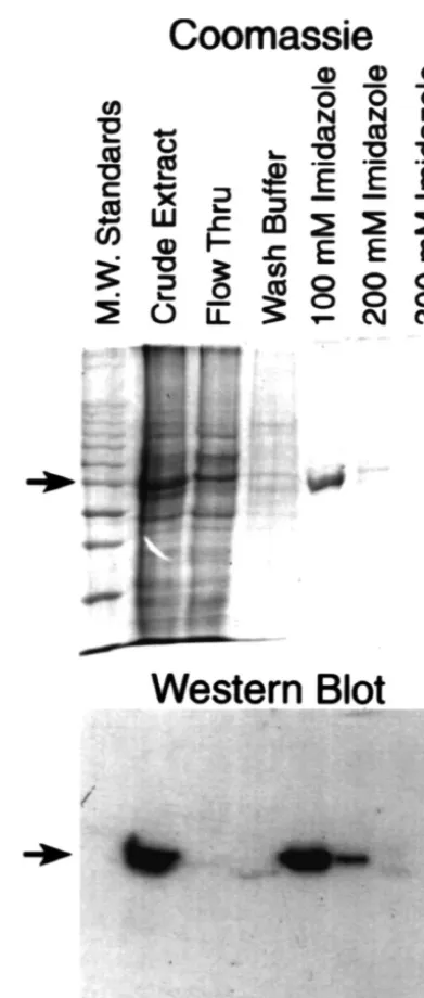
hypothesis, we generated baculoviral expression vectors Fig. 1. bSpIIS1 1–457 is expressed as a fusion protein in
baculoviral-containing the amino-terminal segment ofbSpIIS1. Using infected cells. The amino terminal 457 residues of the bSpIIS1 protein were expressed in a baculovirus system as described in Materials and
PCR we generated a cDNA fragment encompassing the
457 Methods. The spectrin peptide was expressed as a fusion protein ATG methionine-initiation codon through Val of
containing a histidine tag-(His) — at the extreme amino terminus. Crude6
bSpIIS1 and cloned this fragment into a baculovirus extracts and fractions obtained by affinity chromatography were analyzed shuttle plasmid, in frame with a His-tag [(His) ]. As shown6 by SDS–PAGE (Coomassie) and Western analyses (Western blot) using
in Fig. 1, lysates from Sf21 insect cells infected with virus an anti-bSpIIS1 peptide specific antibody, Ab 921. The arrows indicate the 58 KDa recombinant His-taggedbSpIIS1 1–457 protein.
derived from the spectrin shuttle plasmid exhibited marked expression of a|58 KDa peptide (crude extract). This|58
KDa peptide remained bound to a nickel-agarose affinity binding to the|58 KDa peptide found in crude extracts of column (Ni-NTA resin), and was eluted from the column infected SF21 cells and the protein purified on nickel-with buffer containing 100 mM imidazole (100 mM agarose columns (Ni-NTA affinity chromatography). Thus, Imidazole). To confirm that |58 KDa peptide was the these data establish that we have expressed and purified an expected bSpIIS1 amino terminal domain, we performed amino terminal segment of thebSpIIS1 molecule consist-Western blotting analyses utilizing a peptide specific ing of the actin binding domain [18] and the putative antibody directed against amino acids 207 to 221 of the synapsin binding domain [23].
adapted a synapsin blotting assay originally described by Iga et al. [15]. In this assay, baculovirus-infected cells expressing bSpIS1–457, in addition to purified fusion protein, and lysates prepared from erythroid and neural tissues were fractionated by SDS–PAGE and transferred to nitrocellulose. The filters were incubated with synapsin I, isolated from brain tissue, under conditions which allow the binding of synapsin to proteins on the filter paper. Bound synapsin is then visualized using a synapsin-spe-cific antibody. Consistent with our earlier studies and those reported by others [1,12,20,26,29,35], synapsin I exhibits a high degree of non-specific interactions by virtue of its highly charged basic characteristic. At a concentration of 1.5 mg /ml (as used by Iga et al. [15]) synapsin I exhibits non-specific binding to proteins of RBC-membranes as well as standard molecular weight protein controls (data not shown). However under these conditions, we also detect binding to thebSpII 1–457, 58 KDa peptide. Thus, we modified the assay to improve the specificity of synapsin I binding. We found that specific binding occurred quickly, such that after |30–45 min non-specific interactions began to occur raising the background over specific binding, and that non-specific interactions are effectively competed with the addition of other highly charged, basic proteins like histones (data not shown). In addition, we found that reducing the amount of synapsin I in the incubation buffer by|100 fold significantly reduced background interactions thereby enhancing the specificity of binding. To demonstrate the ability of the synapsin blot assay, we utilized it on membranes containing our ex-pressedbSpII 1–457 protein and spectrins from red blood cell and brain tissue (Fig. 2A). As shown by this assay, synapsin I demonstrates specific binding with the beta subunit of brain spectrin and to our baculoviral expressed, amino terminal segment ofbSpIIS1. Therefore, the synap-sin I binding site on brain spectrin resides upon the beta-subunit and is within the amino terminal|450 amino acids of this protein.
3.2. Synapsin binds bSpIIS1 to a region encompassing
amino acid residues211 –235
Fig. 2. Specificity of the synapsin I blotting analysis. A qualitative analysis of synapsin I binding was developed using a modification of the
The demonstration of synapsin I binding to the beta
blotting technique described by Iga et al. [15]. Protein to be examined
subunit of brain spectrin near the actin binding domain is were separated by SDS–PAGE and transferred to nitrocellulose mem-consistent with our earlier studies utilizing by low angle branes. The membranes were incubated in buffer containing synapsin I
rotary shadowing electron microscopy [20]. Since, synap- after which the blots were rinsed and the bound synapsin fixed to the protein on the membrane with 4% paraformaldehyde. The synapsin I was
sin I is the major phosphoprotein on the membrane of
then localized on the blot through Western blotting using a rabbit
synaptic vesicles [7,12,28], its interaction with the beta 125
synapsin I specific antibody and I-Protein A. Panel A shows a
subunit of brain spectrin, specificallybSpIIS1, may play a Coomassie stained gel of molecular weight markers (standards), the
|58
key role in the release of neurotransmitters. To more KDa fusion protein (bSpII 1–457), red blood cell ghosts (RBC) and
precisely localize thebSpIIS1 synapsin I binding site, we purified brain spectrin (Brain Spectrin). Proteins from a parallel gel were transferred to nitrocellulose membranes and incubated in buffer
con-expressed thebSpIIS1 1–457 peptide and COOH-terminal
taining 0.015mg / ml synapsin I after which the bound synapsin I was
truncations of the peptide as GST-fusion proteins in
localized by Western blotting and autoradiography as detailed in Materials
bacteria. Using PCR we cloned the |1.3 Kb bSpIIS1 and Methods (Panel B). The arrows point to the positions of proteins that 1–457 coding sequence in frame with a glutathione-S- specifically bind synapsin I, thebSpIIS1 1–457 fusion protein and the
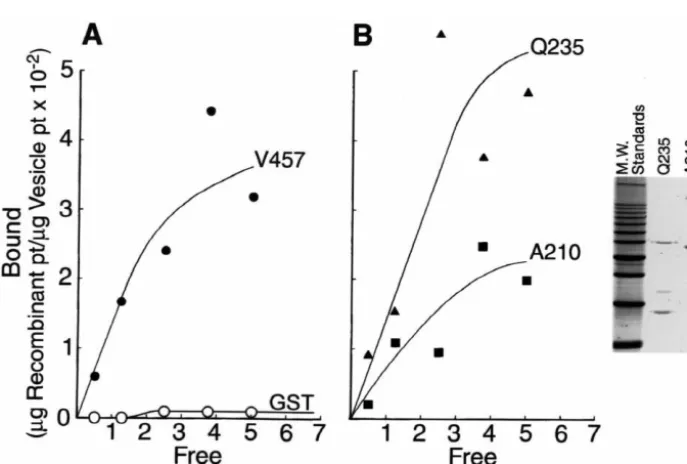
proteins by thio-agarose affinity chromatography. Recent proteins to bind brain synapsin I in blotting assays. GST-experiments from our laboratory [30] demonstrated that spectrin fusion proteins containing the original 457 amino peptide specific antibodies directed to the C-terminal acids specifically bound synapsin I, whereas no binding segment of the putative synapsin binding site (amino acids was observed using GST alone, further demonstrating that 417–428) disrupted synaptic transmission. Therefore we synapsin I binds the amino terminal domain of bSpIIS1. formed multiple bSpIIS1 peptides that are COOH-trunca- Moreover, these data suggest that eukaryotic post-transla-tions starting from the initial synapsin binding V 457 tional modifications ofbSpIIS1 are not critical for synap-peptide. Fig. 3A summarizes our strategy which truncates sin I binding. In addition, examination of synapsin blots peptides by |25 amino acids through the potential synap- such as the one illustrated in Fig. 3B showed that all but sin I binding domain. Each of the bacterially expressed the shortest of the bacterially derived proteins bound bSpIIS1 fragments were successfully expressed and synapsin I (compare Western and synapsin blotting of purified (data not shown). Moreover, Western analyses of bSpII-A210 with adjacent lanes). These qualitative data the expressed proteins using an antibody generated to the suggest that the exact synapsin binding region of the amino terminus of bSpIIS1, within the actin-binding bSpIIS1 molecule is within a small domain of the mole-domain (amino acid residues 8–24), detects bacterially cule between amino acids L211 and Q 235.
derived spectrin peptides of the correct molecular weight
(Fig. 3B). The |30 KDa difference in apparent molecular 3.3. Synaptic vesicle binding capability ofbSpIIS1 is weight of the bacterially synthesized bSpIIS1 fragment conferred by amino acids A210 through Q235 containing amino acids 1–457 and that of the original
bSpII 1–457 baculoviral recombinant protein (Figs. 1 and Blotting experiments (Fig. 3) suggest that synapsin I 2) accounts for the addition of GST to the bacterial binding is mediated primarily through a |25 amino acid
proteins. segment of the bSpIIS1 protein bounded by L211 and
We next examined the ability of the GST-spectrin fusion Q235. As synapsin I has been implicated to play a major
Fig. 3. Localization of the synapsin I binding site on bSpIIS1. The localization of the synapsin I binding site was accomplished using bacterially synthesized truncations of thebSpII 1–457 amino terminus and the synapsin I blotting technique. Panel A diagrams the 10bSpII amino terminal peptides expressed in bacteria. Each successive bacterial peptide was truncated by|25 amino acids so that with the 10 fusion proteins we could examine binding
Table 1
role in linking synaptic vesicles to brain spectrin [28,29],
Binding affinity and maximal binding capacity of synaptic vesicle —
we sought to quantitatively examine the binding of our
spectrin fusion protein interactions
bacterially expressed GST-spectrin fusion proteins with
V457 Q235 A210
isolated synaptic vesicles. We focused upon the two fusion
a
proteins which defined synapsin I binding by blotting Binding (K ) affinityD 5 nM 19 nM 180 nM
b
Binding capacity 4.0 1.1 6.6
analyses (A210, Q235). Recombinant fusion proteins were
a
purified by affinity chromatography (inset, Fig. 4B) and Calculated by least squares analyses of binding isotherms using
125 125
ENZFITTER computer program.
labeled with I using the Bolton–Hunter reagent.
I-b
Values reflectmg recombinant protein /mg vesicle total protein.
labeled recombinant spectrin was incubated with small synaptic vesicles [14,28,29] and vesicle-associated spectrin
separated from unbound proteins by differential centrifuga- binding capacity of these two fusion proteins with synaptic tion. The binding isotherms, for spectrin-vesicle binding, vesicles (Table 1), indicating a difference in the binding were calculated using an ENZFITTER computer program sites upon the vesicles for the recombinant Q235 and A210 (R.J. Leutherburrow, Biosoft, Inc., Milltown, NJ). Full- spectrin fusion polypeptides.
length (V-457) and truncatedbSpIIS1 / Q235 gave
compar-able levels of saturcompar-able binding peptide (KD of 5 nM and 3.4. Peptide specific antibodies to the 25 amino acid 19 nM, respectively). These values agree well with pub- bSpIIS1 synapsin I binding site block synaptic lished values of synaptic vesicle–spectrin interaction (24 transmission in living cells
nM; [28]) and synapsin I–spectrin interactions (9 nM; 27
[15]). This contrasts with the value of 180 nM (1.8310 Following localization of the synapsin I binding domain M) obtained with thebSpIIS1 peptide truncated at amino to amino acids 211 through 235 of thebSpIIS1 molecule acid A210 (Table 1). Thus, removal of 25 amino acids, (Fig. 3) and the demonstration that these amino acids between L211 to Q235, from the bacterial bSpIIS1 comprised the attachment site for synaptic vesicles (Fig. peptides causes a 10-fold reduction in synaptic vesicle 4), we next examined the function of this region in vivo. binding, indicating that the major binding domain of We generated a peptide specific antibody to residues 207 to bSpIIS1 for synapsin I / synaptic vesicles lies between 221 which spans the actual synapsin I attachment site. This these residues. Further, there is a change in the calculated antibody, referred to as Ab 921, was used to confirm the
Fig. 4. Quantitative analysis ofbSpII-GST fusion peptides with synaptic vesicles. Binding of thebSpII-GST fusion proteins with isolated small synaptic vesicles was analyzed by a differential centrifugation assay. The fusion proteins were purified by chromatography upon a glutathione agarose affinity
125 125
column and the purified proteins labeled with I using the Bolton–Hunter reaction. Various amounts of the I labeled proteins were incubated with the synaptic vesicles and then the reaction mixtures centrifuged at 200,0003g for 30 min. The supernatant and pellet were analyzed for labeled peptide and the
Bound (pellet) versus Free (supernatant) data plotted. From these data a K (binding affinity) and binding capacity were derived using the ENZFITTERD
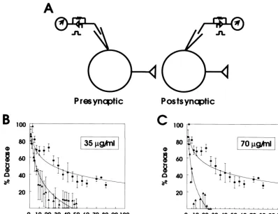
expression of the amino terminal domain in the baculo- were averaged and normalized to the initial maximal virus system (Fig. 1) thus demonstrating the antibody response. The decline of ESPCs developed rapidly and specificity. We reasoned that injection of Ab 921 should maximal suppression was observed after approximately 45 occlude the brain spectrin–synapsin I binding site, thus min. Injection of Ab 921 at 70mg / ml caused a more rapid reducing neurotransmission, if this bSpIIS1 segment was suppression of ESPCs amplitude which reached maximal functionally important for this process. Therefore we effect in about 25 min (Fig. 5C). Therefore, these data examined the effects of Ab 921 in cultured hippocampal illustrate that occlusion of the brain spectrin–synapsin I neurons in which the presynaptic neuron of synaptically interaction causes a specific reduction of synaptic transmis-paired cultured rat hippocampal neurons was injected with sion in coupled hippocampal neurons. The inhibition of different concentrations of Ab 921 IgG, whereas control synaptic transmission is site specific onbSpIIS1 because cells were injected with a similar concentration of rabbit peptide specific antibodies against residues 8–24 (Ab 43) pre-immune IgG. The consequence upon synaptic trans- and 581–597 (Ab 49) have no effect on synaptic transmis-mission of infusing Ab 921 was measured in the amplitude sion [30].
of excitatory postsynaptic currents (ESPCs) recorded from a synaptically-coupled postsynaptic neuron. Fig. 5A
dia-grammatically summarizes our experimental protocol. As 4. Discussion
shown in Fig. 5B, injection of Ab 921 (35mg / ml) caused
a time-dependent reduction of the amplitude of ESPCs It is clear that neuronal spectrin interacts with or binds evoked in synaptically coupled rat hippocampal neurons. synapsin I, both as isolated proteins and when on synaptic ESPCs were evoked every 15 s and each four responses vesicles [1,2,12,20,26–29]. Based upon sequence
homologies, we predicted that the synapsin I binding protein is binding something other than receptor protein on domain of the bSpIIS1 spectrin isoform would reside the synaptic vesicles. Therefore, we suggest that the A210 between residues 207 to 445 [23]. In the studies described recombinant binding represents attachment with the phos-here, an expressed His-fusion recombinant polypeptide pholipids of the vesicle, whereas the Q235 recombinant consisting of the amino terminal |450 amino acids from shows specific, saturable binding with a receptor protein thebSpIIS1 molecule specifically bound synapsin I (Figs. on the synaptic vesicle, namely synapsin I.
1 and 2). Using carboxyl-terminal truncations of this Several studies have implicated synapsin I and spectrin recombinant peptide we localized, in a novel solid phase as key components for synaptic transmission. Electron binding assay, the exact interaction domain ofbSpIIS1 to microscopy of synaptic terminals demonstrate that mor-a segment of 25 mor-amino mor-acids bounded by residues 211 phologically docked vesicles are closely associated with through 235 (Fig. 3). Moreover, this 25 amino acid domain the cytoskeleton, principally actin filaments [21]. In addi-contains the necessary components of functionalbSpIIS1– tion, Landis et al., [21] and Hirokawa and co-workers [13] synapsin I interactions. In vitro analyses demonstrated that demonstrated attachment of synaptic vesicles with 100 nm this segment was the key region of thebSpIIS1 molecule fibers perpendicular to the cellular membrane of the active for binding synapsin I laden synaptic vesicles (Fig. 4) and zone. Based upon this work and studies from our labora-blocking this interaction with peptide-specific antibodies tory, we hypothesized that the 100 nm fibers may represent interrupted synaptic transmission in cultured neuronal cells brain spectrin molecules that tether the vesicles at the (Fig. 5). Thus, our studies directly demonstrate that the active zone, the ‘casting the line hypothesis’ [11,36]. spectrin–synapsin I interaction represents a critical step in Neuronal spectrin is a tetramer of |200 nM in length
neurotransmission. which is arranged in a head to head configuration of
We have previously shown that brain spectrin binds aIIS1 /bIIS1 heterodimers. An attachment may be formed small synaptic vesicles in a synapsin I-dependent manner between the synapsin I on the vesicle membrane and the [28,29]. Here we demonstrate that isolated recombinant brain spectrin on the cellular membrane as the vesicles peptides encoding the amino terminal|450 amino acids of approach the plasma membrane [11,36]. Previously it has the bSpIIS1 molecule retains synapsin I binding, both been shown that synaptic vesicles bind end-on to brain isolated protein and when incorporated into synaptic spectrin at a site near the actin binding domain [20] and vesicles, and our data indicate that the major binding site this binding is dependent upon synapsin I on the vesicle for spectrin binding to synapsin lies between amino acids membrane [28–30]. We have demonstrated here that the L211 and Q 235. Taken together, our data is consistent synapsin I attachment site upon spectrin resides on the beta with previous studies and suggest thatbSpIIS1 specifically subunit and is contained within |25 amino acids (L211– binds synaptic vesicles through synapsin I. Brain spectrin Q235) that are only 25 amino acids C-terminal to the has been shown to bind synapsin proteins fixed to mem- spectrin actin binding domain defined by Karinch et al. branes [1,15,29] and in solution [20]. Moreover, the [18] as amino acids 47–186 of beta spectrin. In ‘the calculated KD of spectrin–synapsin I interactions, 9 nM casting the line hypothesis’ the binding of synaptic vesicles [15] is consistent with the KD value of synaptic vesicle to brain spectrin would cause a release of actin filament binding calculated in our studies with recombinant binding, allowing one half of spectrin to be free from the bSpIIS1 fragments (5–19 nM) and with that of intact membrane. The other end of the spectrin tetramers would brain spectrin (24 nM) [28,29]. Residual binding was be maintained at the membrane via attachments with actin observed with the bSpIIS1 A 210 peptide to the synaptic and ankyrin [2,11] and this arrangement would account for vesicles. This may arise from the remaining amino acids of the electronmicroscopic observation of small synaptic a binding domain, as the homology of spectrins begin at vesicles associated end-on with 100 nm fibers at the active amino acid 207 [23]. However, it has been demonstrated in zone. Further this would suggest that disruption of this a number of laboratories that both erythroid vesicle–spectrin association would be detrimental to [3,16,22,24,25] and neuronal [8] spectrins bind with neurotransmitter release and synaptic function. We have phospholipids in monolayers and bilayers, in addition to shown here that peptide-specific antibodies against the unilammellar and miltilammellar vesicles. Indeed, the KD segment of the spectrin molecule housing the synapsin I of the liposome–spectrin interaction has been demon- attachment site obstructed the release of neurotransmitter strated to be greater than 100 nM (76–200 nM) in these from presynaptic neurons as measured by reducing the earlier studies which is similar to the 180 nM KD we amplitude and frequency of EPSCs in the postsynaptic cell. derived from thebSpIIS1 A210 fragment interacting with Thus, the synaptic vesicle interaction with brain spectrin synaptic vesicles. Further, using the binding data in Fig. 4, via synapsin I is important for regulating synaptic trans-we calculated binding capacities for the bSpIIS1 frag- mission, perhaps by regulating the availability of mor-ments Q235 and A210 as 1.1 mg recombinant protein /mg phologically docked vesicles.
[17] R. Jahn, W. Schiebler, P. Greengard, A quantitative
dot-immuno-presynaptic plasma membrane. We believe that brain
binding assay for proteins on nitrocellulose membranes filters, Proc.
spectrin can then serve as a template on which V-SNARES,
Natl. Acad. Sci. USA 81 (1984) 1684–1687.
T-SNARES, SNAPS and NSF proteins can be arranged in [18] A.M. Karinch, W.E. Zimmer, S.R. Goodman, The identification and 21
the appropriate configuration to allow fusion and Ca sequence of the acting binding domain of human red blood cell
regulated exocytosis. b-spectrin, J. Biol. Chem. 265 (1990) 11833–11840.
[19] R.B. Kelly, The cell biology of the nerve terminal, Neuron 1 (1988) 431–438.
[20] K.E. Krebs, S.M. Prouty, I.S. Zagon, S.R. Goodman, The structural
Acknowledgements and functional relationship of rbc protein 4.1 and synapsin I, Am. J.
Physiol: Cell Physiol. 22 (1987) 6500–6505.
We thank the members of the Goodman and Zimmer [21] D.M.D. Landis, A.K. Hall, L.A. Weinstein, T.S. Reese, The organi-zation of cytoplasm at the presynaptic active zone of a central
laboratories for careful reading and suggestions on this
nervous system synapse, Neuron 1 (1988) 201–205.
manuscript. This work was supported in part by grants
[22] A.E. McKiernan, R.I. McDonald, R.C. McDonald, D. Axelrod,
RO1NS35937 to SRG and RO1 GMS53189 to LAE. Cytoskeletal protein binding kinetics at planar phospholipid
mem-branes, Biophys. J. 73 (1997) 1987–1998.
[23] Y. Ma, W.E. Zimmer, B.M. Reiderer, M.L. Bloom, J.S. Barker, S.R. Goodman, The complete amino acid sequence for brainb-spectrin
References
(b-fodrin): relationship to globin sequences, Mol. Brain Res. 18 (1993) 87–99.
[1] A. Baines, V. Bennett, Synapsin I is a spectrin binding protein [24] K. Michalak, M. Bobrowska, A.F. Sikorski, Interaction of bovine immunologically related to erythrocyte 4.1, Nature 315 (1985) erythrocyte spectrin with amino phospholipid liposomes, Gen.
410–413. Physiol. Biophys. 12 (1993) 163–170.
[2] V. Bennett, Ankyrins, J. Biol. Chem. 267 (1992) 8703–8706. [25] C. Mombers, J. DeGier, R.A. Demel, L.L.M. Van Deenen, Spectrin [3] K. Bialkowska, A. Zembron, A.F. Sikorski, Ankyrin inhibits binding phospholipid interaction: a monolayer study, Biochem. Biophys.
of erythrocyte spectrin to phospholipid vesicles, Biochim. Biophys. Acta 603 (1980) 52–62.
Acta 1191 (1994) 21–26. [26] R.A. Nichols, T.J. Chilcete, A.J. Czernick, P. Greengard, Synapsin I [4] M.B. Clark, Y. Ma, M.L. Bloom, T.E. Barker, I.S. Zagon, W.E. regulates glutamate release from rat brain synaptosomes, J.
Neuro-Zimmer, S.R. Goodman, Braina erythroid spectrin: identification, chem. 58 (1992) 783–785.
compartmentalization, and b-spectrin associations, Brain Res. 663 [27] A. Shartava, C.A. Monteiro, F.A. Bencsath, K. Schneider, B.T. (1994) 223–236. Chait, R. Gussio, L.A. Cassoria-Scott, A.K. Shah, C.A. Heuerman, [5] T.R. Coleman, A.S. Harris, S.M. Mische, M.S. Mooseker, J.S. S.R. Goodman, A post-translational modification of beta-actin Morrow, Beta spectrin bestows protein 4.1 sensitivity on spectrin– contributes to the slow dissociation of the spectrin-4.1–actin com-actin interactions, J. Cell Biol. 104 (1987) 519–526. plex of irreversibly sickled cells, J. Cell Biol. 128 (1995) 805–818. [6] W.M. DeBello, H. Betz, G.J. Augustine, Synaptotagmin and neuro- [28] A.F. Sikorski, S.R. Goodman, The effect of synapsin I phosphoryla-transmitter release, Cell 74 (1993) 947–950. tion upon binding of synaptic vesicles to spectrin, Brain Res. Bull. [7] P. DeCamilli, F. Benfenati, F. Valtorta, P. Greengard, The synapsins, 27 (1991) 195–198.
Annu. Rev. Cell Biol. 6 (1990) 433–460. [29] A.F. Sikorski, G. Terlecki, I.S. Zagon, S.R. Goodman, Synapsin [8] W. Diakowski, A.F. Sikorski, Interaction of brain spectrin (fodrin) I-mediated interaction of brain spectrin with synaptic vesicles, J.
with phospholipids, Biochemistry 34 (1995) 13252–13258. Cell Biol. 114 (1991) 313–318.
[9] L.A. Elferink, M.S. Peterson, R.H. Scheller, A role for synaptotag- [30] A. Sikorski, J. Sangerman, S.R. Goodman, S.D. Critz, Brain spectrin min (p65) in regulated exocytosis, Cell 72 (1993) 153–159. (bSpIIS1) is an essential component of synaptic transmission, Brain [10] S.R. Goodman, K.E. Krebs, C.F. Whitfield, B.M. Riederer, I.S. Res. 852 (2000) 161–166.
Zagon, Spectrin and relate molecules, CRC Crit. Rev. Biochem. 23 [31] T. Sollner, M.K. Bennett, S.W. Whiteheart, R.H. Scheller, J.E. (1988) 171–234. Rothman, A protein assembly–disassembly pathway in vitro that [11] S.R. Goodman, W.E. Zimmer, M.B. Clark, I.S. Zagon, J.E. Barker, may correspond to sequential steps of synaptic vesicle docking,
M.L. Bloom, Brain spectrin: of mice and men, Brain Res. Bull. 36 activation, and fusion, Cell 75 (1993) 409–418.
(1995) 593–606. [32] J.C. Winkelmann, B.G. Forget, Erythroid and non-erythroid spec-[12] P. Greengard, F. Valtorta, A.J. Czernik, F. Benfenati, Synaptic trins, Blood 81 (1993) 3173–3185.
vesicle phosphoproteins and regulation of synaptic function, Science [33] I.S. Zagon, R. Highee, B.M. Riederer, S.R. Goodman, Spectrin 259 (1993) 780–785. subtypes in mammalian brain: an immunoelectron microscopic [13] N. Hirokawa, K. Sobue, K. Kanda, A. Harader, H. Yorifuji, The study, J. Neurosci. 6 (1986) 2977–2986.
cytoskeletal architecture of the presynaptic terminal and molecular [34] W.E. Zimmer, Y. Ma, I.S. Zagon, S.R. Goodman, Developmental structure of synapsin I, J. Cell Biol. 108 (1989) 111–126. expression of brain b-spectrin isoform mRNAs, Brain Res. 594 [14] W.B. Huttner, W. Schiebler, P. Greengard, P. DeCamilli, Synapsin I (1992) 75–83.
(protein I), a nerve terminal specific phosphoprotein III. Its associa- [35] W.E. Zimmer, I.S. Zagon, L.A. Casoria, S.R. Goodman, Identifica-tion with synaptic vesicles studied in highly purified synaptic vesicle tion of an amelin isoform located in axons, Brain Res. 582 (1992) preparation, J. Cell Biol. 96 (1983) 1374–1388. 94–100.
[15] M. Iga, M. Inui, K. Sobue, Characterization of the interaction [36] W.E. Zimmer, S.R. Goodman, Brain spectrin, in: R. Delbello (Ed.), between synapsin I and Calspectin (brain spectrin or Fodrin), 2nd Edition, The Encyclopedia of Human Biology, Vol. 3, Academic Biochem. Biophys. Res. Commun. 231 (1997) 852–855. Press, San Diego, CA, 1996, pp. 1–13.