INTRODUCTION
Human milk oligosaccharides generally represent a large proportion of the total solids with components. Human colostrum and mature milk contain 22–24 g/l and 12–13 g/l of oligosaccharides, respectively; this oligo-saccharide fraction is the third largest solid components, after lactose and lipid (Newburg & Neubauer, 1995). The oligosaccharide content of human milk is about 20-fold higher than that of bovine milk and is even higher than that of bovine colostrum (Veh et al., 1981; Nakamura et al., 1998). Human milk is also considered to be unique in its high content of complex fucosylated and sialylated
REVIEW
Oligosaccharides in Milk: Their Benefi ts and Future Utilization
T. Urashimaa * & E. Taufi ka b
aGraduate School of Animal Husbandry, Animal and Food Hygiene Program, Obihiro University of Agriculture and Veterinary Medicine
Obihiro Shi Inada Cho Nishi 2-11, Hokkaido, 080-8555, Japan
bDepartment of Animal Production Science and Technology, Faculty of Animal Science, Bogor Agricultural University
Jln. Agatis Kampus IPB Darmaga Bogor, 16680, Indonesia (Received 16-11-2010; accepted 28-12-2010)
ABSTRACT
The percentage of carbohydrate in the milk/colostrum of the mammalian is range from trace to over 10%, of which disaccharide lactose (Gal(β1-4)Glc) is usually constitutes the major part. Apart from the lactose (Gal(β1-4)Glc; Gal, D-galactose; Glc, D-glucose), the rest of carbohydrate components is composed of variety of sugars, commonly named as milk oligosaccharides. Human mature milk and colostrum contain 12 ~ 13 g/l and 22 ~ 24 g/l of oligosaccharides, respectively. In contrast, bovine colostrum contains more than 1 g/l oligosaccharides and this concentration rapidly decreases after 48 hr post partum. Most of human milk oligosaccharides (HMO) are resistant to digestion and absorption within the small intestine. Therefore they can reach the infant colon, where they can act as prebiotics that stimulate the growth of benefi cial microorganisms such as various species of Bifi dobacterium. They can also act as receptor analogues that inhibit the a achment of pathogenic microorganisms to the infant’s colonic mucosa. A small part of the milk oligosaccharides is absorbed intact into the circulation and it has been hypothesized that these may act as immunomodulators. Generally, the bovine milk oligosaccharides are believed not to be absorbed by human adults or infants, thus making them available to be utilized as prebiotics or anti-infection materials. The colostrum of cows and other domestic farm animals is a potential source of free oligosaccharides, and oligosaccharides isolated from these natural sources can be utilized as functional foods or animal feedstuff s on the industrial scale.
Key words: milk, oligosaccharides, prebiotic, Bifi dobacterium
* Corresponding author: e-mail: [email protected]
oligosaccharides (Kunz et al., 1999). In this review, we will introduce the structural feature of human milk oligosaccharides, on their fate within the gastrointestinal tract as well as on their possible biological functions as prebiotics, anti infection agents and immunomodulation factors. In addition, we will discuss the possibility of the commercial utilization of bovine milk oligosaccharides and those of other domestic farm animals.
THE CHEMICAL STRUCTURES AND THEIR QUANTITATIVE ASPECTS OF HUMAN
MILK OLIGOSACCHARIDES
The structures of at least 115 human milk oligo-saccharides (HMOs) have been determined to date (Urashima et al., in press), while as many as 200 diff erent oligosaccharides have been separated and studied by microfl uidic high performance liquid chromatography
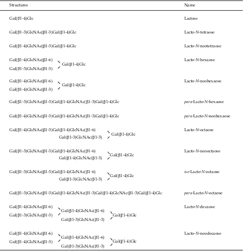
(HPLC) – chip mass spectrometry (MS) (Ninonuevo et al., 2006). The 115 human milk oligosaccharides, the structures of which have been determined to date, can be grouped into 13 series based on their core units as in Table 1 (Urashima et al., in press). The many variations of the oligosaccharides are constructed by the addition of a Neu5Acα2-3/2-6 residue to Gal or GlcNAc, and of Fucα1-2/1-3/1-4 to Gal, GlcNAc or a reducing Glc of the core units.
The main structural features of human milk oligo-saccharides are the presence of oligooligo-saccharides contain-ing the type I unit (Gal(β1-3)GlcNAc), as well as those containing the type II unit (Gal(β1-4)GlcNAc). The milk
oligosaccharides of other species mostly have the type II but not the type I unit (Urashima et al., 2001; Urashima et al., 2007). The many varieties of oligosaccharides in hu-man milk and colostrum are produced by the addition of Neu5Ac and/or Fuc residues to these two units.
The great variety of oligosaccharides in human milk depends on the activity of many diff erent spe-cifi c enzymes in the lactating gland (Urashima et al., in press). For example, a α1-2-fucosyltransferase is expressed in about 77% of all Caucasian women; these are classifi ed as secretors. Therefore, the oligosaccha-rides in the milk from these women are characterized by the presence of α1-2-fucosylated components, e.g.,
Gal( 1-4)Glc
Gal( 1-3)GlcNAc( 1-3)Gal( 1-4)Glc
Gal( 1-4)GlcNAc( 1-3)Gal( 1-4)Glc
Gal( 1-4)GlcNAc( 1-6)
Gal( 1-3)GlcNAc( 1-3) Gal( 1-4)Glc
Gal( 1-4)GlcNAc( 1-6)
Gal( 1-4)GlcNAc( 1-3) Gal( 1-4)Glc
Gal( 1-3)GlcNAc( 1-3)Gal( 1-4)GlcNAc( 1-3)Gal( 1-4)Glc
Gal( 1-4)GlcNAc( 1-3)Gal( 1-4)GlcNAc( 1-3)Gal( 1-4)Glc
Gal( 1-4)GlcNAc( 1-3)Gal( 1-4)GlcNAc( 1-6)
Gal( 1-3)GlcNAc( 1-3) Gal( 1-4)Glc
Gal( 1-4)GlcNAc( 1-3) Gal( 1-3)GlcNAc( 1-3)Gal( 1-4)GlcNAc( 1-6)
Gal( 1-4)Glc
Gal( 1-3)GlcNAc( 1-3)Gal( 1-4)GlcNAc( 1-6)
Gal( 1-3)GlcNAc( 1-3) Gal( 1-4)Glc
Gal( 1-3)GlcNAc( 1-3)Gal( 1-4)GlcNAc( 1-3)Gal( 1-4)GlcNAc( 1-3)Gal( 1-4)Glc
Lactose
Lacto-N-tetraose
Lacto-N-neotetraose
Lacto-N-hexaose
Lacto-N-neohexaose
para-Lacto-N-hexaose
para-Lacto-N-neohexaose
Lacto-N-octaose
Lacto-N-neooctaose
iso-Lacto-N-octaose
para-Lacto-N-octaose
Gal( 1-4)Glc Gal( 1-4)GlcNAc( 1-6)
Gal( 1-3)GlcNAc( 1-3) Gal( 1-4)GlcNAc( 1-6)
Gal( 1-3)GlcNAc( 1-3)
Lacto-N-decaose
Gal( 1-4)GlcNAc( 1-6)
Gal( 1-3)GlcNAc( 1-3)
Gal( 1-4)Glc Gal( 1-4)GlcNAc( 1-6)
Gal( 1-4)GlcNAc( 1-3)
Lacto-N-neodecaose
Structures Name
2)Gal(β1-4)Glc (2’-fucosyllactose: 2’-FL), Fuc(α1-2)Gal(β1-3)GlcNAc(β1-3)Gal(β1-4)Glc(lacto-N -fucopen-taose I : LNFP I), or more complex oligosaccharides, all possessing Fuc(α1-2)Gal(β1-3)GlcNAc residues. In the mammary glands of Lewis (a+b-) individuals, who produce nonsecretor milk, this enzyme activity is absent; as a result, the major fucosylated oligosaccha-ride is Gal(β1-3)[Fuc(α1-4)]GlcNAc(β1-3)Gal(β1-4)Glc (lacto-N-fucopentaose II : LNFP II). This pa ern of HMO is found in about 20% of the population. In about 5% of the population who belong to blood group Lewis (a-b-), LNFP II is absent from the milk because of the absence of α1-4-fucosyltransferase activity (Urashima et al., in press).
The studies on the quantifi cation of each of the oligosaccharides, in terms of their concentration in hu-man colostrum and mature milk, have only recently been undertaken. Milk oligosaccharides can be quanti-fi ed using reverse – phase or normal – phase HPLC subsequent to pre- or post – column labeling techniques. Derivatizations are often performed by condensation with 2-aminopyridine (PA) (Asakuma et al., 2008), 1-methyl-3-phenyl-5-pyrazolone (PMP) (Asakuma et al., 2007, 2008), anthranilic acid (Leo et al., 2009, 2010), or benzoic anhydride (Chaturvedi et al., 2001) to the reducing end of the sugar aldehyde. They can also be quantifi ed by high pH anion exchange chromatography (HPEAC-PAD) without derivatization (Thurl et al., 1996; Coppa et al., 1999; Kunz et al., 1996). Sialyl oligosaccha-rides can be quantifi ed by capillary electrophoresis using phosphate buff er containing sodium dodesyl sulfoxide (SDS) (Shen et al., 2000; Bao et al., 2007).
When the concentrations of representative neu-tral HMO including 2’-FL, Gal(β1-4)[Fuc(α1-3)]Glc (3-FL), LDFT, LNT, LNnT, I, II, LNFP-III, LNDFH-I or LNDFH-II and of representative acidic HMO including Neu5Ac(α2-6)Gal(β1-4)Glc (6’-SL), 3)Gal(β1-4)Glc (3’-SL), Neu5Ac(α2-3)3)GlcNAc(β1-3)4)Glc (LST-a), Gal(β1-3)[Neu5Ac(α2-6)]GlcNAc(β1-3)Gal(β1-4)Glc(LST-b), Neu5Ac(α2-6)Gal(β1-4)GlcNAc(β1-3)Gal(β1-4)Glc (LST-c), Neu5Ac(α2-3)Gal(β1-3)[Neu5Ac(α2-6)]GlcNAc(β1-3)Gal(β1-4)Glc (DSLNT), Fuc(α1-2)Gal(β1-3)[Neu5Ac(α2-6)]GlcNAc(β1-3)Gal(β1-4)Glc(sLNFP-I) and Neu5Ac (α2-3)Gal(β1-3)[Fuc(α1-4)]GlcNAc(β1-3)Gal(β1-4)Glc (sLNFP-II) were quantifi ed in the colostra of Japanese women, the predominant oligosaccharides were shown to be 2’-FL, LNFP-I, LNDFH-I and LNT, in that order (Asakuma et al., 2007, 2008). Assuming that the total oligosaccharide concentration in colostrum was 22~24 g/l (Newburg & Neubauer, 1995), then these four oligosaccharides can be calculated to constitute 25% to 33% of the total. It has been shown that the above four HMO are also predominant oligosaccharides in human mature milk (Chaturvedi et al., 2001; Thurl et al., 1996). The concentrations of each of the acidic oligosaccharides in colostra or in mature milk were lower than those of the four neutral oligosaccharides; the most predominant acidic oligosaccharide was LST-c, followed by DSLNT, 6’-SL, 3’-SL and LST-a in that order (Thurl et al., 1996).
It is notable that three/fourth of the prominent oligosaccharides in human milk or colostra are of type I,
which contain Gal(β1-3)GlcNAc (lacto-N-biose I) at their non-reducing termini (Asakuma et al., 2008). The con-centration of LNT, a type I oligosaccharide, was found to be 3 to 4 times higher than that of LNnT, which is a type II oligosaccharide containing the Gal(β1-4)GlcNAc (N-acetyllactosamine) group at its non-reducing termi-nus (Asakuma et al., 2008). Urashima et al. (2001, 2007) have been characterizing the milk oligosaccharides of many mammalian species; they have found that the milk or colostra of most of these species contain only type II oligosaccharides, notable exceptions being chim-panzee, bonobo and orangutan whose milk or colostrum contains both types I and II. However, type II oligosac-charides predominate over type I in the milk or colostra of these apes (Urashima et al., 2009), suggesting that the predominance of type I milk oligosaccharides is a char-acteristic feature of human milk.
DIGESTION AND ABSORPTION OF MILK OLIGOSACCHARIDES IN THE
GASTROINTESTINAL TRACT
When infants consume milk, the free lactose therein is split into galactose and glucose by intestinal lactase (neutral β-galactosidase, lactose phlorizin hydrolase), an enzyme that is located in the membrane of the microvilli of the brush border of the small intestine. The two monosaccharides are transported into the enterocytes by a specifi c mechanism, whereupon the glucose enters the circulation and is used as an energy source while most of the galactose is converted to glucose in the liver, to be used as an energy source as well.
Much less is known about the exact metabolic fate of human milk oligosaccharides. They are resistant to en-zymatic hydrolysis by the intestinal lactase of the brush border (Engfer et al., 2000), and there is evidence that the major part of oligosaccharides passes through the small intestine without degradation and enters the colon, where they are fermented by colonic bacteria (Brand-Miller et al., 1998; Newburg, 2000). Evidently, the brush border of the small intestine does not contain enzymes, such as sialidase, fucosidase or N-acetylhexosaminidase that can remove sialic acid, fucose, N-acetylglucosamine residues, respectively, from the lactose or other core units of the milk oligosaccharides. A small fraction of human milk oligosaccharides is absorbed intact, perhaps by receptor-mediated endocytosis (Gnoth et al., 2001), some of which are excreted in the urine. It is unclear what proportion and exactly which of the consumed milk oligosaccharides are absorbed, but there is an evi-dence suggesting that circulating oligosaccharides may have immunological eff ects on endothelial cells (Rudloff et al., 1996).
BIOLOGICAL SIGNIFICANCES OF HUMAN MILK OLIGOSACCHARIDES
Bifi dobacterium Growth Stimulation
bo le fed infants even when prebiotic oligosaccharides such as lactulose, galacto oligosaccharides or fructo oligosaccharides are present in the milk replacer (Rotimi & Duerden, 1981; Benno & Mitsuoka, 1986). It was suggested that milk components stimulate the growth of colonic bifi dobacteria. As mentioned above, human milk oligosaccharides are believed to act as prebiotics that stimulate the growth of bifi dobacteria in the lower intestine; this reduces the colonic pH, which would tend to inhibit the growth of pathogenic bacteria such as E. coli.
Which of the more than 100 diff erent human milk oligosaccharides do stimulate the growth of the infant’s colonic bifi dobacteria? Kitaoka et al. (2005) have proposed that milk oligosaccharides which contain lacto-N-biose I (LNB, Gal(β1-3)GlcNAc) are preferential growth factors for some species of Bifi dobacterium. This hypothesis is based on the published genome information relating to bifi dobacteria and the cloning of glycohydrolases, a glycophosphorylase and an ABC transporter from a strain of B. longum. The hypothesis was initially based on the discovery of an enzyme, in cell extracts of B. bifi dum, that phosphorolyses lacto-N-biose, the products being galactose-1-phosphate and N-acetyl-glucosamine (Derensy-Dron et al., 1999): this enzyme was subsequently purifi ed, and its gene, lnp A, from B. longum was cloned (Kitaoka et al., 2005). The lnp A gene was found to be located in a novel operon for galactose metabolism which also includes genes for a mucin desul-fatase and for Gal-1-P uridyl transferase and UDP-Glc-4-epimerase as well as a gene for an ATP-binding casse e sugar transporter (Kitaoka et al., 2005). In addition it was demonstrated that lacto-N-biose phosphorylase can also utilise galacto-N-biose (GNB, Gal(β1-3)GalNAc), which is a mucin core unit, as a substrate, but not N-acetyl-lactosamine (LacNAc, Gal(β1-4)GlcNAc) (Kitaoka et al., 2005).
The high content and increase in the concentrations of LNB containing oligosaccharides such as LNT, LNFP I and LNDFH I in early stage milk/colostrum could aff ect the formation of bifi dus fl ora in the infant colon signifi -cantly. The metabolism of LNFP I, which is a dominant human milk oligosaccharide, by B. bifi dum JCM1254 was clarifi ed by the cloning of an α-fucosidase (Katayama et al., 2004), which catalyses the release of non reducing fucose from LNFP I, and a lacto-N-biosidase (Wada et al., 2008), which catalyses the hydrolysis of LNT to produce LNB and lactose, and by the purifi cation and crystallization of an ABC type transporter (Suzuki et al., 2008) which delivers LNB through the cell membrane. Since lacto-N-biose phosphorylase was purifi ed from an extract of this bifi dobacterial strain, it has been sug-gested that its α-fucosidase produces LNT from LNFP I, while its lacto-N-biosidase liberates LNB from LNT, which is followed by the uptake of LNB through the cell membrane, the LNB is then metabolized to galactose-1-phosphate and N-acetylglucosamine. A possible meta-bolic pathway for the N-acetylglucosamine, produced from LNFP I or LNT (see above) is suggested, based on the analysis of gene products of the galactose operon of B. longum ssp. longum NCC2705; N-acetylglucosamine is converted to N-acetylglucosamine-1-phosphate by the
novel enzyme N-acetylhexosamine 1-kinase and then to UDP-N-acetylglucosamine by UDP-glucose hexose 1-phosphate uridyltransferase, fi nally entering the meta-bolic pathway of amino sugars (Nishimoto & Kitaoka, 2007).
On the other hand, growth tests with B. longum ssp infantis ATCC 15697, B. longum ssp. longum ATCC 15707, B. adolescentis ATCC 15703, B. breve ATCC 27539 or B. bifi dum ATCC 29521 were performed using a medium containing 1% human milk oligosaccharides but no lactose, which was incubated at 37˚C for 350 hr (Ward et al., 2007). This resulted in a cell density of B. longum ssp. infantis strains that was three times higher than that of other strains. When oligosaccharide and monosac-charide profi les in the medium containing human milk oligosaccharides were studied using high performance thin layer chromatography, a clear change was observed after incubation with B. longum ssp. infantis, when com-pared with the control, which was the medium with no inoculation. This change was not found in the medium after incubation with B. breve and adolenscentis. These re-sults showed that B. longum ssp. infantis is able to grow with human milk oligosaccharides as the only carbon source.
The degradation of human milk oligosaccharides was studied during the growth of B. longum ssp. infantis ATCC15697, B. breve ATCC15700 or B. longum ssp. lon-gum DJO010A in broth containing oligosaccharides but no lactose by observation of the molecular mass of ions in FTICR-MS (LoCascio et al., 2007). In this work, tri to heptasaccharides were completely degraded during the growth of B. longum ssp. infantis ATCC 15697 over 25 and 50 hr of incubation, while higher oligosaccharides were partially degraded.
The genome of B. longum ssp. infantis ATCC15697 was fully sequenced to permit investigations into the genetic basis and molecular mechanisms underlying this phenotype. Many of its genomic features encode enzymes that are active on human milk oligosaccharides and include a novel 43 kbp region dedicated to oligosac-charide utilization (Sela et al., 2008). Subsequent bio-chemical and molecular characterization of human milk oligosaccharide – related glycosidases and transport proteins have further resolved the mechanism by which this strain imports and catabolizes milk oligosaccha-rides. The glycosidases were identifi ed to be sialidase, fucosidase, β-galactosidase, β-hexosaminidase and LNB phosphorylase, but lacto-N-biosidase was not found. This approach suggested the existence of a pathway for the metabolism of milk oligosaccharides in which they are transported into the bifi dobacterial cell and then degraded from the non-reducing end by the above glycosidases.
Anti-pathogenic Agents
sugar chains of glycoconjugates, which are found on the surface of epithelial cells. It was known that infection by many bacteria and viruses starts by binding to particular sugar chains of glycoconjugates on the surface of cells of the mucous epithelium of the digestive and respiratory tracts. Therefore, human milk oligosaccharides might be useful for elucidating the structures of the sugar chains on the surface of epithelial cells that are the targets of specifi c bacteria or viruses.
Ruiz-Palacios et al. (2003) found that HMO contain-ing Fuc(α1-2)Gal(β1-4)GlcNAc (H-2 antigen epitope) at their non-reducing termini inhibit the a achment of Campylobacter jejuni to colonic epithelial cells. This organism causes diarrhea and paralysis of motor nerves in infants. A universal correlation has been observed between the breast milk concentration of 2’-FL, which is usually the most prominent oligosaccharide in secretor donor’s milk, and the frequency of diarrhea in breast fed infants, supporting the view that 2’-FL reduces the pathogenicity of C. jejuni.
An interaction of Helicobacter pylori with sialylated glycans has been reported. H. pylori is a Gram-negative bacterium, resides in the gastric mucosa and adheres to the epithelial cells lining the stomach. This organ-ism infects around 50% of the world population, with a higher incidence in developing countries. H. pylori is associated with the development of peptic ulcers, mu-cosa-associated lymphoid-tissue (MALT) lymphoma and gastric adenocarcinoma. The preferred interaction is with α3-linked sialic acid; glycans having α6-linked Neu5Ac are non-binding. For example, 50% inhibition by H. pylori of hemagglutination of human erythrocytes was observed at a low concentration of some sialylated saccharides. The data showed that S-3 PG (3)Gal(β1-4)GlcNAc(β1-3)Gal(β1-4)Glc-Cer), Neu5Ac(α2-3)Gal(β1-4)[Fuc(α1-3)]GlcNAc, 3’-N -acetylneuraminyl-N-acetyllactosamine (Neu5Ac(α2-3)Gal(β1-4)GlcNAc) as well as 3-N-acetylneuraminyl-lacto-N-neotetraose (Neu5Ac(α2-3)Gal(β1-4)GlcNAc(β1-3)Gal(β1-4)Glc) all bound to H. pylori CCUG17874 at similar strength. 3’-SL also bound to this organism but its binding ability was somewhat weaker than that of the above saccharides. It has also been reported that LST a, a human milk oligo-saccharide, was able to bind to another strain, H. pylori J99 (Johansson et al., 2005).
The binding of 3’-SL to H. pylori CCUG17874 is noteworthy because this saccharide is found in human milk and bovine colostrum. Asakuma et al. (2007) found that at the start of lactation the concentration of 3’-SL in human colostrum was 360 mg/L, similar to that of 6’-SL. However, the concentration of 3’-SL decreased during the subsequent two days of lactation, whereas that of 6’-SL did not. This suggests that, very early stage in lac-tation, 3’-SL may be more signifi cant in the prevention of transmission of H. pylori from mother to infant than later on.
Recent studies on the ability of various fractions of human milk oligosaccharides to inhibit the adhesion of three intestinal microorganisms (enteropathogenic E. coli serotype 0119, Vibrio cholerae and Salmonella fyris) to diff erentiated Caco-2 cells have shown that the acidic fraction had an anti-adhesive eff ect on all three
patho-genic strains. The neutral high molecular weight fraction signifi cantly inhibited the adhesion of E. coli 0119 and V. cholerae, but not that of S. fyris; the neutral low molecular weight fraction was eff ective toward E. coli 0119 and S. fyris but not V. cholerae (Coppa et al., 2006). This demon-strated that human milk oligosaccharides inhibit the ad-hesion to epithelial cells not only of common pathogens such as E. coli but also of other aggressive bacteria such as V. choleraeand S. fyris. Thus, oligosaccharides may be important factors in human milk that defend against acute diarrhea in breast-fed infants.
Furthermore, it has been reported that the human milk oligosaccharides fraction, at a concentration of 0.5 g/L, reduced the binding of the HIV-1 envelope glyco-protein gp120 to dendritic cell – specifi c ICAM3 – grab-bing non – integrin (DC-SIGN) in human dendritic cells by more than 60%. In addition, the binding of gp120 to Raji cells, which expressed DC-SIGN, was reduced by more than 60%. It is worth noting that mother to child transmission accounts for more than 40% of all HIV-1 infections in children, with breast-feeding being the predominant postnatal transmission route, especially in developing countries. However, a majority of breast-fed infants born to HIV-positive mothers remain uninfected despite continuous exposure to the virus over many months. Viral entry across the infant’s mucosal barrier is partially mediated by binding of gp120 to DC-SIGN on human dendritic cells (DC). It has been hypothesized that human milk oligosaccharides, when they reach DC in the colon, bind to DC-SIGN and inhibit the transfer of HIV to CD4 + T lymphocytes. It has also been suggested that this inhibition of HIV-gp120 binding to DC-SIGN is caused by oligosaccharides carrying multiple Lewis epitopes (Hong et al., 2009).
Immuno – modulating Eff ect
It is thought that the immuno – modulating eff ect of human milk oligosaccharides in infants is acquired while the oligosaccharides are circulating after their absorption, or by immuno stimulation in the colon produced by growth-stimulated benefi cial bacteria. Although unequivocal detection of human milk oli-gosaccharides in the blood of infants has not yet been reported (Bode, 2006), it nevertheless seems very likely on the basis of their observed urinary excretion (Rudloff et al., 1996; Obermeier et al., 1999) that small amounts of intact milk oligosaccharides are normally absorbed from the gastrointestinal tract, and that they are transported into the systemic circulation.
in-fl ammation. P-selectin is also involved in the formation of platelet-neutrophil complexes (PNC), a sub-popula-tion of highly activated neutrophils primed for adhe-sion, phagocytosis and enhanced production of reactive oxygen species. Recent studies suggest that oligosaccha-rides containing sialyl Lex or its stereoisomer sialyl Lea, which resemble the P-selectin ligand, inhibit the binding of selectin ligands to the surface of endothelial cells and platelets; this interferes with the formation of PNC, the eff ect of which is anti-infl ammatory. The following oligosaccharide fractions were tested in vitro to establish whether they reduce leukocyte deceleration on U937 cells, which express the P-selectin ligand: total human milk oligosaccharides, neutral oligosaccharides, total acidic oligosaccharides, neutral oligosaccharides with a polymerization degree of 4, fucosylated oligosaccharides and disialyl lacto-N-tetraose. The acidic oligosaccharide fraction produced a slight but defi nite reduction of P-selectin ligand binding, similar to that of standard sialyl Lex, whereas the total neutral oligosaccharides and neutral fucosylated oligosaccharides fractions did not (Schumacher et al., 2006). These results support the notion of anti-infl ammatory eff ects of acidic human milk oligosaccharides.
MILK OLIGOSACCHARIDES OF THE COW AND OTHER DOMESTIC FARM ANIMALS AND THEIR
UTILIZATION IN THE FUTURE INDUSTRY
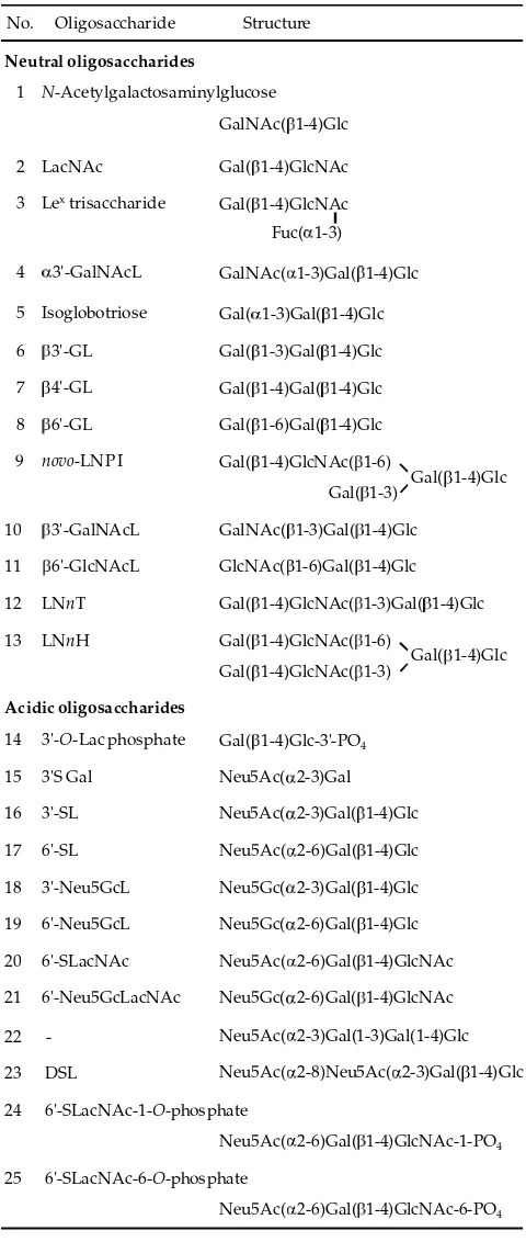
Nakamura et al. (2003) and Gopal & Gill (2000) stated that bovine colostrum collected immediately after parturition contains more than 1 g/L of oligosaccharides, but the mature milk contains only small amounts. Although 25 bovine oligosaccharides structures isolated from the colostrum have been completely characterized (Table 2), as many as 39 oligosaccharides have been de-tected using a combination of nanoelectrospray Fourier transform ion cyclotron resonance (nESI-FTICR) mass spectrometry and matrix-assisted laser desorption/ ionization Fourier transform ion cyclotron resonance (MALDI-FTICR) mass spectrometry (Tao et al., 2008). Some bovine oligosaccharides include those that have N -acetyllactosamine (Gal(β1-4)GlcNAc) instead of lactose at their reducing ends, such as free N-acetyllactosamine or 6’-N-acetylneuraminyl-N-acetyllactosamine (6’-SLN, Neu5Ac(α2-6)Gal(β1-4)GlcNAc) and others. There is a diff erence in this respect between bovine and human milk oligosaccharides, insofar as almost all HMO con-tain a lactose unit at their reducing ends.
Most of the acidic oligosaccharide fraction of bo-vine colostrum consists of 3’-SL, 6’-SL, 6’-SLN and DSL (Neu5Ac(α2-8)Neu5Ac(α2-3)Gal(β1-4)Glc), with 3’-SL constituting 70% of this total (Tao et al., 2008). There is a diff erence between human and cow in the ratio of 3’-SL to 6’-SL, in that 6’-SL predominates over 3’-SL in human milk/colostrum. The levels of the acidic oligosaccharides were maximal immediately after parturition, rapidly decreasing by 48 h post-partum (Nakamura et al., 2003).
Since bovine colostrum contains some oligosaccha-rides, which are mainly acidic, and mature bovine milk also contains them, although at lower concentrations, it can be expected that oligosaccharides derived from
bovine colostrum or mature milk will be utilized by industry as functional foods, animal feeds or biomedical products. For example, several reports have suggested that bovine milk oligosaccharides can be used as anti infection materials.
It is thought that adhesion of Neisseria meningitides, a human-specifi c pathogen causing meningitis and speticemia, is mediated by type IV pili (Hakkarainen et al., 2005). A mictotiter well pili-binding assay was used Table 2. Bovine milk oligosaccharides
No. Oligosaccharide
Neutral oligosaccharides
Structure
1 N-Acetylgalactosaminylglucose GalNAc( 1-4)Glc
2 LacNAc Gal( 1-4)GlcNAc
3 Lextrisaccharide Gal( 1-4)GlcNAc
4 3'-GalNAcL GalNAc( 1-3)Gal( 1-4)Glc Fuc( 1-3)
5 Isoglobotriose Gal( 1-3)Gal( 1-4)Glc 6 3'-GL Gal( 1-3)Gal( 1-4)Glc 7 4'-GL Gal( 1-4)Gal( 1-4)Glc 8 6'-GL Gal( 1-6)Gal( 1-4)Glc
novo-LNP I
9 Gal( 1-4)GlcNAc( 1-6)
Gal( 1-4)Glc Gal( 1-3)
10 3'-GalNAcL GalNAc( 1-3)Gal( 1-4)Glc
Acidic oligosaccharides
14 3'-O-Lac phosphate Gal( 1-4)Glc-3'-PO4
15 3'S Gal Neu5Ac( 2-3)Gal
16 3'-SL Neu5Ac( 2-3)Gal( 1-4)Glc 17 6'-SL Neu5Ac( 2-6)Gal( 1-4)Glc 18 3'-Neu5GcL Neu5Gc( 2-3)Gal( 1-4)Glc 19 6'-Neu5GcL Neu5Gc( 2-6)Gal( 1-4)Glc 20 6'-SLacNAc Neu5Ac( 2-6)Gal( 1-4)GlcNAc 21 6'-Neu5GcLacNAc Neu5Gc( 2-6)Gal( 1-4)GlcNAc 11 6'-GlcNAcL GlcNAc( 1-6)Gal( 1-4)Glc 12 LNnT Gal( 1-4)GlcNAc( 1-3)Gal( 1-4)Glc 13 LNnH Gal( 1-4)GlcNAc( 1-6)
Gal( 1-4)GlcNAc( 1-3) Gal( 1-4)Glc
22 23 DSL
24 6'-SLacNAc-1-O-phosphate
25 6'-SLacNAc-6-O-phosphate
Neu5Ac( 2-3)Gal(1-3)Gal(1-4)Glc Neu5Ac( 2-8)Neu5Ac( 2-3)Gal( 1-4)Glc
Neu5Ac( 2-6)Gal( 1-4)GlcNAc-1-PO4
-to investigate the binding of type IV pili isolated from N. meningitides to diff erent glycoproteins. Inhibition of pili binding to bovine thyroglobulin and human salivary ag-glutinin by fractionated human and bovine milk oligo-saccharides was demonstrated. The binding of Neisseria pili to bovine thyroglobulin was more eff ective and was clearly inhibited by neutral or acidic bovine milk oligosaccharides at concentrations of 1~2 g/l, suggesting that these fractions had the potential ability to inhibit the a achment of this bacterium to the colonic mucosa (Hakkarainen et al., 2005).
Fractions containing milk oligosaccharides, in the form of supernatants that had been separated from co-lostrum and from transitional, mature and late lactation milk of Spanish brown cows by ethanol precipitation and subsequent centrifugation, were used to investigate the inhibition of hemagglutination by seven enterotoxi-genic E. coli strains (K99, FK, F41, F17, B16, B23 and B64). These strains had been isolated from diarrheal calves. The fractions from the transitional and mature milk in-hibited hemagglutination by all of these strains, whereas those from colostrum and late lactation milk produced weaker inhibition (Martin et al., 2002). It was suggested that this inhibition was due to 3’-SL, 6’-SL, 6’-SLN and DSL. The fractions from transitional and mature milk, in which the ratio of 6’-SL to 3’-SL was higher than in the fractions from colostrum and late lactation milk, had a stronger eff ect than the others
It can be expected that milk oligosaccharides of other domestic farm animals, such as goats, sheep, and camels, will also be used as biofunctional materials. The milk oligosaccharide content of goat milk is 0.25~0.39 g/l; this is higher than that of bovine (0.03~0.06 g/l) or ovine (0.02~0.04 g/l) milk. In addition, the variety of oligosaccharides in goat milk is greater than that in bovine or ovine milk, as shown by the profi les from HPEAC analysis (Martinez-Ferez et al., 2006; Mehra & Kelly, 2006). Colostrum from the Japanese Saanen breed contains more 6’-SL than 3’-SL; it also contains 6’-N-glycolylneuraminyllactose (Neu5Gc(α2-6)Gal(β1-4)Glc), Gal(α1-3)Gal(β1-4)Glc, Gal(β1-3)Gal(β1-4)Glc, Gal(β1-6)Gal(β1-4)Glc and 2’-FL (Urashima et al., 1994, 1997). Another study has shown that mature milk from Spanish goats contains 6’-SL, 3’-SL, disialyllactose, N -glycolylneuraminyllactose, 3’-galactosyllactose, N-acetyl glucosaminyllactose, LNH and additional high molecu-lar oligosaccharides, as demonstrated by analysis with FAB-MS, but no fucosyl oligosaccharides (Martinez-Ferez et al., 2006). Ovine colostrum contains more 3’-N-glycolylneuraminyllactose than 3’-SL and 6’-SL (Nakamura et al., 1998) and, notably, contains Neu5Gc in preference to Neu5Ac.
A recent study on rats showed that goat milk oligo-saccharides have an anti-infl ammatory eff ect in the colon (Daddaowa et al., 2006). In this study, colitis was induced by the hapten, trinitrobenzenesulfonic acid (TNBS). The experimental rats (OS) were fed a diet containing 500 mg/kg per day of goat milk oligosaccharides, from 2 days prior to the induction until day 6, after which all the rats were weighed and then killed, the entire colon was removed, opened and scored for visible damage and then divided into several pieces for biochemical
determinations. When the OS rats were compared with control rats in which colitis had been induced by TNBS but that had not been treated with oligosaccharides, it was found that the OS rats showed decreased anorexia, reduced loss of body weight, reduced bowel wall thick-ening and less necrosis of the colon. Biochemically, the OS rats had lower colonic levels of inducible nitric oxide synthase (iNOS), cyclooxygenase 2 (COX2), interleukin-1β and mucin 3, as well as increased trefoil factor 3. These results showed that goat milk oligosaccharides are anti-infl ammatory when administered as a pretreat-ment in the TNBS model of rat colitis, most likely due to their action as prebiotics resulting in favorable changes in the colonic bacterial fl ora. Since TNBS-induced colitis is widely used as a preclinical model of infl am-matory bowel disease in humans, it was suggested that goat milk oligosaccharides might be useful in the man-agement of this disease.
CONCLUDING REMARKS
As the development and improvement of methods for structural determination by using smaller amount of samples as presently required still going on, the new structure of milk oligosaccharides will be still discovered. More studies on the functional biological characteristics of the milk oligosaccharides are still needed. Those study results are needed to confi rm milk oligosaccharides role in the various biological processes, as well as to overcome the problems especially in the synthesis of milk oligosaccharides-like compound at the industrial level. The isolation of milk oligosaccharides from the colostrum of domestic animals at industrial scale, for their utilization as biofunctional food and feedstuff s as well as for drugs is still in the early stage. Although still in its infancy, milk oligosaccharides industrial utilization can be expected in the near future.
REFERENCES
Asakuma, S., M. Akahori, K. Kimura, Y. Watanabe, T. Nakamura, M. Tsunemi, I. Arai, Y. Sanai, & T. Urashima. 2007. Sialyl oligosaccharides of human
colos-trum: Changes in concentration during the fi rst three days
of lactation. Biosci. Biotech. Biochem. 71: 1447-1451. Asakuma, S., T. Urashima, M. Akahori, M. Ohbayashi, T.
Nakamura, K. Kimura, Y. Watanabe, I. Arai, & Y. Sanai. 2008. Variation in neutral oligosaccharide levels in human colostrums. Eur. J. Clin. Nutr. 62: 488-494.
Bao, Y., L. Zhu, & D. S. Newburg. 2007. Simultaneous
quan-tifi cation of sialyloligosaccharides from human milk by
capillary electrophoresis. Anal. Biochem. 370: 206-214. Benno, Y. & T. Mitsuoka. 1986. The development of
gastroin-testinal micro-fl ora in humans and animals. Bifi dofact. Microfl ora 5: 13-25.
Bode, L., S. Rudloff , C. Kunz, S. Strobel, & N. Klein. 2004. Human milk oligosaccharides reduce platelet-neutrophil
complex formation leading to a decrease in neutropil β2
integrin expression. J. Leukoc Biol. 76: 820-826.
Bode, L. 2006. Recent advances on structure, metabolism and function of human milk oligosaccharides. J. Nutr. 136: 2127-2130.
Pediatr. 133: 95-98.
Chaturvedi, P., C. D. Warren, M. Altaye, A. L. Morrow, G. Ruiz-Palacios, L. K. Pickering, & D. S. Newburg. 2001. Fucosylated human milk oligosaccharides vary between individuals and over the course of lactation. Glycobiology 11: 365-370.
Coppa, G. V., P. Pierani, L. Zampini, I. Carloni, A. Carlucci, C. Catacci, & O. Gabrielli. 1999. Oligosaccharides in human
milk during diff erent phases of lactation. Acta Paediat.
Suppl. 430: 89-94.
Coppa, G. V., L. Zampini, T. Galeazzi, B. Facinelli, L. Ferrante,
R. Carpe i, & G. Orazio. 2006. Human milk oligosac-charides inhibit the adhesion to caco-2 cells of diarrhea pathogens: Escherichia coli, Vibrio cholerae, and Salmonella fyris. Pediatr. Res. 59: 377-382.
Daddaowa, A., V. Puerta, P. Requena, A. Martinez-Ferez, E. Guadix, Sanchez de Mediza, A. Zarzuelo, M. Dolerez Svarez, J. Josa Boya, & O. Martinez-Augustin. 2006. Goat
milk oligosaccharides are anti-infl ammatory in rats with
hapten-induced colitis. J. Nutr. 136: 672-675.
Derensy-Dron, D., F. Krzewinski, C. Brassart, & S. Bouquelet.
1999. β-1,3-Galactosyl-N-acetylhexosamine phosphorylase from Bifi dobacterium bifi dum DSM 20082: characterization,
partial purifi cation and relation to mucin degradation.
Biotechnol. Appl. Biochem.29: 3-10.
Engfer, M. B., B. Stahl, B. Finke, G. Sawa ki, & H. Daniel.
2000. Human milk oligosaccharides are resistant to enzy-matic hydrolysis in the upper gastrointestinal tract. Am. J. Clin. Nutr. 71: 1589-1596.
Gnoth, M. J., S. Rudloff , C. Kunz, & R. K. H. Kinne. 2001. Investigations of the in vitro transport of human milk oligosaccharides by Caco-2 monolayer using a novel high performance liquid chromatography-mass spectrometry technique. J. Biol. Chem. 276: 34363-34370.
Gopal, P. K. & H. S. Gill. 2000. Oligosaccharides and glyco-conjugates in bovine milk and colostrums. Br. J. Nutr. 84: S69-S74
Hakkarainen, J., M. Toivanen, A. Leinonen, L. Frangsmyr, N. Stronberg, S. Lapinjoki, X. Nassif, & C. Tikkanen-Kaukanen. 2005. Human and bovine milk oligosaccha-rides inhibit Neisseria meningitidis pili a achment in vitro, J. Nutr. 135: 2445-2448.
Hong, P., M. R. Ninonuevo, B. Lee, C. Lebrilla, & L. Bode. 2009. Human milk oligosaccharides reduce HIV-1-gp120
binding to dendritic cell-specifi c ICAM3-grabbing
non-integrin (DC-SIGN). Br. J. Nutr. 101: 482-486.
Johansson, P., J. Nilsson, J. Angstrom, & H. Miller-Pandraza. 2005. Interaction of Helicobacter pylori with sialylated
car-bohydrates: the dependence on diff erent parts of the bind-ing trisaccharide Neu5Acα3Galβ4GlcNAc. Glycobiology
15: 625-636.
Katayama, T., A. Sakuma, T. Kimura, Y. Makimura, J. Hiratake, K. Sakata,, T. Yamanoi, H. Kumagai, & K. Yamamoto. 2004. Molecular cloning and characterization
of Bifi dobacterium bifi dum 1,2-α-L-fucosidase (AfcA), a
novel inverting glycosidase (glycoside hydrolase family 95). J. Bacteriol.186: 4885-4893.
Kitaoka, M., J. Tian, & M. Nishimoto. 2005. Novel putative galactose operon involving lacto-N-biose phosphorylase
in Bifi dobacterium longum. Appl. Environ. Microbiol. 71:
3158-3162.
Klein, A., A. Schwertman, M. Peters, C. Kunz, & S. Strobel.
2000. Immunomodulatory eff ects of breast milk oligo-saccharides. In: Short and Long Term Eff ects of Breast Feeding on Child Health (B. Kole ki, ed.), pp. 251-259,
Kluwer Academic/Plenum Publishers, New York.
Kunz, C., S. Rudloff , A. Hintelmann, G. Pohlen , & H. Egge.
1996. High-pH anion-exchange chromatography with pulsed amperometric detection and molar response
fac-tors of human milk oligosaccharides. J. Chromatogr. B 685: 311-221.
Kunz C, G. M. Rodriguez-Palmero, B. Kole ko, & R. Jensen.
1999. Nutritional and biochemical properties of human milk, Part I: General aspects, proteins, and carbohydrates. Clin Perinatol 26, 307–333.
Leo, F., S. Asakuma, T. Nakamura, K. Fukuda, A. Senda, & T. Urashima. 2009. Improved determination of milk oligosaccharides using a single derivatization with anthranilic acid and separation by revsersed-phase high-performance liquid chromatography. J. Chromatogr. A. 1216: 1520-1523.
Leo, F., S. Asakuma, K. Fukuda, A. Senda, & T. Urashima. 2010. Determination of sialyl and neutral oligosaccharide levels in transition and mature milks of Samoan women, using anthranilic derivatization followed by reverse phase high performance liquid chromatography. Biosci. Biotech. Biochem. 74: 298-303.
LoCascio, R. G., M. R. Ninonuevo, S. L. Freeman, D. A. Sela., R. Grimm, C. B. Lebrilla, D. A. Mills, & J. B. German.
2007. Glycoprofi ling of bifi dobacterial consumption
of human milk oligosaccharides demonstrates strain
specifi c, preferential consumption of small chain glycans
secreted in early human lactation. J. Agric. Food. Chem. 55: 8914-8919.
Martin, M. J., S. Martin-Sosa, & P. Hueso. 2002. Binding of milk oligosaccharides by several enterotoxigenic Escherichia coli strains isolated from calves. Glycoconj. J. 19: 5-11.
Martinez-Ferez, A., S. Rudloff , A. Guadix, C. A. Henkel, G. Pohlen , J. J. Boza, E. M. Guadix, & C. Kunz. 2006. Goat’s milk as a natural source of lactose-derived oligo-saccharides isolation by membrane technology. Int. Dairy J. 16: 173-181.
Mehra, R. & P. Kelly. 2006. Milk oligosaccharides: Structural and technological aspects. Int. Dairy J. 16: 1334-1340. Messer, M. & T. Urashima. 2002. Evolution of milk
oligosac-charide and lactose. Trends Glycosci. Glycotech. 14: 153-176. of sialyl oligosaccharides in bovine colostrum and milk during the prepartum and early lactation. J. Dairy Sci. 86: 1315-1320.
Newburg, D. S. & S. H. Naubauer. 1995. Carbohydrates in
milks: Analysis, quantities and signifi cance. In: Handbook
of Milk Composition (R, G. Jensen ed.), pp. 273-349, Academic Press, San Diego.
Newburg, D. S. 2000. Oligosaccharides in human milk and bacterial colonization. J. Pediatr. Gastroenterol. Nutr. 30: S8-S17. acetylhexosamine 1-kinase in the complete lacto-N-biose I/galacto-N-biose metabolic pathway in Bifi dobacterium longum. Appl. Environ. Microbiol.73: 6444-6449.
Obermeier, S., S. Rudloff , S. Pohlen , M. J. Len e, & C.
Kunz. 1999. Secretion of 13C-labelled oligosaccharides into human milk and infants urine after an oral [13C]galactose load. Isotopes Environ. Haelth Stud. 35: 119-125.
Rotimi, V. O. & B. I. Duerden. 1981. The development of the
51-58.
Rudloff , S., G. Pohlen , L. Diekmann, H. Egge, & C. Kunz.
1996. Urinary excretion of lactose and oligosaccharides in preterm infants fed human milk or infant formula. Acta Paediatr. 85: 598-603.
Ruiz-Palacios, G.M., L. E. Cervantes, P. Ramos, B. Chavez-Munguia, & D. S. Newburg. 2003. Campylobacter
jejuni binds intestinal H(O) antigen (Fuc α 1, 2Gal β 1,
4GlcNAc), and fucosyloligosaccharides of human milk inhibit its binding and infection, J. Biol. Chem. 278: 14112–14120.
Schumacher, G., G. Bendas, B. Stahl, & C. Beermann. 2006.
Human milk oligosaccharides aff ect P-selectin binding
capacities: In vitro investigation. Nutr. 22: 620-627. Sela, D. A., J. Chapman, A. Adeuya, J. H. Kim, F. Chen, T. R.
Whitehead, A. Lapidus, D. S. Rokhsar, C. B. Lebrilla, & J. B. German. 2008. The genome sequence of Bifi dobacterium
longum subsp. infantis reveals adaptations for milk utiliza-tion within the infant microbiome. Proc. Natl. Acad. Sci. U S A105: 18964-18969.
Shen, Z., C. D. Warren, & D. S. Newburg. 2000. High-perfor-mance capillary electrophoresis of sialylated oligosaccha-rides of human milk. Anal. Biochem. 279: 37-45.
Suzuki, R., J. Wada, T. Katayama, S. Fushinobu, T. Wakagi, H. Shoun, H. Sugimoto, A. Tanaka, H. Kumagai, H. Ashida, M. Kitaoka, & K. Yamamoto. 2008. Structural and thermodynamic analyses of solute-binding Protein from
Bifi dobacterium longum specifi c for core 1 disaccharide and
lacto-N-biose I. J. Biol. Chem.283: 13165-13173.
Tao, N., E. J. DePeters, S. Freeman, J. B. German, R. Grimm, & C. B. Lebrilla. 2008. Bovine milk glycome. J. Dairy Sci. 91: 3768-3778.
Thurl, S., B. Muller-Wermer, & G. Sawa ki. 1996.
Quantifi cation of individual oligosaccharide compounds
from human milk using high-pH anion exchang chroma-tography. Anal. Biochem. 235: 202-206.
Urashima, T., W. A. Bubb, M. Messer, Y. Tsuji, & Y. Taneda. 1994. Studies of the neutral trisaccharides of goat (Capra hircus) colostrum and of the one- and two-dimensional 1H
and 13C NMR spectra of 6’-N-acetylglucosaminyllactose. Carbohydr. Res. 262: 173-184.
Urashima, T., S. Murata, & T. Nakamura. 1997. Structural determination of monosialyl trisaccharide obtained from caprine colostrum. Comp. Biochem. Physiol. 116B: 431-435.
Urashima, T., T. Saito, T. Nakamura, & M. Messer. 2001. Oligosaccharides of milk and colostrum in non-human mammals. Glycoconj. J. 18: 357-371.
Urashima, T., S. Asakuma, & M. Messer. 2007. Milk oli-gosaccharides. In: J.P. Kamerling, G.-J. Boons, Y.C. Lee, A. Suzuki, N. Taniguchi & A.G.J. Voragen (eds.). Comprehensive Glycoscience. Elsevier, Amsterdam. Vol. 4, pp. 695-724.
Urashima, T., G. Odaka, S. Asakuma, Y. Uemura, K. Goto, A. Senda, T. Saito, K. Fukuda, M. Messer, & O. T. Oftedal. 2009. Chemical characterization of oligosac-charides in chimpanzee, bonobo, gorilla, orangutan, and siamang milk or colostrum. Glycobiology 19: 499–508 doi:10.1093/glycob/cwp006.
Urashima, T., M. Kitaoka, T. Terabayashi, K. Fukuda, M. Ohnishi, & A. Kobata. (in press). Milk oligosaccha-rides. In: N.S. Gordon (ed.). Oligosaccharides: Sources, Properties and Applications. Nova Science Publishers, Inc. New York. pp. 1-77.
Veh, R. W., J. C. Michalski, A. P. Corfi eld, M. Sander-Wewer,
D. Gies, & R. Schauer. 1981. New chromatographic sys-tem for the rapid analysis and preparation of colostrums sialyloligosaccharides. J Chromatogr 212: 313–322. Ward, R. E., M. Ninonuevo, D. A. Mills, C. B. Lebrilla, & J.
B. German. 2007. In vitro fermentability of human milk
oligosaccharides by several strains of bifi dobacteria. Mol.
Nutr. Food Res. 51: 1398-1405.