Research Article
Characterization of
Pantoea ananatis
Isolated from Garlic and Shallot
Karakterisasi
Pantoea ananatis
yang Diisolasi dari Bawang Putih dan Bawang Merah
Nanik Nurjanah1)
, Tri Joko 2), & Siti Subandiyah2)* 1)Agricultural Quarantine Agency Class 1 Ambon
Jln. Rurehe, Waihaong, Ambon, Maluku 97111
2) Department of Crop Protection, Faculty of Agriculture, Universitas Gadjah Mada
Jln. Flora 1, Bulaksumur, Sleman, Yogyakarta 55281
*Corresponding author. E-mail: [email protected]
ABSTRACT
The new disease on garlic (Allium sativum) and shallot (A. cepaL. aggregatumgroup) have been found in several production centers of garlic and shallot in Tawangmangu and Temanggung, Central Java. The infected plants showed symptoms of leaf blight accompanied by chlorosis. The objective of this study was to determine the pathogen that causes leaf blight and chlorosis based on the phenotypic characterization andgyrBgene sequences analysis. The research started from the isolation of pathogen, physiological and biochemical test, DNA extraction, and sequence analysis ofgyrBusinggyrB 01-F and gyrB02-R primer. The results showed that the isolated bacterial pathogen have a yellow pigment, slimy colonies with regular borders, convex, gram-negative, non-spore, facultative anaerobic, motile, catalase production, indole production, and acid production from D-glucose, D-mannitol, sucrose, and lactose. From the pathogenicity test, it was found that the bacteria produced the typical symptom of leaf blight. Characterization of pathogens based on gyrB gene sequence revealed that the pathogen was placed in the group of Pantoea ananatis.
Keywords: Alium cepa, Alium sativum, gyrB, leaf blight, Pantoea ananatis
INTISARI
Penyakit baru pada bawang putih (Allium sativum) dan bawang merah (A. cepaL. aggregatum group) telah ditemukan di beberapa sentra produksi bawang putih dan bawang merah di Tawangmangu dan Temanggung, Jawa Tengah. Tanaman yang terinfeksi menunjukkan gejala hawar daun disertai klorosis. Tujuan penelitian untuk mengetahui karakter patogen berdasarkan fenotipik dan sekuen gen gyrB. Penelitian dimulai dengan isolasi bagian tanaman yang sakit, uji fisiologi dan biokimia, ekstraksi DNA dengan metode CTAB/NaCl dan amplifikasi gen gyrBmenggunakan primer gyrB 01-F and gyrB 02-R. Hasil uji menunjukkan koloni berlendir, cembung, pigmen berwarna kuning, gram negative, tidak berspora, aerob fakultatif, motil, produksi katalase, indol, membentuk asam dari D-glukosa, D-monnitol, sukrosa dan laktosa, dan patogenesitas positif. Karakterisasi patogen berdasarkan sekuen gen gyrB, menunjukkan patogen hawar daun berkerabat dekat dengan Pantoea ananatis.
Kata kunci: Alium cepa, Alium sativum, gyrB, leaf blight, Pantoea ananatis
INTRODUCTION
Pantoea ananatisis an important bacterial plant pathogen in the world and has a very wide host range.
P. ananatiscan survive in a various ecosystem as the saprofit, endophytes, epiphytes and pathogens. P. ananatisis reported to cause center rot disease of onion, and accounted for 100% loss in some fields (Gitaitis & Gay, 1997). While maize white spot (MWS) caused by this pathogen could cause up to 60% yield loss (Miller et al., 2016).
In Indonesia, P. ananatishas never been reported to infect plants until recently, although the tropical
climate conditions in Indonesia are also suitable for the development ofP. ananatis. The risks and possibility of the pathogen to spread in Indonesia is highly considered by the import commodities from countries that have reported the presence of these bacteria. According to Carr et al.(2010), the development of center rot symptoms in onion bulbs during storage poses a significant problem. The onion is often stored for months prior to grading and marketing will result in a more severe infection. However, onion bulbs that are infected solely by P. ananatisremain firm and exhibit subtle or no external symptoms, they can be
difficult to detect on grading lines. The onion bulbs can be a source of inoculum for other plants, although the onion is not cultivated in Indonesia.
The metabolic pattern of P. ananatisfrom crop debris is similar to those recovered directly from lesions or from healthy leaves, suggesting that these bacteria in crop debris could act similarly to epiphytic isolates, being a source of inoculum for further infections. P. ananatis can survive as epiphytes in the leaves of healthy maize plants, non-host plants and in crop debris, and possibly multiply there (Sauer et al.,
2015). The bacteria infect host through flowers, mechanical injury, wound insect bite and friction injuries plants with the current crop of strong winds (Azad et al.,2000). Tobacco thrips of Frankliniella fusca are a vector of P. ananatis caused center rot in onion (Gitaitis et al.,2003).
The gyrBgene codes for the b-subunit of DNA gyrase, a type II DNA topoisomerase, which introduces negative supercoils into closed circular DNA molecules. One of the reasons why the gyrB gene is selected for phylogenetic studies is that, as horizontal gene transfer (HGT) occurs infrequently in informational genes that are involved in transcription and translation, it is assumed not to undergo HGT (Harayama & Kasai, 2006). GyrB gene sequences have been widely used for the identification of bacterial species. GyrBgene sequence is more suitable for determining genetic relationships and the identification of bacterial than the 16S rRNA (Parkinson et al.,2009; Takeda et al.,
2010).
The objectives of this study was to characterize bacterial pathogen isolated from onion and garlic based on phenotypic properties and gyrBgene sequences analyses.
MATERIALS AND METHODS
Survey and sampling were carried out based on purposive random sampling method (Sumardiyono
et al., 2011; Windari et al.,2015; Ismiyatuningsih et al., 2016). Symptomatic tissue showing leaf blight and chlorosis diseases were collected from several fields of garlic and shallot production area in Central Java.
Isolation of Bacterial Pathogen
Bacteria were isolated from the affected tissue according to Joko et al. (2011a) with slight modification. A one gram of the sample was crushed in a small tube 500 μl sterilized ddH2O. One loopful of the
suspension was streak onto YP agar (yeast extract 5 g, peptone 10 g, agar 15 g, water 1000 ml, pH 6.8) plate medium and it was incubated for 2 days at 28oC
(Wibowo et al., 2010; Wardhika et al., 2014). Yellow colonies were formed on YP agar plate, and single colonies were subcultured onto YP agar slants.
Physiological and Biochemical Characterization
The bacterial isolates were then characterized as follows (Lelliot & Stead, 1987; Joko et al., 2000; Schaad et al., 2001):
Gram reaction with 3% KOH is to differentiate bacteria based on the structure of the cell wall. Gram-negative bacterial will become gummy upon mixing with a loop, while gram-positive bacterial will not.
Catalase test is to detect the presence of catalase enzyme in bacteria that is able to hydrolyze hydrogen peroxide (H2O2) into water and oxygen. If the bacterium has a catalase enzyme, it will form gas bubbles.
Anaerobic growth test aims to determine whether the bacteria can grow in aerobic or anaerobic conditions in media containing bromotimol blue covered with sterile oil paraffin (anaerobic) and without paraffin (aerobic). A color change from blue to yellow in both tubes is recorded as positive for anaerobic growth/ fermentation.
Indole test uses Kovac’s method, that is reagents which contains hydrochloric acid and p-dimethyl aminobenzaldehyde in amyl alcohol. The Bacteria that produce the enzyme tryptophanase can convert the amino acid tryptophan to by-products that include indole. The indole which is produced was detected by adding Kovac’s reagent which produced cherry red (Aneja, 2003).
Oxidase test is to detect the presence of pathogenic bacteria cytochromeoxidase. The bacteria are streaked onto filter paper that has been containing tetramethyl-p-phenylenediamine dihydrochloride 1%, The strain was rated oxidase-positive if a purple color develops within 10s, delayed positive if coloration develops within 10-60s and negative if no color develops after 60 seconds.
acid in 150ml of 30% acetic acid). The color change occurs in the culture of bacteria, nitrate is reduced to form a perfect nitrogen gas (N2) or ammonium gas (NH3), a positive reaction happens when the red color is formed about 30 minutes and medium cracks. Arginine dihydrolase test is to detect the condition of the growth of anaerobic bacteria in Thornley media (arginine medium with phenol red dye), and covered with liquid paraffin to create anaerobic conditions and incubated at 28oC for 4 days. The
positive reaction was occurred when there was a change in the medium from pink to red indicates arginine compound is hydrolyzed to urea and ornithine. Gelatin hydrolysis test is to detect the presence of proteolytic enzymes. The bacteria are grown in gelatin medium and incubated for 7-14 days at room temperature. Before being observed, the tube was cooled at 4°C for 30 min (until control is gelled) every day to check for gelatin liquefaction. The nutrient gelatine medium inoculated with a gelatine negative bacterium will remain solid after the cold treatment (Leboffe & Pierce, 2010).
H2S production. The bacteria are grown in SIM medium, that is medium containing peptone and sodium thiosulfate as a sulfur source. The presence of H2S is indicated by a formation of a black precipitate at the stabbing side.
The presence of urease is detectable when the organisms are grown in a urea broth medium containing the pH indicator phenol red. An increase in alkalinity is indicated by a magenta red color (pH approx. 9.0) was evidence of urease activity.
Hypersensitivity reaction (HR) on tobacco leaves. The suspension of P. ananatis were grown for 48 h in YP broth (yeast peptone medium at pH 6.8) by shaking at 120 rpm, then diluted to approximately 108 cfu/ml (Kidoet al., 2010) and injected in the
mesophyll which is located between the bones of tobacco leaves.
Pathogenicity test using 108−109cfu/ml concentration,
the suspension was injected and sprayed under the leaf epidermis and sterile dH2O as negative control.
DNA Extraction
Bacterial genomic DNA was extracted using mini preparation DNA isolation technique with slight modification (Joko et al., 2007a; 2007b; Danaatmadja
et al., 2009). As much of 1.5 ml of cell culture was centrifuged at 5,000 g for 2 min. The pellet of DNA was diluted with 540 µl of TE buffer (0.1 M
Tris-HCl, 0.1 M EDTA pH 8), added with 30 µl 10% SDS and then incubated at 37oC for 60 min. Afterwards,
the pellet was added with 100 µl of 5 M NaCl and 80 µl of CTAB/NaCl and then incubated at 65oC for
10 min prior to addition of 750 µl of chloroform isoamyl alcohol (24:1) and centrifuged at 12,000 g for 5 min. The upper layer was transferred into 1.5 ml Eppendorf tube, added with 600 µl of phenol/ chloroform isoamylalcohol (25:24:1) and then centrifuged at 12,000 g for 5 min. The supernatant was transferred again into new 1.5 ml Eppendorf tube. As much of 0.6 times volume of isopropanol was added and centrifuged at 12,000 g for 5 min. The pellet was rinsed with ethanol 70%, air-dried and then diluted with 20 µl of TE buffer.
gyrBGen Sequence Analysis
The amplification of gyrBgene ofP. ananatiswas done using primer set of gyrB 01-F (5’-TAARTT Y G A Y G A YA A C T C Y T A YA A A G T - 3 ’ ) (R=A/G;Y=C/T); and gyrB 02-R (5’-CMCCYTC CACCARGTAMAGTTC-3’) (M=A/C) (Pérez-y-Terrón et al., 2009). PCR Master mix consisted of 30 μl Taqready mix PCR kit (KAPA), 6 μl forward primer, 6 μl reverse primer, 12 μl nuclease free water. The DNA sample is then added 1 μl according to the number of bacterial isolates. DNA amplification was done in a Bio-Rad thermocycler as follows: predenaturation at 95°C for 5 min, denaturation at 95°C for the 30 s, annealing at 55°C for 1 min, extension at 72°C for 1 min, and a final extension at 72°C for 10 min. The cycle is repeated 35 times (Joko et al., 2011b). Samples were run on a 2% agarose gel stained with ethidium bromide and visualised under a UV light for the presence of amplified products (Joko et al., 2012).
DNA Sequencing was carried out by submitting the PCR products to 1stBASE company. Nucleotide
sequence was edited using Genetyx program 7thversion
al., 2017). An unrooted phylogram was obtained by the neighbor joining (NJ) method. The stability of the tree was assessed by 1000 bootstrap replications with the neighbor-joining method and Jukes-Cantor distance analysis. An interior branch test was done (heuristic option, 1000 replications) to check the tree topology for robustness. Some reference strains with similarity close to 100% were determined. Additionally, the Poisson correction was applied NJ for distance estimation, and the complete deletion option was used in handling gaps or missing data obtained from alignments (Kumar et al., 2016).
RESULTS AND DISCUSSION
The results of a survey on the highland revealed that a new disease of garlic and shallot has been found which is showing a dry white spot symptom with chlorosis extending from the middle to the base of the leaves of garlic and shallots (Figure 1). The incidence of the disease becomes more severe when the chlorosis line becomes gray and dry, causing plants to die. In shallot, there was a leaf blight growing to the base of the leaf, causing the leaves turned to dry. Connet al.
(2012) reported that the infection of P. ananatisand
P. agglomeransusually starts with leaf spot growing down to the bulb neck and causes a disease known as the center rot.
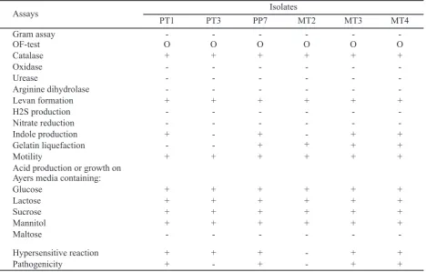
The bacterial pathogen isolates obtained in this study are: 1 garlic isolate of Temanggung (PP), 2 garlic isolates of Tawangmangu (PT), 3 shallots isolates of Tawangmangu (MT). From the physiological and biochemical characterization, it was shown that the bacterial pathogens have a yellow pigment, slimy colonies with regular borders, gram-negative, non-spore, facultative anaerobic bacterium, motile, producing acid from D-glucose, D-mannitol, sucrose,
and lactose, and catalase production. The bacterial isolates were able to form indole from tryptophan as a source of carbon and hydrolyzed gelatin, with the exception of isolate PT3. All isolates reacted negatively in the production of oxidase, arginine dihydrolase, urease, and nitrate reduction (Table 1). Isolates of PT1, PP7, MT3 and MT4 possess the same characteristics with reportedP. ananatis,but garlic isolates of Tawangmangu (PT3) and shallot isolates of Tawangmangu (MT2) does not have the ability to produce indole. In pathogenicity tests, isolate PT3 and MT2 does not cause symptoms in the shallot and garlic. According to Walcott et al.
(2002) the ability to produce indole distinguished P. ananatis fromP. stewartiisubsp.stewartii.
Egorovaet al. (2015) reported that P. ananatis
isolates causing leaf blight of rice in Russia were shown to have similar physiological and biochemical characteristics. These Russian isolates were found to react positively to the β-galactosidase, produced acid from D-glucose, D-mannitol, D-melibiose, arabinose, sucrose, meso-inositol, glycerol, d-sorbitol, amygdalin, and utilize citrate and tartrate.
Phylogenetic Analysis of Leaf Blight Bacterial Isolates
From the amplification of PCR based on the gyr B
gene sequences with gyrB01-F and gyrB02-R primer, all the samples resulted in single band at 970 bp. Phylogenetic analysis using Jukes-Cantor algorithm in Mega 7.0 revealed that the bacterial isolates causing leaf blight of garlic from Tawangmangu (PT1), Temanggung (PP7) and shallot of Tawangmangu (MT3 and MT4) were closely related to P. ananatis
PA13 isolated from rice plants (Choi et al.,2011) with a 99% degree of homology and bootstrap value 99. PT3 and MT2 isolates are closely related to P. stewartii
subsp. stewartii CFBP 3614 (Rezzonico et al.,2009) with 99% homology and bootstrap value 89 (Figure 2).
Figure 1. Symptoms of leaf blight and discoloration in shallot (A) and garlic (B) as shown by the arrow
Assays Isolates
PT1 PT3 PP7 MT2 MT3 MT4
Gram assay - - -
-OF-test O O O O O O
Catalase + + + + + +
Oxidase - - -
-Urease - - -
-Arginine dihydrolase - - -
-Levan formation + + + + + +
H2S production - - -
-Nitrate reduction - - -
-Indole production + - + - + +
Gelatin liquefaction - - + + + +
Motility + + + + + +
Acid production or growth on Ayers media containing:
Glucose + + + + + +
Lactose + + + + + +
Sucrose + + + + + +
Mannitol + + + + + +
Maltose - - -
-Hypersensitive reaction + + + - + +
Pathogenicity + - + - + +
Description: PP (garlic isolate of Temanggung), PT (garlic isolate of Tawangmangu), MT (shallot isolate of Tawangmangu). + : indicated positive reaction; - : indicated no reaction; O : indicated oxidative
Table 1. Physiological and biochemical analyses of bacterial isolates from garlic and shallot
Figure 2. Phylogenetic tree showing the relationship of the leaf blight bacterial isolates and the closely related strains available in the GenBank; on the basis of the alignment of gyrBgene sequences, a phylogenetic tree was constructed using the neighbor-joining method; the stability of the tree was assessed by 1000 bootstrap replications with the neighbor-joining method and Jukes-Cantor distance analysis; the sequence of
CONCLUSION
The use of other phylogenetic markers beside 16S rRNA is necessary to achieve unambiguous identification at the species level or below. In the present study we describe a method for identifying
Pantoea ananatis using partial sequence of gyrBas a phylogenetic marker. Since gyrB is a universally distributed gene in all prokaryotes, we are currently extending our work to similar studies of other bacterial families with the aim of establishing the use of gyrB
as a universal phylogenetic marker.
ACKNOWLEDGEMENT
The authors with to thank Australian Center for International Agricultural Research (ACIAR) for supporting this work trough research grant (ACIAR HORT 2009/056) to SS.
LITERATURE CITED
Aneja, K.R. 2003. Experiments in Microbiology, Plant Pathology and Biotechnology. Fourth Edition. New Age International Publishers, New Delhi. 607 p.
Azad, H.R., G.J. Holmes, & D.A. Cooksey. 2000. A New Leaf Blotch Disease of Sudan Grass Caused by Pantoea ananas and Pantoea stewartii. Plant Disease84: 973–979.
Carr, E.A., J.M. Bonasera, A.M. Zaid, J.W. Lorbeer, & S.V. Beer. 2010. First Report of Bulb Disease of Onion Caused by Pantoea ananatis in New York. Plant Disease94: 916.
Choi, O., J.Y. Lim, Y.S. Seo, I. Hwang, & J. Kim. 2012. Complete Genome Sequence of the Rice Pathogen Pantoea ananatisStrain PA13. Journal of Bacteriology194: 531.
Conn, K., J. Lutton, & S. Rosenberger. 2012. Onion Disease Guide. Seminis Vegetable Seeds, California, USA. 72 p.
Danaatmadja, Y., S. Subandiyah, T. Joko, & C.U. Sari. 2009. Isolasi dan Karakterisasi Ralstonia syzygii. Jurnal Perlindungan Tanaman Indonesia
15: 7−12.
Dwimartina, F., T. Arwiyanto, & T. Joko. 2017. Potential of Endophytic and Rhizobacteria as an Effective Biocontrol for Ralstonia syzygiisubsp.
syzygii. Asian Journal of Plant Pathology 11: 191–198.
Egorova, M., E. Mazurin, & A.N. Ignatov. 2015. First Report of Pantoea ananatis Causing Grain Discolouration and Leaf Blight of Rice in Russia.
New Disease Reports32: 21.
Gitaitis, R.D. & J.D. Gay. 1997. First Report of Leaf Blight, Seed Stalk Rot, and Bulb Decayof Onion by P. ananatis in Georgia. Plant Disease 81: 1096.
Gitaitis, R.D., R.R. Walcott, M.L. Wells, J.C. Diaz Perez, & F.H. Sanders. 2003. Transmission of P. ananatis,Causal Agent of Center Rotof Onion, by Tobacco Thrips, Frankliniella fusca. Plant Disease87: 675–678.
Harayama, S. & H. Kasai. 2006. Bacterial Phylogeny Reconstruction from Molecular Sequences. InE. Stackebrandt (ed.) Molecular Identification, Systematics, and Population Structure of Prokaryotes, Chap. 5. Springer, Berlin.
Ismiyatuningsih, T. Joko, & S. Hartono. 2016. Survey and Detection of Pectobacterium atrosepticum
in Major Potato-Growing Areas in Central Java Province, Indonesia. Ilmu Pertanian1: 1−6.
Joko, T., S. Subandiyah, & S. Somowiyarjo. 2000. The Role of Extracellular Protein on the Pathogenicity of Xanthomonas campestris pv. citri. Jurnal Perlindungan Tanaman Indonesia6: 32−38.
Joko, T., H. Hirata, & S. Tsuyumu. 2007a. Sugar Transporter (MfsX) of Major Facilitator Super-family is Required for Flagella-Mediated Patho-genesis in Dickeya dadantii 3937. Journal of General Plant Patholology73: 266−273.
Joko, T., H. Hirata, & S. Tsuyumu, 2007b. A Sugar Transporter (MfsX) is also Required by Dickeya dadantii 3937 for in Planta Fitness. Journal of General Plant Patholology73: 274−80.
Joko, T., D. Kiswanti, S. Subandiyah, & Hanudin. 2011a. Occurence of Bacterial Soft Rot of
Phalaenopsis Orchids in Yogyakarta and West Java, Indonesia, p. 255–265. In Y. Koentjoro (ed.), Proceeding of Internasional Seminar on “Natural Resources, Climate Change, and Food Security in Developing Countries”, 27−28 June 2011. Surabaya, Indonesia.
Joko, T., N. Kusumandari, & S. Hartono. 2011b. Optimasi Metode PCR untuk Deteksi Pectobacterium carotovorum, Penyebab Penyakit Busuk Lunak Anggrek. Jurnal Perlindungan Tanaman Indonesia
17: 54–59.
Joko, T., M.P. Koentjoro, S. Somowiyarjo, M.S. Rohman, A. Liana, & N. Ogawa. 2012. Response of Rhizobacterial Communities in Watermelon to Infection with Cucumber Green Mottle Mosaic Virus as Revealed by Cultivation-Dependent RISA. Archives of Phytopathology and Plant Protection45: 1810−1818.
of Orchid. Archives of Phytopathology and Plant Protection47: 1239−1250.
Kido, K., M. Hasegawa, H. Matsumoto, M., Kobayashi, & Y. Takikawa. 2010. Pantoea ananatisStrains are Differentiated Into Three Groups Based on Reactions of Tobacco and Welsh Onion and on Genetic Characteristics. Journal of General Plant Pathology76: 208–218.
Kumar, S., G. Stecher, & K. Tamura. 2016. MEGA7: Molecular Evolutionary Genetics Analysis Version 7.0 for Bigger Datasets. Molecular Biology and Evolution33: 1870–1874.
Leboffe, M.J. & B.E. Pierce. 2010. Microbiology Laboratory Theory and Application, 3rd ed. Morton Publishing Company, Englewood, Co. 656 p.
Lelliot, R.A. & D.E. Stead. 1987. Methods in Plant Pathology. Vol. 2. Methods for the Diagnosis of Bacterial Diseases of Plants. British Society for Plant Pathology by Blackwell Scientific Publication, Oxford. 216 p.
Mahfut, T. Joko, B.S. Daryono, & S. Somowiyarjo. 2016a. Survei Odontoglossum Ringspot Virus (ORSV) yang menginfeksi anggrek alam tropis di Indonesia. Jurnal Perlindungan Tanaman Indonesia17: 54–59.
Mahfut, T. Joko, & B.S. Daryono. 2016b. Molecular Characterization of Odontoglossum Ringspot Virus (ORSV) in Java and Bali, Indonesia. Asian
Journal of Plant Pathology10: 9−14.
Miller, A.M., J.E.F. Figueiredo, C.L. Chaves, E.A. Ruas, M.I. Balbi-Peña, N.B. Colauto, & L.D. Paccola-Meirelles. 2016. Genomic Variability of
P. ananatis in Maize white Spot Lesions Assessed by AFLP Markers. Genetics and Molecular Research15: 1–13.
Parkinson, N., C. Cowie, J. Heeney, & D. Stead. 2009. Phylogenetic Structure of XanthomonasDetermined by Comparison of gyrBSequences. International Journal of Systematic and Evolutionary Microbiology
59: 264– 274.
Pérez-y-Terrón, R., M.C. Villegas, A. Cuellar, J. Muñoz-Rojas, M. Castañeda-Lucio, I. Hernández-Lucas, R. Bustillos-Cristales, C.L. Bautista-Sosa, J.A. Munive, R. Caicedo-Rivas, & L.E. Fuentes-Ramírez. 2009. Detection of P. ananatis, Causal Agent of Leaf Spot Disease of Maize, in Mexico.
Australasian Plant Disease Notes4: 96–99.
Rezzonico, F., T.H. Smits, E. Montesinos, J.E. Frey, & B. Duffy. 2009. Genotypic Comparison of
Pantoea agglomeransPlant and Clinical Strains.
BMC Microbiology9: 204.
Sauer, A.V., K.R. Rocha, R.M. Gonçalves, & W.F. Meirelles. 2015. Survival of Pantoea ananatis, Causal Agent of Maize White Spot Disease in Crop Debris. Agronomy Science and Biotechnology
1: 21–24.
Schaad, N.W., J.B. Jones, & W. Chun. 2001.
Laboratory Guide for Identification of Plant Pathogenic Bacteria. Third Edition. Minnesota: The American Phytopathological Society. 373 p.
Suharti, T., T. Joko, & T. Arwiyanto. 2017. Deteksi Bakteri Patogen Terbawa Benih Akor (Acacia auriculiformisA. Cunn. ex Benth.). Jurnal Hama dan Penyakit Tumbuhan Tropika17: 19–36.
Sumardiyono, C., T. Joko, Y. Kristiawati, & Y.D. Chinta. 2011. Diagnosis dan Pengendalian Penyakit Antraknosa pada Pakis dengan Fungisida.
Jurnal Hama dan Penyakit Tumbuhan Tropika
11: 194–200.
Takeda, K., Y.Q. Kang, K. Yazawa, T. Gonoi, & Y. Mikami. 2010. Phylogenetic Studies of Nocardia Species Based on gyrB Gene Analyses. Journal of Medical Microbiology59: 165–171.
Walcott, R.R, R.D. Gitaitis, A.C. Castro, F.H. Sanders, & J.C. Diaz-Perez. 2002. Natural Infestation of Onion Seed by P. ananatis, Causal Agent of Center Rot.Plant Disease86: 106–111.
Wardhika, C.M., Suryanti, & T. Joko. 2014. Eksplorasi Bakteri Agens Pengendali Hayati Fusarium solani dan Meloidogyne incognitapada Lada.
Jurnal Perlindungan Tanaman Indonesia 18: 90−95.
Wibowo, A., T. Joko, S. Subandiyah, I. Mariska, Y. Supriyati, Y. Suryadi, & I. Roostika. 2010. Peningkatan Ketahanan Tanaman Pisang Kepok Kuning Terhadap Penyakit Darah Melalui Variasi Somaklonal dan Simbiosis Endofitik.
Jurnal Perlindungan Tanaman Indonesia 16: 15–21.
Widyaningsih, S., S.N.H. Utami, T. Joko, & S. Subandiyah. 2017. Development of Disease and Growth on Six Scion/Rootstock Combinations of Citrus Seedlings under Huanglongbing Pressure.
Journal of Agricultural Science9: 229−238.