ISSN: 1511-3701 © Universiti Putra Malaysia Press
TROPICAL AGRICULTURAL SCIENCE
Journal homepage: http://www.pertanika.upm.edu.my/
E-mail addresses: [email protected];
[email protected] (Nesti Fronika Sianipar), [email protected] (Ragapadmi Purnamaningsih) * Corresponding author
Article history:
Received: 28 March 2017 Accepted: 27 November 2017 ARTICLE INFO
Enhancement of the Contents of Anticancer Bioactive
Compounds in Mutant Clones of Rodent Tuber (Typhonium
flagelliforme
Lodd.) based on GC-MS Analysis
Nesti Fronika Sianipar1,2* and Ragapadmi Purnamaningsih3
1Department of Food Technology, Faculty of Engineering, Bina Nusantara University, 11480 Jakarta,
Indonesia
2Research Interest Group Food Biotechnology, Bina Nusantara University, 11480 Jakarta, Indonesia 3Indonesian Center for Agricultural Biotechnology and Genetic Resources Research and Development
(BB-Biogen), 16111 Bogor, Indonesia
ABSTRACT
Rodent tuber (Typhonium flagelliforme Lodd.), which is a well known herbal plant from the Araceae family, is know for its anticancer activities. The genetic variation of rodent tuber is low due to its commonly applied vegetative propagation methods. Thus, its genetic variation has to be increased to obtain a new and superior plant containing a high amount of anticancer compounds. The aim of this study was to analyse the chemical compounds of the leaves and tubers of rodent tuber’ mutant and non irradiated (control) plants by GC-MS method. In this study, in vitro calli of rodent tuber was irradiated with 6 Gy of gamma ray which produced mutant plantlets which was genetially different from non irradiated (control) plants. Mutant plantlets had been acclimated and propagated in the greenhouse to obtain the 6th generation vegetative mutant clones (MV6), which are stable superior
mutants. The results indicated that MV6 contained six new anticancer compounds in the leaves and four new anticancer compounds in the tubers which have not been detected in
control. The new anticancer compounds present in leaves and tubers were identified by
GC-MS. They are hexadecanoic acid methyl ester, octadecadienoic acid, phytol¸ gamma-sitosterol, eicosane, geranylgeraniol, squalene, octacosane and 7-pentadecyne. MV6, is the new superior variety and a potential source of anticancer drugs.
Keywords: Typhonium flagelliforme Lodd., new
superior mutant, anticancer bioactive component,
INTRODUCTION
Rodent tuber (Typhonium flagelliforme
Lodd.) is a medicinal herbal plant from Indonesia that belongs to the Araceae family (Essai, 1986). Biologically active chemicals in this plant are alkaloids, saponins, steroids, and glycosides (Syahid, 2007). A study has documented the effectiveness of ethanolic fraction of the rodent tuber’ extract in inhibiting the growth of T47D breast cancer cell line (Nurrochmad et al., 2011)
while another has show the efficacy of its
dichloromethane fraction against MCF-7 breast cancer cell line (Putra & Winarto, 2012). The rodent tuber’s extract has also been found toinhibit the proliferation of human T4 lymphoblastoid (Mohan et al., 2008) and NCI-H23 non-small lung carcinoma cell line (Lai, et al., 2008). Anticancer compounds could be found in all parts of the plant, such asthe roots, tubers, stems, and leaves (Choo et al., 2001). The hexane extract of rodent tuber was proven to be cytotoxic against Artemia salina larvae (Sianipar et al., 2013a). This plant also has antibacterial and antioxidant properties (Mohan et al., 2008).
The main obstacle in the development of rodent tuber into drugs are its low genetic diversity and low organic compounds content (Syahid, 2008). The low genetic variation is due to commonly practiced vegetative propagation methods, mainly through conventional buds separation (Essai, 1986). Although vegetative propagation methods could produce seedlings, these methods rarely cause genetic recombination. Thus, the genetic variation in species level
is low and reduce the creation of new genotypes (Syahid & Kristina, 2007). Genetic variation of rodent tuber has to be increased in order to obtain superior mutant clones which contain a high amount of anticancer compounds. Mutation induction is an effective way to its increase genetic diversity (Wulan, 2007). The mutation could be induced by irradiating the respective sample with physical mutagens, such as gamma ray (Poespordasono, 1988). In vitro embryogenic somatic cell population or calli of rodent tuber has been induced, proliferated, and regenerated with single node culture method (Sianipar et al., 2011). In an earlier study, the rodent tuber calli were irradiated with gamma ray. They were regenerated into in vitro plantlets that showed various growth responses based on the observation of the number of shoots, number of leaves, and plant’s height (Sianipar et al., 2013b). Rodent tuber calli was irradiated at many doses to induce mutation. Rodent tuber mutation induction successfully done at the dose 6 Gy. The 1st generation plantlet of rodent tuber
through gamma rays irradiation can detect genetic changes of mutant by using RAPD (Sianipar et al., 2015b).
307 Pertanika J. Trop. Agric. Sci. 41 (1): 305 - 320 (2018)
greenhouse to obtain the 6th generation of
vegetative mutant clones (MV6). The 6th
generation of vegetative stable superior mutant population (MV6) from Bogor (Sianipar et al., 2013), is in the patenting process. There were 8 mutant MV6 clones (6-3-3-6, 6-1-1-2, 6-3-2-5, 6-9-1, 6-2-5-2, 6-1-2, 6-9-4, 6-2-6-3) and 1 (non-irradiated) for control. Each sample had 9 replicates. The leaves and tubers of rodent tubers underwent metabolite extraction.
Preparation of extract from rodent tuber
The rodent tuber was dried and macerated in 96% ethanol overnight. The solvent
was removed after it was filtered through Whatman filter paper No. 1. The concentrated
extract was collected and used for GC-MS analysis.
Identification of chemical content with
GC-MS
The concentrated ethanol extracts were injected into the GC column. Injection volume was 5µl with 5:1 split ratio and the injection temperature was 250°C. Helium was used as carrier gas with velocity 0.8 µl per minute. Column temperature was set at 70°C with 5°C per minute increment. At 200°C, the temperature was kept constant for 1 minute before it was increased to 280°C at the rate of 20°C per minute. The temperature remained constant for another 28 minutes. Mass spectrometer wasused in electron impact ionisation mode with 70 eV voltage.
propagated and regenerated into the 6th
generation of vegetative mutant (MV6). Extraction involves isolation or
purification of chemicals from raw samples
(Mohrig, 2010). Mutant clones contain a lot of bioactive compounds with various functional groups and polarities (Hota, 2007). Hydrophilic substances were extracted with polar solvents, such as ethanol (Rostagno et al., 2013). Genetic mutation could affect the relative abundances of plant’s bioactive compounds. Gas Chromatography-Mass Spectrometry (GC-MS) is able to analyse
the metabolomic profile of an organism.
GC uses gas as its mobile phase to separate various chemicals. The MS could separate chemicals based on their molecular weight (Kayser & Quax, 2007). The GC-MS is a powerful device to identify chemicals by referring to their databases (Kayser & Quax, 2007) and it has been applied to analyse phytochemical and bioactive compounds of herbal plants, such as Melia orientalis (Marimuthu, 2013) and Maranta arudinacea L. (Nishaa, 2013). The GC-MS has also been employed to identify chemical compounds in the nonpolar fraction of rodent tuber from Malaysia (Mohan et al., 2011). The aim of this study is to analyse the bioactive anticancer compound content in the polar fraction of rodent tuber stable superior mutant clones by using GC-MS.
MATERIALS AND METHODS
Plant material
The 1st generation of mutant plantlets
The mass spectrum of GC-MS was identified by referring to the National Institute Standard Technique (NIST)
database with ≥ 90% fit factor. The content of
the compound was calculated by comparing its average peak area to the total area.
RESULTS AND DISCUSSION
The results of fresh and dry weight for each clone (Table 1) are different. Mutant clones 6-1-2, 6-9-4, 6-2-6-3, and 6-2-5-2 had higher stem, leaves and tuber weight than control. Mutant clone 6-2-5-2 had the highest fresh and dry weight compared with control. The highest tuber dry weight was obtained by clones 6-1-2 and 6-2-5-2. The differences in fresh and dry weight are due to genetic changes or mutation, but the increase in fresh or dry weight did not occur in all mutant clones. Genetic changes are not always followed by morphological changes.
Sianipar et al. (2013) have shown irradiated calli can lead to genetic changes. These genetic changes are evidenced by genetic variation in 1st generation mutant of the
rodent tuber (MV1), 3rd generation mutant
(MV3), 4th generation mutant (MV4) by
using RAPD (Sianipar et al., 2015a; 2016; 2017).
In vitro culture treatment and gamma ray irradiation could induce chromosomal a b b e r a t i o n i . e . t h e m o d i f i c a t i o n o f chromosomal number and structure (Surya & Soeranto, 2006; Pillay & Tenkouano, 2011). These modifications can alter morpho-physiological properties of mutant clones (Table 1). The DNA mutation will lead to the generation of new genotypes and affect transcription, translation, protein synthesis and enzyme expressions that leads to production of secondary metabolites (Gorbunova & Levy, 1997; Kovacs & Keresztes, 2002).
Table 1
Fresh and dry weights of rodent tuber’s control and mutant clone
Plant Total fresh weight (g) Dry weight (g) Total Leave and stem Tuber
Field control 150 38,8 8,7 32
In vitro control 100 22,7 NM 19
6-9-1 100 17,3 7,4 13
6-3-3-6 100 26 8,4 20
6-3-2-5 50 20,1 2,5 20
6-1-1-2 50 23 5,7 21
6-1-2 162,5 34,48 14 37
6-9-3 50 17,4 NM NM
6-9-4 100 38,2 8,9 30
6-1-3-4 50 22 NM NM
6-2-6-3 200 34,5 13,5 29
6-2-4-1 150 22 NM NM
6-2-5-2 300 54,8 17,9 37
309 Pertanika J. Trop. Agric. Sci. 41 (1): 305 - 320 (2018)
Chemical composition of the leaves, tubers of rodent tuber’s control and mutant plants were successfully identified with GC-MS (Figure 1, Tables 2-5), which showed that there were phytochemical profile differences between control and mutant clones as well as between each of the mutant clones. Leaves and tubers of control plants contained 19 and 26 different chemicals respectively (Tables 2 and 3). Five most abundant chemicals in the leaves of
control plant were 9,12,15-octadecatrienoic acid or linolenic acid, hexadecanoic acid, stigmasterol, (2E)-3,7,11,15-tetramethyl-2-hexadecen-1-ol, and campesterol
(ergost-5-en-3-ol). Meanwhile, five most abundant
compounds in the tubers of control plant were (9E,12E)-9,12-octadecadienoic acid, hexadecanoic acid/palmitic acid, methyl (9z,12z)-9,12-octadecadienoate, stigmasterol and ergost-5-en-3-ol.
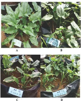
Figure 1. Control and stable superior mutant clones of rodent tuber at 8th week in greenhouse
Note: A = Control; B = 6-2-5-2; C = 6-2-6-3; D = 6-3-3-6
A B
Table 2
Chemical compounds in leaves of rodent tuber control plant based on GC-MS
RT Relative abundance (%) Chemical compound
30,051 2,98 2,6,10-trimethyl,14-ethylene-14, pentadecne (neophytadiene) 30,34 0,94 2,6,10-trimethyl,14-ethylene-14, pentadecne (neophytadiene) 30,534 1,24 2,6,10-trimethyl,14-ethylene-14,pentadecne (neophytadiene) 31,54 0,2 Hexadecanoic acid, ethyl ester
31,678 20,77 Hexadecanoic acid 32,216 0,36 9,17-octadecadienal
32,257 0,86 9,12,15-octadecatrienoic acid, methyl ester (linolenic acid methyl ester)
32,375 5,87 (2E)-3,7,11,15-Tetramethyl-2-hexadecen-1-ol 32,602 1,05 Ethyl (9z,12z)-9,12-octadecadienoate
32,795 41,16 9,12,15-octadecatrienoic acid (linolenic acid) 33,278 0,9 Methyl 8,11,14-heptadecatrienoate
35,043 0,12 Oleic acid (9-octadecadienoic acid)
35,746 0,6 2,6,10,14,18,22-Tetracosahexaene, 2,6,10,15,19,23-hexamethyl (squalene; spinacene)
36,146 0,36 Octacosane
39,587 3,16 Campesterol (ergost-5-en-3-ol)
39,939 7,67 Stigmasterol
40,746 1,96 Beta-sitosterol
44,8 2,87 Pyridine-3-carboxamide, oxime,
N-(2-trifluoromethylphenyl)-50,158 0,25 Pyridine-3-carboxamide, oxime,
N-(2-trifluoromethylphenyl)-Note: Compounds were identified by comparing retention time data with authentic standard database of
NIST/EPA/NIH fit factor ≥ 90%. Relative abundance was determined based on area percentage of each
compound
Table 3
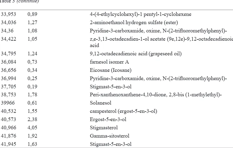
Chemical compounds in tubers of rodent tuber control plant based on GC-MS
RT Relative abundance (%) Chemical compound 31,168 0,08 Farnesol, methyl ether
31,954 22,08 Hexadecanoic acid (palmitic acid) 32,526 0,73 Hexadecanoic acid (palmitic acid)
32,581 0,22 Hexadecanoic acid (palmitic acid)
32,616 0,26 Heptadecanoic acid (potassium heptadecanoate) 32,699 0,2 n-hexadecanoic acid (palmitic acid)
32,919 0,85 linoleic acid ethyl ester (ethyl linoleate) 33,078 38,37 (9E,12E)-9,12-octadecadienoic acid 33,616 4,34 Methyl (9z,12z)-9,12-octadecadienoate
311 Pertanika J. Trop. Agric. Sci. 41 (1): 305 - 320 (2018)
33,953 0,89 4-(4-ethylcyclohexyl)-1 pentyl-1-cyclohexene 34,036 1,27 2-aminoethanol hydrogen sulfate (ester)
34,36 1,08 Pyridine-3-carboxamide, oxime,
N-(2-trifluoromethylphenyl)-34,422 1,05 z,e-3,13-octadecadien-1-ol acetate (9e,12e)-9,12-octadecadienoic acid
34,795 1,24 9,12-octadecadienoic acid (grapeseed oil) 36,084 0,73 farnesol isomer A
36,656 0,34 Eicosane (Icosane)
36,994 0,25 Pyridine-3-carboxamide, oxime,
N-(2-trifluoromethylphenyl)-37,705 0,19 Stigmast-5-en-3-ol
38,753 1,78 Peri-xanthenoxanthene-4,10-dione, 2,8-bis
(1-methylethyl)-39966 0,61 Solanesol
40,532 1,55 campesterol (ergost-5-en-3-ol) 40,573 2,38 Ergost-5-en-3-ol
40,966 4,05 Stigmasterol
41,876 1,92 Gamma-sitosterol
41,945 1,63 Stigmast-5-en-3-ol
Note: Compounds were identified by comparing retention time data with authentic standard database of NIST/
EPA/NIH fit factor ≥ 90%. Relative abundance was determined based on area percentage of each compound
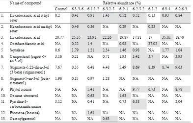
One of the clones with highest amount of anticancer compounds was 6-1-2 (Tables 4 and 5). Either in the leaves or tubers of this clone, the most abundant compound was 9,12-octadecadienoic acid (Figure 2). Octadecadienoic acid could induce apoptosis of various cancer cells (Yoo et al., 2007). The other most abundant compounds in leaves of 6-1-2 were 9,12-octadecadienoic acid, hexadecanoic acid, phytol, neophytadiene, and stigmasterol. While in its tubers were (9E,12E)-9,12-octadecadienoic acid, hexadecanoic acid, hexadecanoic acid ethyl ester, and ethyl (9Z,12Z)-9,12-octadecadienoate.
The amount of some anticancer bioactive compounds in mutant clones
were higher than control. Mutant clones also contained several new anticancer compounds which were not found in control (Tables 6 and 7). Leaves of mutant clones had at least six anticancer compounds and in greater quantities compared with control. The amount of hexadecanoic acid was the highest in mutant clone 6-9-4, about 15.04% compared with control. Also known as palmitic acid, it has cytotoxic effect against MOLT-4 leukimia (cancer cell line) by interacting with DNA topoisomerase I to induce apoptosis (Kwan et al., 2014). Palmitic acid has also been reported to exhibit antitumour activity in vivo (Harada et al., 2002).
Table 4
Chemical compounds in leaves of rodent tuber mutant clone 6-1-2 based on GC-MS
RT Relative abundance (%) Chemical compound
30,147 5,43 2,6,10-trimethyl,14-ethylene-14, pentadecne (neophytadiene) 30,416 1,04 2,6,10-trimethyl,14-ethylene-14,pentadecne (neophytadiene) 30,602 1,89 (2E)-3,7,11,15-Tetramethyl-2-hexadecen-1-ol
31,051 0,23 Hexadecanoic acid, methyl ester
31,588 0,13 Hexadecanoic acid, ethyl ester 32,023 14,44 Hexadecanoic acid
32,278 0,94 Hexadecanoic acid
32,319 1,62 Hexadecanoic acid
32,478 6,73 Phytol
32,678 1,03 9,12-octadecadienoic acid, ethyl ester
33,085 36,01 9,12-octadecadienoic acid 33,354 1,96 13-tetradece-11-yn-1-ol
33,443 1,84 Methyl 8,11,14-heptadecatrienoate 33,692 1,01 (9E, 12E)-9,12-Octadecadienoic acid 34,588 0,16 8-(2-octylcyclopropyl)octanal
36,263 0,27 Nonacosane
39,966 3,7 Ergost-5-en-3-ol
40,387 3,71 Stigmasterol
40,408 4,68 Stigmasterol
41,249 4,32 Cholest-5-en-3-ol,23-ethyl-, (3 beta 23s) 45,489 2,97 5,5-dimethyl-7,8-epoxyspiro (3.5) nonan-1-one 51,737 0,03 5,9-dimethyl-4,10-octadecadiene
Note: Compounds were identified by comparing retention time data with authentic standard database of NIST/
EPA/NIH fit factor ≥ 90%. Relative abundance was determined based on area percentage of each compound
Table 5
Chemical compounds in tubers of rodent tuber mutant clone 6-1-2 based on GC-MS
RT Relative abundance (%) Chemical compound
31,912 8,26 Hexadecanoic acid, ethyl ester
31,968 22,8 Hexadecanoic acid
32,568 1,41 9,12-octadecadienoic acid, methyl ester (linoleic acid, methyl ester/ methyl linoleate)
32,954 6,67 Ethyl (9Z,12Z)-9,12-octadecadienoate 33,03 24,29 9,12-octadecadienoic acid
33,112 23,4 (9E,12E)-9,12-octadecadienoic acid
33,616 1,4 Tricosane
313 Pertanika J. Trop. Agric. Sci. 41 (1): 305 - 320 (2018)
Figure 2. GC-MS chromatogram of leaves and tubers of mutant clones. X-axis represents retention time while Y-axis represents relative abundance. The chemical structure of 9,12-octadecadienoic acid, which has the highest relative abundances are shown (chemical structure obtained from NIST database) here
Note: A = leaves of 6-1-2, B = tubers of 6-1-2
34,505 0,56 Eicosane
35,464 0,89 Eicosane
36,263 1,05 (6E,10E,14E,18E)-2,6,10,15,19,23-hexamethyl-2,6,10,14,18,22-tetracosahexaene
36,705 1,71 Heptacosane, 1-chloro
38,456 0,83 Nonacosane
41,063 1,03 Stigmasterol
41,152 0,04 Stigmasterol
Note: Compounds were identified by comparing retention time data with authentic standard database of NIST/
EPA/NIH fit factor ≥ 90%. Relative abundance was determined based on area percentage of each compound
Leaves of mutant clones contained six new anticancer compounds, namely hexadecanoic acid methyl ester, octadecadienoic acid, phytol¸ gamma-sitosterol, eicosane, and geranylgeraniol. Hexadecanoic acid methyl ester has been known to to inhibit the growth and induce apoptosis of human gastric cancer cells (Yu et al., 2005). Phytol is an antitumour chemical which could induce the apoptosis of human gastric adenocarcinoma (Song & Cho, 2015) and hepatocellular carcinoma cells. Therefore, it has good potential as a medicine for liver cancer (Kim et al., 2015). Additionally,
Table 6
Comparison of the relative abundances of anticancer compounds in the rodent tuber’s control and mutant plants based on GC-MS analysis
Name of compound Relative abundance (%)
Control 6-3-3-6 6-1-1-2 6-3-2-5 6-9-1 6-2-5-2 6-1-2 6-9-4 6-2-6-3 1. Hexadecanoic acid ethyl
ester
0,2 0,41 0,91 1,43 0,52 0,52 0,13 0,93 0,84
2. Hexadecanoic acid methyl ester
NA 0,46 0,36 NA 0,29 NA 0,23 NA NA
3. Hexadecanoic acid 20,77 25,35 23,91 22,26 19,87 17,81 17 35,81 18,79
4. Octadecadienoic acid NA 0,22 1,4 NA 0,98 NA 37,02 NA NA
5 Squalene 0,6 1,79 1,21 2,34 1,46 0,98 NA 1,77 1,04
6. Campesterol (ergost-5-en-3-ol)
3,16 0,21 NA 0,71 1,93 3,42 3,7 NA 3,93
7. Stigmasta-5,22-dien-3-ol (3 beta) (stigmasterol)
7,67 0,35 6,48 4,48 5,49 8,69 8,39 8,74 9,63
8. Stigmast-5-en-3-ol (beta-sitosterol)
1,96 0,11 0,97 1,28 NA NA NA NA NA
9. Phytol isomer NA NA 5,41 NA NA 9,77 6,73 NA 8,78
10. Gamma sitosterol NA NA 0,68 NA 1,63 NA NA NA NA
11. Pyridine-3-carboxamide,oxime
3,12 NA 0,41 NA 0,73 6,38 NA NA 2,04
12. Eicosane (Icosane) NA NA 1,61 NA NA NA NA NA NA
13. Geranylgeraniol NA NA NA 0,63 NA NA NA NA NA
Note: Chemicals were identified by comparing the retention time with authentic standard database of NIST/
EPA/NIH (fit factor ≥ 90%). NA is not available, which means that the quantity of the compound is too low
to be detected by GC-MS. The quantities of anticancer bioactive compounds in MV6 mutant clones which are higher than control are indicated by the grey highlights
315 Pertanika J. Trop. Agric. Sci. 41 (1): 305 - 320 (2018)
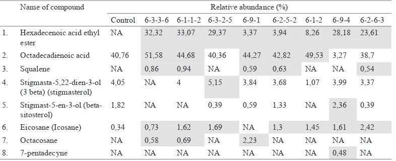
adenocarcinoma (Yoshikawa et al., 2009). Mutant clones’ tubers contain greater amounts of at least 5 different anticancer compounds compared with control. The highest increase from control was observed in the chemical profile of clone 6-3-3-6 which contained octadecadienoic acid, 10.82% higher compared with control plant. This compound could induce apoptosis of colon cancer cells (Yoo et al., 2007).
Tubers of mutant clones contain four new anticancer compounds, namely hexadecanoic acid ethyl ester, squalene, octacosane, 7-pentadecyne, which were not found in control plant. Hexadecanoic acid ethyl ester has antioxidant and antimicrobial properties (Bodoprost & Rosemeyer, 2007). In addition, it could reduce the risk of
Table 7
Comparison of the relative abundances of anticancer compounds in the rodent tuber’s control and mutant plants based on GC-MS analysis
Name of compound Relative abundance (%)
Control 6-3-3-6 6-1-1-2 6-3-2-5 6-9-1 6-2-5-2 6-1-2 6-9-4 6-2-6-3 1. Hexadecenoic acid ethyl
ester
NA 32,32 33,07 29,37 3,37 3,94 8,26 28,18 23,61
2. Octadecadienoic acid 40,76 51,58 44,68 40,36 44,27 42,82 49,53 3,27 38,7
3. Squalene NA 0,86 0,94 NA 0,59 0,63 NA NA 0,54
4. Stigmasta-5,22-dien-3-ol (3 beta) (stigmasterol)
4,05 NA 4 5,15 3,84 3,68 1,07 3,99 3,37
5. Stigmast-5-en-3-ol (beta-sitosterol)
1,82 NA NA 0,39 0,59 1,33 NA 2,36 0,39
6. Eicosane (Icosane) 0,34 0,73 1,62 1,69 NA 1,3 1,45 1,61 2,42
7. Octacosane NA 0,58 0,69 NA 2,23 NA NA NA NA
8. 7-pentadecyne NA NA NA NA NA NA NA 0,48 NA
Note: Chemicals were identified by comparing the retention time with authentic standard database of NIST/
EPA/NIH (fit factor ≥90%). NA means it is not available, which means that the quantity of a compound is
negligible and cannot be detected by GC-MS. The quantities of anticancer bioactive compounds in MV6 mutant clones which are higher than control are indicated by the yellow highlights
coronary heart disease and cancer (Lai et al., 2008; Bodoprost & Rosemeyer, 2007). Squalene has been proven to be able to inhibit the carcinogenesis of various cancer cell lines, such as colon cancer (Rao et al., 1998). Octacosane was cytotoxic against B16F10-Nex2 skin cancer cells based on in vitro experiment (Figueiredo, 2014). The chemical structure of 7-pentadecyne was similar to protein kinase C activator, so it has the potential to be developed into an effective anticancer drugs (Block, 2012). The anticancer activity of 7-pentadecyne
has also been confirmed by (Kozikowski
et al., 2001).
plants. Stigmast-5-en-3-ol (3.beta,24s) (beta-sitosterol) is a phytosterol with various biological activities, such as reducing cholesterol level in cells, modify membrane lipid profile (Awad, 1996), anti-diabetic (Sujtha et al., 2010), and inhibit cancer cells (Fraille et al., 2012; Von et al., 1998). Ergost-5-en-3-ol (3 beta) (campesterol) is a phytosterol that could prevent carcinogenesis in lung (Mendilaharsu et al., 1998), gastric (De et al., 2000) and ovarium (Mccann et al., 2003).
Stigmasta-5,22-dien-3-ol (3 beta) (stigmasterol) is antiproliferative against PC3 prostate cancer cells by inducing its apoptosis (Traber & Atkinson, 2007). Stigmasterol could reduce the number of Ehrich Ascites Carcinome (EAC) and is an antioxidant because it could reduce lipid peroxidation and increase glutathione, superoxide dismutase, and catalase in the liver of EAC mice (Ghosh et al., 2011). Another anticancer bioactive compounds of rodent tuber are phenolic compounds (Mohan et al., 2011), pyridine carboxamide (Surjana et al., 2012), octadecanoic acid (Habib et al., 1987), and geranygeraniol (Fernandes et al., 2013).
Rodent tuber’s MV1 has undergone protein expression changes compared with control based on 1D and 2D SDS-PAGE analysis (Sianipar et al., 2016). The greater amount of anticancer compounds in mutant clones compared with control has also been observed in MV1 (Sianipar et al., 2016). Leaves and tubers of MV1 contain greater amounts of 3 and 4 anticancer compounds
compared control, respectively. Leaves and tubers of MV1 each contains four new anticancer compounds.
According to (Yaycili & Alikamanoglu,
2012), genetic modification of potato plants
(alteration of DNA sequence induced by irradiation) can be different between one somatic cell to another. This also happened to rodent tuber MV6 clones. Although they originated from one mother plant, they came from different somatic cells with different genetic make-up. Genetic variation between mutant clones might be due to the difference in DNA repair mechanism or random mutation induced by gamma irradiation (Pillay & Tenkouano, 2011). Genetic variations between each of the mutant clones can result in differences in the chemical contents.
Table 1 shows five mutant clones which
had a higher dry weight than control, i.e. clone 6-1-2, 6-9-4, 6-2-6-3, 6-2-4-1, and 6-2-5-2. Four of them, i.e, clone 6-1-2, 6-9-4, 6-2-6-3, dan 6-2-5-2, contained higher amounts of anticancer compounds in their leaves and tubers compared with control (Tables 6 and 7). Those four clones have a potential to be developed into new superior varieties because they have a fast propagation rate and contain a higher amount of valuable anticancer compounds.
CONCLUSION
317 Pertanika J. Trop. Agric. Sci. 41 (1): 305 - 320 (2018)
were detected in the 6th generation of
stable superior mutant clones (MV6) of rodent tuber. Of these, six new anticancer compounds were detected in the leaves and four were detected in tubers. Among the mutant clones, the mutant clone 6-1-1-2 produced the highest amount of new anticancer bioactive compounds. This is the first study of this nature to identify anticancer compound of the 6th generation
stable superior mutant clones of rodent tuber using GC-MS method. Therefore, this study should be continued to develop purified bioactive compounds as anticancer drugs.
ACKNOWLEDGEMENT
T h e a u t h o r s t h a n k B i n a N u s a n t a r a University who funded this research through competitive grant (Hibah BINUS) project. Gratitude is also due to Prof. Dr. Ika Mariska for reviewing this manuscript.
REFERENCES
Awad, A. B., Chen, Y. C., Fink, C. S., & Hennessey, T. (1996). Beta-sitosterol inhibits HT-29 human colon cancer cell growth and alters membrane lipids. Anticancer Research, 16(5A), 2797-2804.
Block, R., Kakinami, L. S,. Liebman, G. C., Shearer, Kramer, H., & Tsai, M. (2012). Cis-vaccenic acid and the Framingham risk score predict chronic kidney disease: the multi-ethnic study of atherosclerosis (MESA). Prostaglandins, Leukotrines, and Essential Fatty Acids. 86(4-5), 175-182.
Bodoprost, J., & Rosemeyer, H. (2007). Analysis of Phenacylester derivatives fatty acids from human skin surface sebum by reversed-phase HPTLC: chromatographic mobility as a function of physicochemical properties. International Journal of Molecular Sciences, 8(11), 1111-1124.
Choo, C., Chan, K., Takeya, K., & Itokawa, H. (2001). Cytotoxin activity of Typhonium flagelliforme (Araceae). Phytotheraphy Research, 15(3), 260-262.
De Stefani, E., Boffetta, P., Ronco, A. L., Brennan, P., Deneo-Pellegrini, H., Carzoglio, J. C., & Mendilaharsu, M. (2000). Plant sterols and risk of stomach cancer: a case-control study in Uruguay. Nutrition and Cancer, 37(2), 140-144.
Essai. (1986). Medicinal herbs index in Indonesia.
Indonesia: PT Essai Indonesia.
Fernandes, N. V., Yeganehjoo, H., Katuru, R., DeBose-Boyd, R. A., Morris, L. L., Michon, R., … & Mo, H. (2013). Geranylgeraniol suppresses the viability of human DU145 prostate carcinoma cells and the level of HMG CoA reductase.
Experimental Biology Medicine (Maywood), 238(11), 1265-1274.
Figueiredo, C. R., Matsuo, A. L., Pereira, F. V., Rabaça, A. N., Farias, C. F., Girola, N., … & Silva, R. M. (2014). Pyrostegia venusta heptane extract containing saturated aliphatic hydrocarbons induces apoptosis on B16F10-Nex2 melanoma cells and displays antitumor activity in vivo. Pharmacognosy Magazine, 10(2), S363-S376
Fraile, L., Crisci, E., Córdoba, L., Navarro, M. A., Osada, J., & Montoya, M. (2012). Immunomodulatory properties of beta-sitosterol in pig immune responses. International Immunopharmacology, 13(3), 316-321.
Ghosh, T., Maity, T. K., & Singh, J. (2011). Evaluation of antitumor activity of stigmasterol, a constituent isolated from Bacopa monnieri Linn aerial parts against Ehrlich Ascites Carcinoma in mice.
Oriental Pharmacy and Experimental Medicine, 11(1), 41-49.
Gorbunova, V., & Levy, A. A. (1997). Non-homologous DNA end joining in plant cells isassociated with
Habib, N. A., Wood, C. B., Apostolov, K., Barker, W., Hershman, M. J., Aslam, M., ... & Jenkins, W. E. (1987). Stearic acid and carcinogenesis. British Journal of Cancer, 56(4), 455-458.
Harada, H., Yamashita, U., Kurihara, H., Fukushi, E., Kawabata, J., & Kamei, Y. (2002). Antitumor activity of palmitic acid found as a selective cytotoxic substance in a marine red alga.
Anticancer Research, 22(5), 2587-2590.
Hota, D. (2007). Bioactive Medical Plant. New Delhi: Global Media.
Kayser, O., & Quax, W. (2007). Medicinal Plant Biotechnology. Germany: Wiley VCH.
Kim, C. W., Lee, H. J., Jung, J. H., Kim, Y. H., Jung, D. B., Sohn, E. J., … & Kim, S. H. (2015). Activation of Caspase-9/3 and Inhibition of Epithelial Mesenchymal Transition are Critically Involved in Antitumor Effect of Phytol in Hepatocellular Carcinoma Cells. Phytother Res, 29(7), 1026-1031.
Kovacs, E., & Keresztes, A. (2002). Effect of gamma and UV-B/C radiation on plant cells. Micron, 33(2), 199-210.
Kozikowski, A. P., Wang, S. M., & Qiao, L.X. (2001). Substituted 2-Pyrrolidone Activators of PKC. US Patent Number 6,284,784, Issue date: September 4.
Kwan, H. Y., Fu, X., Liu, B., Chao, X., Chan, C. L., Cao, H., … & Yu, Z.L. (2014). Subcutaneous adipocytes promote melanoma cell growth by activating akt signaling pathway: role of palmitic acid. Journal of Biological Chemistry, 289(44), 30525-30537.
Lai, C. S., Mas, R. H. M. H., Nair, N. K., Majid, M. I. A., Mansor, S. M., & Navaratnam, V. (2008). Typhonium flagelliforme inhibits cancer cell growth in vitro and induces apoptosis: An evaluation by the bioactivity guided approach.
Journal of Ethnopharmacology, 118(1), 14-20.
Marimuthu, S., Padmaja, B., & Nair, S. (2013). Phytochemical screening studies on Melia orientalis by GC-MS analysis. Pharmacognosy Research, 5(3), 216-218.
Mendilaharsu, M., Stefani, E. D., Deneo-Pellegrini, H., Carzoglio, J., & Ronco, A. (1998). Phytosterols and risk of lung cancer: A case-control study in Uruguay. Lung Cancer, 21(1), 37-45.
McCann, S. E., Freudenheim, J. L., Marshall, J. R., & Graham, S. (2003). Risk of human ovarian cancer is related to dietary intake of selected nutrients, phytochemicals and food groups.
Journal of Nutrition, 133(6), 1937-1942.
Mohan, S., Bustamam, A., Ibrahim, S., Al-Zubairi, A. S., & Aspollah, M. (2008). Anticancerous Effect of Typhonium flagelliforme on Human T4-Lymphoblastoid Cell Line CEM-ss. Journal of Pharmacology and Toxicology, 3(6), 449-456.
Mohan, S., Abdul, A., Abdelwahab, S., Al-Zubairi, A., Sukari, M., Abdullah, R., … & Syam, S. (2010).
Typhonium flagelliforme induces apoptosis in
CEMss cells via activation of caspase-9, PARP cleavage and cytochrome c release: Its activation coupled with G0/G1 phase cell cycle arrest.
Journal of Ethnopharmacology, 131(3), 592-600.
Mohan, S., Bustamam, A., Ibrahim, S., Al-Zubairi, A., Aspollah, M., Abdullah, R., & Elhassan, M. M. (2011). In vitro ultramorphological assessment of apoptosis on CEMss induced by linoleic acid-rich fraction from Typhonium flagelliforme tuber. Evidence-based Compelementary and Alternative Medicine, 2011, 421894.
Mohrig, J. R., Hammond, C. N., Schatz, P., & Myers, A. (2010). Techniques in Organic Chemistry.
319 Pertanika J. Trop. Agric. Sci. 41 (1): 305 - 320 (2018)
Nishaa, S., Vishnupriya, M., Sasikumar, J. M., & Gopalakrishnan, V. K. (2013). Phytochemical screening and GC-MS analysis of ethanolic extract of rhizomes of Maranta arudinacea L.
Research Journal of Pharmaceutical Biological and Chemical Science, 4(2), 52-59.
Nurrochmad, A., Lukitaningsih, E., & Meiyanto E. (2011). Anti-cancer activity of rodent tuber
(Thyphonium flagelliforme (Lodd.) Blume on
human breast cancer T47D cells. International Journal of Phytomedicine, 3(1),138-146.
Pejin, B., Kojic, V., & Bogdanovic, G. (2014). An insight into the cytotoxic activity of phytol at in vitro conditions. Natural product research, 28(22), 2053-2056.
Pillay, M., & Tenkouano, A. (2011). Banana Breeding Progress and Challenges. New York: CRC Press.
Putra, A., & Winarto, T. (2012). Efektivitas ekstrak umbi Typhonium flagelliforme fraksi diklorometanolik dalam menghambat proliferasi sel mcf-7 kanker payudara. Journal Indonesia Medicine Association, 62(1), 10-15.
Poespordasono, S. (1988). Dasar-dasar ilmu pemuliaan tanaman (p. 168). PAU IPB dan LSI IPB. Bogor
Rostagno, M., Prado, J., & Kraus, G. (2013). Natural Product Extraction. UK: Royal Society of Chemistry.
Rao, C. V., Newmark, H. L., & Reddy, B. S. (1998). Chemopreventive effect of squalene on colon cancer. Carcinogenesis, 19(2), 287-290.
Sianipar, N. F., Rustikawati, Maarisit, W., Wantho, A., Sidabutar, D.N.R,. (2011). Embryogenic calli induction, proliferation and regeneration of rodent tuber plant (Thyphonium flagelliforme Lodd) by single node culture. In Proceeding International Conference on Biological Science BIO-UGM (pp. 84-92). Yogyakarta.
Sianipar, N. F., Wantho, A., & Maarisit, W. (2013). Effects of Gamma Irradiation and Mutant Morphology of In Vitro Culture of Rodent Tuber
(Typhonium flagelliforme Lodd.). Hayati Journal
of Biosciences, 20(2), 51-56.
Sianipar, N. F., Maarisit, W., & Valencia, A. (2013a). Toxic activities of hexane extract and column chromatography fractions of rodent tuber (Typhonium flagelliforme Lodd.) on Artemia salina. Indonesian Journal of Agricultural Science, 14(1), 1-7.
Sianipar, N. F., Wantho, A., & Rustikawati, M. W. (2013b). The Effect of Gamma Irradiation on Growth Response of Rodent Tuber (Typhonium
flagelliforme Lodd.) Mutant in In Vitro Culture.
HAYATI Journal of Bioscience, 20(2), 51-56.
Sianipar, N. F., Laurent, D., Purnamaningsih, R., & Darwati, I. (2013c). The effect of Gamma Irradiation and somaclonal variation on morphology variation of mutant rodent tuber (Typhonium flagelliforme Lodd) Lines.
Proceeding International Conference on Biological Science ICBS UGM. Indonesia.
Sianipar, N. F., & Ariandana, M. W. (2015a). Detection of Gamma-Irradiated Mutant of Rodent Tuber (Typhonium flagelliforme Lodd.)
In vitro Culture by RAPD Molecular Marker.
Procedia Chemistry, 14, 285 – 294.
Sianipar, N. F., Laurent, D., Purnamaningsih, R., & Darwati, I. (2015b). Genetic Variation of the First Generation of Rodent Tuber (Typhonium
flagelliforme Lodd.) Mutants Based on RAPD
Molecular Markers. HAYATI Journal of Biosciences, 22 (2), 98-104.
Sianipar, N. F., Purnamaningsih, R., Gumanti, D. L., & Rosaria, V. M. (2017). Analysis of gamma irradiated-third generation mutants of rodent tuber (Typhonium flagelliforme Lodd.) based on morphology, RAPD, and GC-MS markers.
Pertanika Journal of Tropical Agricultural Science, 40(1), 185-202.
Sujatha, S., Anand, S., Sangeetha, K. N., Shilpa, K., Lakshmi, J., Balakrishnan, A., & Lakshmi, B. S.
(2010). Biological evaluation of
(3β)-stigmast-5-en-3-ol as potent anti-diabetec agent in regulating glucose transport using in vitro model.
International Journal of Diabetes Melitus, 2(2), 101-109.
Surjana, D., Halliday, G. M., Martin, A. J., Moloney, F. J., & Damian, D. L. (2012). Oral Nicotinamide Reduces Actinic Keratoses in Phase II Double-Blinded Randomized Controlled Trials. Journal of Investigative Dermatology, 132(5), 1497– 1500.
Song, Y., & Cho, S. K. (2015). Phytol Induces Apoptosis and ROS-Mediated Protective Autophagy in Human Gastric Adenocarcinoma AGS Cells. Biochemistry and Analytical Biochemistry, 4(4), 211.
Syahid, S. F. (2007). Perbanyakan keladi tikus
(Typhonium flagelliforme Lodd) secara in vitro.
Warta Puslitbangbun, 13(3), 19-20.
Syahid, S. F. (2008). Keragamanmorfologi, pertumbuhan, produksi, mutu dan fitokimia keladi tikus (Typonium flagelliforme Lodd.) Blume asal variasi soma klonal. Jurnal Littri, 14(3), 113-118.
Syahid, S., & Kristina, N. (2007). Induksi dan regenerasi kalus keladi tikus (Typhonium flagelliforme. Lodd.) secara in vitro. Jurnal Littri, 13(4), 142-146.
Traber, M. G., & Atkinson, J. (2007). Vitamin E, antioxidant and nothing more. Free Radical Biology Medicine, 43(1), 4-15.
Von Holtz, R. L., Fink, C. S., & Awad, A. B. (1998). Beta-sitosterol activates the sphingomyelin cycle and induces apoptosis in LNCaP human prostate cancer cells. Nutrition and Cancer, 32(1), 8-12.
Wulan, M. T. (2007). Peningkatan keragaman bunga sepatu (Hibiscus rosasinensis Linn.) melalui induksi iradiasi sinar gamma. Skripsi. Bogor, Departemen Budidaya Tanaman, Fakultas Pertanian, IPB.
Yaycili, O., & Alikamanoglu, S. (2012). Induction of salt-tolerant potato (Solanum tuberosum
L.) mutants with gamma irradiation and characterization of genetic variations via RAPD-PCR analysis. Turkey Journal Biology, 36(4), 405-412.
Yoo, Y. C., Shin, B. H., Hong, J. H., Lee, J., Chee, H. Y., Song, K. S., & Lee, K. B. (2007). Isolation of fatty acids with anticancer activity from Protaetia brevitarsis larva. Archives of Pharmacal Research, 30(3), 361-365.
Yu, F., Lian, X., Guo, H., Mc Guire, P., Li, R., Wang, R., & Yu, F. (2005). Isolation and characterzation of methyl esters and derivatives from Euphorbia kansui (Euphorbiaceae) and their inhibitory effects on the human SGC-7901 cells. Journal of Pharmacy and Pharmaceutical Sciences, 8(3), 528-535.
Yoshikawa, N. I., Yamada, J., Tsuno, N. H., Okaji, Y., Kawai, K., Tsuchiya, T., … & Takahashi K. (2009). Plaunotol and geranylgeraniol induce caspase-mediated apoptosis in colon cancer.