INFANT FACE ANALYSIS FOR PAIN
IDENTIFICATION WITH NONCLASSICAL
RECEPTIVE FIELD AND NEONATAL FACIAL
CODING SYSTEM
Name of Student : Mira Chandra KiranaNRP : 2212205006
Supervisor I : Prof. Dr. Ir. Mauridhi Hery Purnomo M.Eng. Supervisor II : Mochamad Hariadi, ST. MSc., PhD.
Abstract
Infant mortality is the death that is occurred after born until exactly before one year old baby. The number of Infant Mortality Rate (IMR) is always being a medical concern in the world, especially in developing countries. It is influenced by many factors, however in broad outline, there are two causes of this mortality; endogenous and exogenous. Preventing diagnosis mistake of infant pain is one of any efforts to reduce the infant mortality rate. The difference of infant’s face expression in pain is able to be identified not only by seeing and watching, but also assessing any characteristic parameters. This problem encourages research on the pain identification of infant facial expres-sions. This study is aim to distinguish infant’s face that showing the pain or not by analyzing some change in their face using Ganglion Cells working principles with nCRF mechanism and Neonatal Facial Coding System (NFCS) indicators.
The research design was an early phase and exploratory study. There was two groups, pre-operative infants and post-operative infants. There are several ways of infant pain identification in medical, Neonate Facial Coding System (NFCS) is one which focuses in face. Pain assessment in infants with NFCS has ten indicators, but the last indicator did not happen to full term infants. Five of nine indicators are selected in this study, include brow lowering, eyes squeezed shut, mouth stretch, and lip pursuing. This research find that GC’s nCRF is able to generate some features of NFCS pain assessment in five facial actions that commonly occur in pain infants.
PREFACE
The work outlined in this thesis was carried out in the Electrical Engineering Department, Faculty of Industrial Technology, Institut Teknologi Sepuluh Nopember, over the past year. This thesis is the fruits of my labor and I could not have completed this thesis without the kindness of others. Firstly, I am grateful to my supervisor 1, Professor Dr. Ir. Mauridhi Hery Purnomo, M. Eng., for his patience, for being inspirational, and for teaching me the importance of using curiosity as the driving force behind research. I would like to thank him for invaluable lessons about the importance of guidance and for the freedom he granted to me during my work in his lab. My supervisor 2, Mochamad Hariadi, ST., M.Sc., Ph.D., for his wisdom and a high level of intellect in the middle of a very busy time, he was willing to provide the time. I am also indebted to Dr. Ir. Yoyon Kusnendar Suprapto, M.Sc. Regardless of the growing responsibility for his own lab, he was always willing to take time for adding new ideas to my project, teaching me writing techniques. I owe a deep debt of gratitude to dr. Hanindito to his time and support, especially for providing data research for the succesful of this thesis. I also would like to thank all lecturers and my colleagues of the Multimedia Intelligent Network 2012 for creating a friendly and supportive environment. Finally, I thank my parents and family for their support. Special thanks go to my lovely husband and children.
The work described in this thesis would not have been possible without the generous financial support from the Dirjen Dikti 2012-2014 towards my scholarship.
CONTENTS
CHAPTER IV RESULT AND ANALYSIS 25 4.1 Exploratory of six point pairs in pain and not-pain conditions 25 4.1.1 Point Pair 1 . . . 25
CHAPTER V CONCLUSION AND FUTURE WORK 41 5.1 Conclusion . . . 41 5.2 Future Work . . . 41
BIBLIOGRAPHY 43
LIST OF FIGURES
Figure 2.1 (a) Neutral Face, (b) Reacted Face . . . 7
Figure 2.2 The cross section of human eye . . . 10
Figure 2.3 Ganglion Cells of Human . . . 12
Figure 2.4 Linear Receptive Field Ganglion Cell Model . . . 13
LIST OF TABLES
Table 3.1 Corresponding of facial actions and point pairs . . . 23
CHAPTER I
INTRODUCTION
1.1 Background
A newborn infant, or neonate, is a child under 28 days of age. During these first 28 days of life, the child is at highest risk of dying. It is thus crucial that appropriate feeding and care are provided during this period, both to improve the child’s chances of survival and to lay the foundations for a healthy life. Pass the neonate age, will entering infant age from two until eight month. This period is more survive and saver than neonate, instead they still have high risk of dying due to the lack of verbal communications so that reduce self-report in varies uncomfortable circumstances.
Infant mortality is the death that is occurred after born until exactly before one year old baby. It is influenced by many factors, however in broad outline, there are two causes of this mortality; endogenous and exogenous. Endogenous infant mortality is occurring in first month after birth and is generally caused by factors that brought from birth, which was obtained from the parents at the time of conception or during pregnancy. While exogenous infant mortality happens after the first month until one year age that is caused by factors related to the influence of the external environment. Infant Mortality Rate (IMR) is the probability of infants dying before reaching one year of age per thousand births. Population Projection of Provinces in Indonesia 2005-2015, BPS presents the highest IMR was in West Nusa Tenggara province, 43.51, while the lowest IMR in Jakarta there are only 10.95 (BPS, 2009). In 2014, Indonesia has 71st Rank in the world with total 25.16 deaths/1000 live births consist of Male 29.45 deaths/1000 live births and Female 20.66 deaths/1000 live births (CIA, 2014).
neurons. It is caused by external stimuli that gained by sensory receptors in part of body. Pain sensory neuron generates uncomfortable and suffering circumstances. This reaction stated in any actions, such as face expression, voice and another limb movement. Crying and changing of face expression significantly are common infant’s reactions of pain. Most infants and babies express their pain by crying due to have not abilities in communicating as adults. The difference of infant’s face expression in pain is able to be identified not only by seeing and watching, but also assessing any characteristic param-eters. Previous research (medical research) about pain analysis on infant’s immunized using three indications of pain. Purpose of this study is deter-mining reliability, validity and practicality from three measures of acute pain in infant, Modified Behavioral Pain Scale (MBPS), Neonatal Infant Pain Scale (NIPS) and Face Legs Activity Cry Consolability Scale (FLACC) (Taddioa, Hogana, Moyera, Girgisa and Gergesa, 2011). However, it is focused on infant immunized for pain identification.
post-operative. Generally, the patients have digestive disease which is carried from born, that surgery is the only choice for medical world to heal.
1.2 Problem
Infants have inability to express the pain, as normally as children or adult, use verbal communication. The disparity of communication lead to various allegations that have the possibility of diagnosis mistake obtained as of the treatment is not appropriate or even impact. Research of infant’s pain by Martin Schiavenato (1997) used NFCS and point pair calculation by manually observe the video recording. Hence, need image processing to get more accurate parameters to be assessed.
1.3 Purpose of Research
The main purpose of this research is to distinguish normal or pain infant through face expression with GC’s nCRF that approach the working principal of human visual system and NFCS that have validity from medical field in infant’s pain assessment.
1.4 Benefit of Research
Contribution of this study: Provide the appropriate parameters/distance and differences of facial actions alterations, between pre and post-operative infants that are generated from image processing (GC’s nCRF) and pain scale measurement from medical site (NFCS) with expectation in greater assist of infant’s diagnosis and treatment.
1.5 Originality
CHAPTER II
LITERATURE
2.1 Paint in Infant
Pain is a subjective experience and, therefore, defies complete under-standing of another’s suffering. When assessing a patient in pain, acknowl-edging that pain is what the patient says it is, is strongly advocated (Agency for Health Care Policy and Research 1992). However, the question of how to assess pain in non-communicating individuals is paramount. Until an addendum was published in 2003, the International Association for the Study of Pain (IASP) codified a bias towards non-verbal populations’ experiences of pain in their definition (IASP Task Force on Taxonomy 1994, 2003, Anand and Craig 1996). The 2003 addendum expanded the IASP pain definition by noting that ‘the inability to communicate verbally in no way negates the possibility that an individual is experiencing pain and is in need of appropriate pain relieving treatment’ (IASP Task Force on Taxonomy 2003). This revised definition equated the importance of non verbal indicators, and allowed for a more inclusive definition of pain in infancy and across the lifespan in non-communicating populations.
emotional response, add further definition to the overall concept of pain. The prevalence of pain in infants, children and adolescents are often unappreciated and do not get sufficient care.
Pain is defined in several ways, including definition from International Association for the Study of Pain (IASP), an interdisciplinary organization that was founded in 1973 to study pain and develop pain management through research, education and communication. IASP defines pain as unpleasant sensory and emotional experience and associated with actual and potential tissue damage, or described in terms of such damage (Ceelie, 2011). Second edition of Guide to Physical Therapist defines as ”sensation disorders that cause suffering or distress” (Guide to Physical Therapist Practice. 2nd ed., 2001). Suffering is an affective or emotional reaction to pain, while pain behavior is the response of individual behavior that can be observed.
Pain identification in infant is one of hard challenges for doctors, researchers and parents. This difficulty occurred as a result of the inability of infant in verbal communication to tell about their pain. A cry is the infant’s first verbal communication. It can be interpreted as a message of urgency or distress. The sound is nature’s way of ensuring that adults attend to the baby as quickly as possible, because few people can simply listen to a crying baby. Almost everyone recognizes that infants cry for many reasons and that crying is a normal part of infancy. However, the stress and anxiety that parents experience in response to frequent or constant crying can be considerable. The sound is perceived as an alarm, and it is very frustrating not to be able to figure out what’s wrong and soothe the baby. Parents, especially first-time parents, begin to question their ability to cope if the child frequently cannot be comforted.
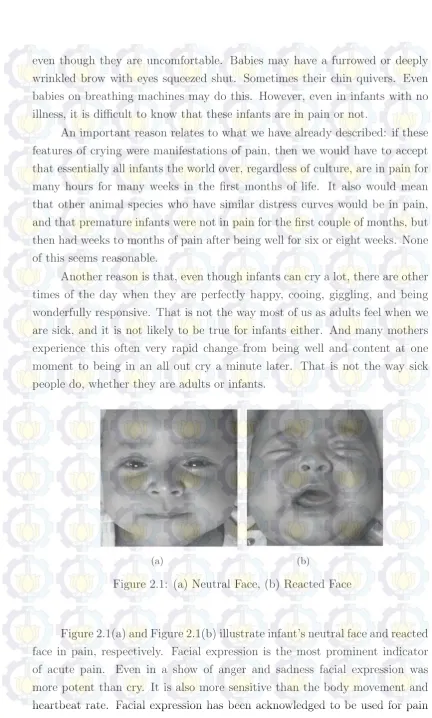
even though they are uncomfortable. Babies may have a furrowed or deeply wrinkled brow with eyes squeezed shut. Sometimes their chin quivers. Even babies on breathing machines may do this. However, even in infants with no illness, it is difficult to know that these infants are in pain or not.
An important reason relates to what we have already described: if these features of crying were manifestations of pain, then we would have to accept that essentially all infants the world over, regardless of culture, are in pain for many hours for many weeks in the first months of life. It also would mean that other animal species who have similar distress curves would be in pain, and that premature infants were not in pain for the first couple of months, but then had weeks to months of pain after being well for six or eight weeks. None of this seems reasonable.
Another reason is that, even though infants can cry a lot, there are other times of the day when they are perfectly happy, cooing, giggling, and being wonderfully responsive. That is not the way most of us as adults feel when we are sick, and it is not likely to be true for infants either. And many mothers experience this often very rapid change from being well and content at one moment to being in an all out cry a minute later. That is not the way sick people do, whether they are adults or infants.
(a) (b)
Figure 2.1: (a) Neutral Face, (b) Reacted Face
assessment and few research study about postoperative pain facial response (Anand K J S, Bonnie J and J, 2007).
Post-operative is very risky condition if the identification of pain not properly. It will cause a variety of effects, if it does not get proper treatment. This study use action face as the parameters to be explored to determine differences in the condition that occurs in infants before and after surgery. The alteration of face expression is the most common changes and easier to be observed because it contains a variety of parameters, including the movement of the mouth, eyelids and eyebrows. Pain assessment using the baby’s face as reference parameters including Face, Leg, Activity, Cry and Consola-bility(FLACC), the Neonatal Infant Pain Scale(NIPS) and the Neonatal Facial Coding System(NFCS). Among these three methods are only focused on the face is NFCS (Taddioa dkk., 2011), therefore, this study refers to the method to obtain the various parameters that can be achieved using image processing.
2.2 Neonatal Facial Coding System (NFCS)
The Neonatal Facial Coding System (NFCS; Grunau & Craig 1987, 1990) originally comprised ten precisely defined facial actions, adapted from compre-hensive anatomically based facial coding of human infants (Baby FACS; Oster & Rosenstein, 1993), to specifically identify facial actions related to infant pain. Reliability and validity have been well established, and the NFCS has been utilized widely internationally (348 citations Web of Science July 2010). The NFCS was validated for use with full-term and preterm neonates during procedural pain from birth to 18 months, at bedside, and during prolonged or post-operative pain. The cumulative evidence indicates fewer than 10 facial actions fully capture the facial expression of infant pain. Earlier we dropped three of the original NFCS facial actions (Lip Purse, Chin Quiver, Tongue Protrusion) that were not indicative of pain across infancy. Subsequently it was demonstrated that 5 NFCS facial actions were sufficient (Peters, Jeroen W B, Hans M, Ruth E, Josien, Marieke J, Dick and J, 2003). The revisions in this Manual reflect shortening the NFCS to 5 facial actions.
(i.e. extremely preterm to post-full term neonates). Thus, the NFCS can be adapted to suit the specific situation of the study or clinical circumstances.
Neonatal Facial Coding System (NFCS) has been widely used to measure pain or acute pain. It is an action based on anatomy using 10 different codes individually, which are facial actions. Not only for using in premature neonate and term-born, but also infants aged 18 to 22 months. NFCS has ten facial actions which are monitored, they are (Grunau, Ruth Eckstein, Tim, Liisa and F, 1998):
1. Brow lowering (lowering and drawing together of the brow can result in brow bulge)
2. Eyes squeezed shut
3. Deepening of the naso-labial furrow (fold)
4. Open lips (any separation of the lips is an occurrence)
5. Vertical mouth stretch
6. Horizontal mouth stretch
7. Taut tongue (cupping of the tongue)
8. Chin quiver (high frequency vibration of the chin and lower jaw)
9. Lip pursing (tightening the muscles around the lips to form an ”oo”)
10. Tongue protrusion (this is a ”no pain” response in full term infants)
Point 10 is especially for pre-term infants, therefore remain nine indicators that available use for full-term infans. Some studies did not use all of indicators of NFCS, Rushforth, Lavene (1994) and Ramenghi et al (1996) used 4 facial actions, lower brow, eye shut, naso-labial furrow and open mouth. Stevens et al (1996) used three actions.[8] Logical reason to reduce the indicators is that some pain responds, such as open mouth and eyes shut more often occur then others in neonates or infants. The score just provided in three scales: 0 for do not occur, 1 for occur and 0.5 for partially occur. Hence, there is maximum 10 score for pre-term and maximum 9 score for full-term.
subset. The NFCS can be coded at the bedside or from videotapes. Observing infants at the bedside (e.g., in the NICU) provides reliable data (Grunau et al, 1998), as does coding from videotape using real time, slow motion and stop frame feedback. These two different coding environments affect the type of observations the coders are required to make. No matter which method is applied, the principle is the same that coders are required to watch the infant and code only the presence or absence of the NFCS facial actions. Each facial action is recorded as “occurring” (score 1) or “not occurring” (score 0), or “out of view” (score not visible NV)
2.3 Human Visual System
The human visual system consists of two functional parts, the eye and (part of the) brain. The brain does all of the complex image processing, while the eye functions as the biological equivalent of a camera. Figure 2.2Figure 2.2 shows a cross section of the human eye and identifies its most important parts. What our eyes perceive of a scene is determined by the light rays emitted or reflected from that scene.
Figure 2.2: The cross section of human eye
brain through the optic nerve. When a light ray hits the eye, it will first pass through the cornea, then subsequently through the aqueous humor, the iris, the lens, and the vitreous humor before finally reaching the retina. The cornea is a transparent protective layer, which acts as a lens and refracts the light. The iris forms a round aperture that can vary in size and so determines the amount of light that can pass through. Under dark circumstances the iris is wide open, letting through as much light as possible. In normal daylight, the iris constricts to a small hole. The lens can vary its shape to focus the perceived image onto the retina.
Visual processing begins in the retina, where light enters and strikes the photoreceptors (rods and cone). A great deal of information processing and convergence occurs in the retina, with inputs from 100 million rods and 4 million cones contacting 1 million ganglion cells. Ganglion cells are the cells comprising masses of nerve tissues in the body. These masses are known as ganglia. The cells themselves consist of axon and dendrite structures that send and receive nerve impulses. The two most common types of ganglion cells are found within the adrenal glands and within the eye’s retina, although cells can also be found in other parts of the nervous system. The ganglion cells transmit the information to the brain via optic nerve.
2.3.1 Retinal Ganglion Cells
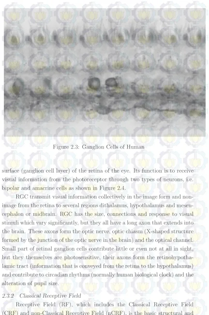
Figure 2.3 shows the ganglion cells of human. Ganglion cells are the final output neurons of the retina. The ganglion cell collects the electrical messages concerning the visual signal from the two layers of nerve cells preceding it in the retinal wiring scheme. A great deal of preprocessing has been accomplished by the neurons of the vertical pathways (photoreceptor to bipolar to ganglion cell chain), and by the lateral pathways (photoreceptor to horizontal cell to bipolar to amacrine to ganglion cell chain) before presentation to the ganglion cell and so it represents the ultimate signaller to the brain of retinal information.
Figure 2.3: Ganglion Cells of Human
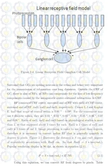
surface (ganglion cell layer) of the retina of the eye. Its function is to receive visual information from the photoreceptor through two types of neurons, i.e. bipolar and amacrine cells as shown in Figure 2.4.
RGC transmit visual information collectively in the image form and non-image from the retina to several regions dithalamus, hypothalamus and mesen-cephalon or midbrain. RGC has the size, connections and response to visual stimuli which vary significantly, but they all have a long axon that extends into the brain. These axons form the optic nerve, optic chiasm (X-shaped structure formed by the junction of the optic nerve in the brain) and the optical channel. Small part of retinal ganglion cells contribute little or even not at all in sight, but they themselves are photosensitive, their axons form the retinohypotha-lamic tract (information that is conveyed from the retina to the hypothalamus) and contribute to circadian rhythms (normally human biological clock) and the alteration of pupil size.
2.3.2 Classical Receptive Field
Figure 2.4: Linear Receptive Field Ganglion Cell Model
Surround that GCs are spiking neurons in the retina and hence very important for the transmission of information over long distances. Outside the CRF of GC, there is also nCRFs. nCRFs can compensate for the loss of low-frequency components caused by the antagonistic center-surround interactions in CRF.
RF consists of CRF center, surround and nCRF with radii of CRF center, surround and nCRF, rad1, rad2 and rad3, respectively. Croner L.J and Kaplan E. find that scope of central retina is 0.01 o
– 0.08o
. For the convenient may use 8 discrete values, they are 0.01 o
, 0.02 o
. Ratio of rad1, rad2 and rad3 based on physiological studies is more than 2, so that supposed about 3 times for each. Rad2 is 3 times of rad1 and rad3 is 3 times of rad 2. Image processing is easier to use pixel than degrees, therefore it is necessary to convert radius RF that is generally quantify in degrees to pixels. Figure 2.5 illustrates the distance is S cm and rad1 degrees of eccentricity accordance with Rad1 cm. So that Rad1 = S ∗ tan(rad1). Popular resolution display is 96 dpi, as shown in equation (2.1).
P =S∗tan(rad1)∗37.795 (2.1)
Figure 2.5: Calculation the amount of pixel associated with radius CRF center
I shows the number of pixels covered by a CRF center with a radius r1 at different distances. Combining Table I with r2 = 3∗r1 and r3 = 3∗r2, we can calculate the size, in pixels, covered by the GC surround and nCRF (Wei, Hui, Xiao-Mei and Lei, 2012).
Based on previous studies, GC response profiles can be stimulated by three Gaussian functions as shown in equation (2.2).
W3 = A3 2σ2
1
e
(x−x0)2+(y−y0)2
2σ23 (2.5)
CHAPTER III
RESEARCH METHODOLOGY
The purpose of this study was to distinguish infant’s face that showing the pain or not by analyzing some change in their face using GC’s array and NFCS indicators. Specific aim: to determine the points of interest from each expression for calculating the different and finding the pattern. The hypothesis is feasible to do three steps to obtain certain result that will be analyzed.
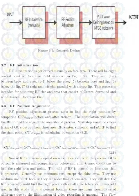
3.1 Research Design
The research design was an early phase and exploratory study. There was two groups, pre-operative infants and post-operative infants as each patient acted as their own control, in other words each subject’s change (or lack there-of) was according to their condition. Pain assessment in infants with Neonatal Facial Coding System (NFCS) has ten indicators, but the last indicator did not happen to full term infants. Five of nine indicators are selected in this study, those are: 1) brow lowering (lowering and drawing together of the brow can result in brow bulge), 2) eyes squeezed shut, 3) vertical mouth stretch, 4) horizontal mouth stretch, and 5) lip pursing (tightening the muscles around the lips to form an ”oo”). Four of them were not used because of commonly reaction and geometrically reason. Each facial actions that will be analyzed, should be obtain from two types face image of infants, pre and post-operative condition.
Figure 3.1: Research Design
3.2 RF Initialization
RF initialization is performed manually on face area. There will be eight central point of Receptive Field as shown in Figure 3.2. They are: (1-2) between brow and eyes, (3-4) below the eyes, (5) between nose and lip, (6) below the lip, (7-8) right and left chic parallel with narrow lip. This process is intended for obtaining RF size and area that consist of Centre, Surround and non-Classical Receptive Field.
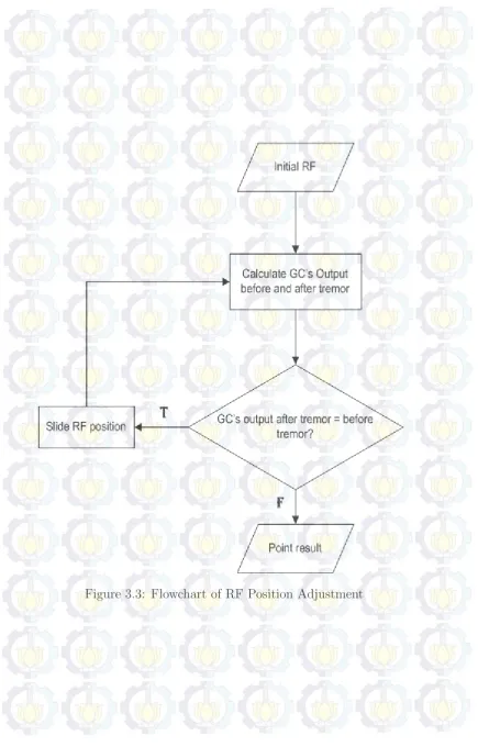
3.3 RF Position Adjusment
RF position adjustment process aims to find the right position by comparing GC′s
output before and after tremor. The stimulation will direct
the RF to find the edge of the area should process. Next step would be calcu-lating of GC’s output from three area RF, center, surround and nCRF to find the right point. GC′s
output is calculating by equation (3.2).
GC′s
output =GC′soutputCRF center−GC′soutputCRF surround+GC′soutputnCRF
Figure 3.2: RF Initialization
process is illustrated by Figure 3.3 with at least one iteration to gain the right position.
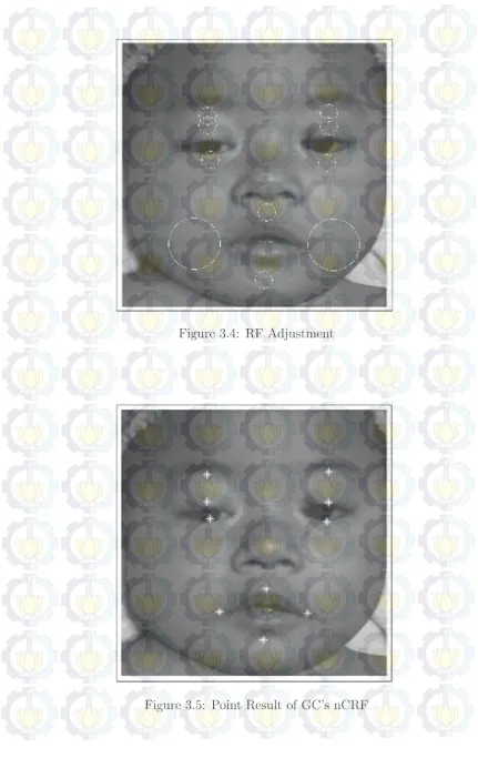
The iterations are performed repeatedly until obtain proper location as seen in Figure 3.4. Following the calculating of GC’s output, it can be seen that there are ten GC’s array to obtain ten points on each face. There are two arrays between brow and eyes to get two points above and below in order for getting the distance of eyebrow and eyes, first indicators that are needed. Below the eyes are took for checking eyes shut or open. Vertical and horizontal mouth stretch are possible to gain from above, below, left and right side of mouth. Moreover, lip pursing is available to be identified by comparing vertical and horizontal mouth stretch. Those points is illustrated by Figure 3.5.
The distance (D) between two point, eg. (x1, y1) and (x1, y1) will be calculating by equation 3.2.
Figure 3.4: RF Adjustment
3.4 Point Value Defining Based On NFCS
The distance between two points is described in Figure 3.6, with yellow circle as point pairs and red line as distance that will be the indicators of facial action based on NFCS assessment. The indicators 1 and 2 are used to determine lowering brow, then 3 and 4 to specify eye shut, afterwards 5 and 6 to define vertical and horizontal mouth stretch, respectively. Last facial action, lip pursuing, will be specified by comparing indicators 5 and 6.
Figure 3.6: Point Pair Indicators NFCS
Table 3.1: Corresponding of facial actions and point pairs
P.Pair1 P.Pair2 P.Pair3 P.Pair4 P..Pair5 P..Pair6
brow lowering X X
eyes squeezed shut X X
vertical mouth stretch X
horizontal mouth stretch X
CHAPTER IV
RESULT AND ANALYSIS
The results of data analysis are presented in this chapter. The findings are discussed in two sections: 1) Exploratory of six point pairs pain and not-pain conditions, and 2) Comparing each facial actions on not-pain and not-not-pain infants.
4.1 Exploratory of six point pairs in pain and not-pain conditions Exploration of six point pairs in pain (post-operative) and not-pain (pre-operative) conditions are shown in figure 4.1 to figure 4.6. Each point pair has own characteristic for pain and not-pain infants as seen on every graph.
4.1.1 Point Pair 1
This parameter is obtained from distance between right eyebrow and eye that show the difference of not-pain and pain reaction. Figure 4.1 illustrates the data spread of point pair 1 value with red circle is not-pain infants and blue diamond is pain infants. Most of not-pain has higher value than pain. It can be seen from the difference of both averages, not-pain is 26.48 pixels and pain is 25.75 pixels. Besides, change of the point pair value in not-pain condition presents 15 pixels from maximum 35 pixels to minimum 20 pixels. On the other hand, pain shows 16 pixels of change, form maximum 38 pixels to minimum 22 pixels. This number presents that pain infants tend to lowering brow than not-pain that is appropriate with NFCS scale to assess the infants in pain.
4.1.2 Point Pair 2
Figure 4.1: Point Pair 1 on Pain and Not-Pain Infants
4.1.3 Point Pair 3
Third scale is height of right eye, which is presented by??. The averages of both condition, generally shows that not-pain infants, about 21.53 pixels, have higher value than pain infants, about 11.29 pixels. It shows more than ten pixels of range, with maximum 34.79 pixels and minimum 4 pixels of not-pain condition, and also 28 pixels and 1 pixels for maximum and minimum of pain condition, respectively. The difference shows that commonly, infants in pain have eyes narrowing or eyes shut in facial reaction, as shown in the graphics that most pain infants have smaller distance of opening eyes.
Figure 4.3: Point Pair 3 on Pain and Not-Pain Infants
4.1.4 Point Pair 4
Frequently, next point pair is resembled with previous scale, except that the side taken is left. Figure 4.4 illustrates the left vertical eye distance. It can be seen that the result is not far from the right side, except that the left side of some infants have the same value of not-pain infants which is showed in maximum of pain condition and not-pain condition are 36.01 pixels.
4.1.5 Point Pair 5
Figure 4.4: Point Pair 4 on Pain and Not-Pain Infants
and not-pain have the similar spread, despite of the averages of pain is higher than not-pain with 53.22 pixels and 47.09 pixels, respectively. According to maximum value, approximately 83.15 pixels for pain and 73.11 for not-pain, indicates that infants in pain generally open their mouth wider than not-pain to express their feeling.
4.1.6 Point Pair 6
The last distance is width of the horizontal mouth stretch as shown in Figure 4.6. It presents the related scale of mouth distance that consistent with vertical distance (previous point pair). Pain condition shows the same evidence that pain average is higher than not-pain, with values 92.45 pixels for pain and 84.14 pixels for the other. Maximum and minimum values still denote that pain is generally open their mouth wider than not-pain as their way to express their pain. This hypothesis taken from maximum of pain is about 138.00 pixels that higher than not-pain, about 122.00 pixels and minimum value, 64.63 pixels of pain condition that still higher than minimum not-pain in 46.10 pixels.
Figure 4.5: Point Pair 5 on Pain and Not-Pain Infants
of averages than not-pain in most distances except distance five and six that indicates the vertical and horizontal mouth stretch. Distance one until four in pain have values of range between 11.29 to 26.78 pixels, less than not-pain have values between 21.01 to 26.48 pixels. Differ from distance five and six in pain have 53.22 and 92.45 pixels of averages, higher than not-pain with 50.02 and 75.91 pixels, respectively.
Table 4.1: Not-Pain: Max, Min and Mean of Point Pairs
Min Max Range Average Point Pair 1 20.00 35.00 15.00 26.48 Point Pair 2 20.00 46.00 46.00 27.30 Point Pair 3 4.00 34.79 30.79 21.53 Point Pair 4 4.12 36.01 31.89 21.01 Point Pair 5 23.09 73.11 50.02 47.09 Point Pair 6 46.10 122.00 75.91 84.14
Table 4.2: Pain: Max, Min and Mean of Point Pairs
Min Max Range Average Point Pair 1 22.00 38.00 16.00 25.75 Point Pair 2 20.00 36.00 16.00 26.78 Point Pair 3 1.00 28.00 27.00 11.29 Point Pair 4 1.00 36.01 35.01 13.37 Point Pair 5 14.32 83.15 68.83 53.22 Point Pair 6 64.63 138.00 73.37 92.45
4.2 Comparing each facial actions on pre and post-operative patients
Facial actions which are used this research have four indicators in pixel, they are lowering brow as facial action 1, eye shut as facial action 2, vertical mouth stretch as facial action 3, and horizontal mouth stretch as facial action 4. One more facial action that shows in score 1 : occur and 0 : did not occur. Figure 4.9 and Figure 4.8 illustrate first actions with forty reactions for each conditions, not-pain and pain respectively.
hand, not-pain have 37.50 pixels and 21.50 pixels for maximum and minimum value respectively. This outcome indicates that brow lowering, as facial action 1, occur at pain condition according to the average and the change comparison.
Figure 4.7: Facial Action 1 in Not-Pain
Subsequently, second action is eyes squeeze shut or eyes narrowed. This parameter is one of commonly facial reaction in pain. ?? and Figure 4.10 depicts that pain average is lower than not-pain with 12.33 pixels and 21.27 pixels respectively. This evidence shows that eyes shut or eyes narrowed most occur in pain condition, in spite of the range between highest and lowest value is over the other condition. Range of pain is 30.80 pixels from highest (32.01 pixels) and lowest (1.21 pixels). On the other hand, not-pain have range about 30.45 from highest (34.51 pixels) and lowest (4.06 pixels).
Figure 4.8: Facial Action 1 in Pain
side, pain have range at 68.83 pixels from 83.15 pixels as maximum and 14.32 pixels as minimum distance.
Horizontal mouth stretch has the similar result with vertical as impact, because both actions is influenced each other. Opening mouth is increase the distance of vertical and horizontal mouth, even though there are some special case that change only one size, vertical or horizontal. The average still consistent with 92.45 pixels in pain is higher than not-pain, about 84.14 pixels. Nonetheless, the range give the opposite with not-pain (75.91 pixels) from 122.00 as highest value and 46.10 as lowest value), higher than pain (73.37 pixels) that is obtained by maximum at 138.00 pixels and minimum at 64.63 pixels.
Those outcomes show that horizontal and vertical is commonly occur in pain infants, appropriate with NFCS scale to assess the pain. This opinion arises from the average comparison of pain and not-pain conditions from 40 infants reaction on each.
Figure 4.10: Facial Action 2 in Pain
CHAPTER V
CONCLUSION AND FUTURE WORK
5.1 Conclusion
Each type of patient, pre (not-pain) and post (pain) has their own charac-teristic even though in several ways they have similar results. The change of point pair distance show several react in pain and not-pain. Infants in pain show more change than not pain, especially in lowering brow and eyes shut, otherwise vertical and horizontal mouth stretch in pain have maximum stretch higher than not pain infants. The change of mouth stretch most occur in not pain infants, because of pain infants more often crying so that the eye brow is lower than not pain and eyes shut most occur. This is evidenced by max and min value of point pair show that most difference of pain is lower than not pain. Five facial actions on each type of patients show that pain condition tends to NFCS indicators, such as lowering brow, eyes shut and mouth stretch. Pain infants show more than half that lowering their brow and most eyes shut, so does the mouth stretches have higher and longer stretch than not-pain. On the other hand, most infants in both pain and not-pain did not perform lip pursuing, only one infants on each condition. This research find that GC’s nCRF is able to generate some features of NFCS pain assessment in five facial actions that commonly occur in pain infants.
5.2 Future Work
BIBLIOGRAPHY
Anand K J S, S., Bonnie J, M. and J, P. (2007), ‘Assessment of pain in neonates and infants’, Elsevier Health Sciences Chapter 6.
BPS (2009), ‘Trends of the Selected Socio-Economic Indicators of Indonesia’, Catalogue BPS 3101015 .
Ceelie, I. (2011), Postoperative Analgesia in Infants and Neonates, PhD thesis, Erasmus University Rotterdam.
CIA (2014), ‘People and Society:: Indonesia’. last checked: 28.05.2014. <URL: https://www.cia.gov/library/publications/
the-world-factbook/geos/id.html>
Grunau, Ruth Eckstein, O., Tim, H., Liisa, W. and F, M. (1998), ‘Bedside appli-cation of the Neonatal Facial Coding System in pain assessment of premature neonates’, Elsevier Science B.V., Pain76, 277–286.
Guide to Physical Therapist Practice. 2nd ed.(2001), Phys Ther. 81, 9–74.
Peters, Jeroen W B, PhD, K., Hans M, G., Ruth E, d. B., Josien, v. D., Marieke J, T., Dick, D. and J, H. (2003), ‘Neonatal Facial Coding System for Assessing Postoperative Pain in Infants: Item Reduction is Valid and Feasible’, The Clinical Journal of Pain 19, No. 6.
Schiavenato, M., Byers, J. F., Scovanner, P., McMahon, J. M., Xia, Y., Lu, N. and He, H. (2008), ‘Neonatal pain facial expression: Evaluating the primal face of pain’, Pain138.
Taddioa, A., Hogana, M. E., Moyera, P., Girgisa, A. and Gergesa, S. (2011), ‘Evalu-ation of the reliability, validity and practicality of 3 measures of acute pain in infants undergoing immunization injections’, Vaccine 29, 1390–1394.
BIOGRAPHY
I. Personal Data
1. Name : Mira Chandra Kirana
2. Place/Date of Birth : Surabaya, May 30th 1979
3. Gender : Female
4. Region : Islam
5. Address : Jl. Jojoran IV No.12X Gubeng Surabaya, East Java
email : [email protected]
II. Education
No. Level Name of School Study Program
4. D3 EEPIS - ITS Surabaya Electrical Engineering
Electronics 2000
5. S1 FTI - ITS Surabaya Electrical Engineering
Computer System Engineering
2006
6. S2 FTI - ITS Surabaya Electrical Engineering
Multimedia Intelligent Network
III. Work Experience
1. Lecturer of Informatics Engineering in Politeknik Negeri Batam, 2009-now
IV. Publication
1. Kirana, Mira C., Purnama, I Ketut E., Suprapto, Yoyon K., Hariadi, Mochamad, and Purnomo, Mauridhi H. (2013) ”Facial Feature Extraction on Pre and Post-operative Infant With NFCS and nCRF”, International Conference on Instrumentation, Communication, Infor-mation Technology and Biomedical Engineering, November 7th-8th, 2013, Bandung, Indonesia
Surabaya, June 2014