Coronary Circulation and Hemodynamics
Hendry Purnasidha Bagaswoto, Erika Maharani, Budi Yuli Setianto
Department of Cardiology and Vascular Medicine
Faculty of Medicine Universitas Gadjah Mada - Dr. Sardjito General Hospital, Yogyakarta, Indonesia
Introduction
Coronary circulation is the circulation of blood in the blood vessels of the heart muscle (myocardium). The vessels that deliver oxygen-rich blood to the myocardium are known as coronary arteries. The vessels that remove the deoxygenated blood from the heart muscle are known as cardiac veins.
As the left and right coronary arteries run on the surface of the heart, they can be called epicardial coronary arteries. These arteries, when healthy, are capable of autoregulation to maintain coronary blood fl ow at levels appropriate to the needs of the heart muscle. These relatively narrow vessels are commonly affected by atherosclerosis and can become blocked, causing angina or a heart attack. The coronary arteries that run deep within the myocardium are referred to as subendocardial.
The coronary arteries are classifi ed as “end circulation”, since they represent the only source of blood supply to the myocardium; there is very little redundant blood supply, which is why blockage of these vessels can be so critical.
Anatomy of Coronary Blood Flow
Oxygen and nutrients needs of the heart muscles come from the right and left coronary
arteries, which both came from the aorta artery.1
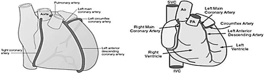
Left main coronary artery (LM) branched into the left anterior descending (LAD) artery, which will run in interventricular groove on the surface of the anterior wall of the heart and left circumfl exa (LCx) artery, which will run in the posterior along the groove located between the left atrium and ventricle. While the main right coronary artery (RCA), runs between the right atrium and ventricle toward the posterior part of the heart (Figure 1). 2 In
most people, the two branches of the Cx and RCA contribute to provide blood fl ow to the left ventricle posterior part, which is referred to as a balanced circulation. While 10% of the normal heart, RCA its small size, and the posterior descending artery (PDA) are derived from LCx and provide a single blood supply to the posterior part of the left ventricle (left dominant circulation). There is also a less common right dominant circulation when LCx is small and left ventricular posterior part is supplied mainly by the left ventricular branch of RCA.3 Most of the coronary venous blood
[image:1.595.83.527.564.687.2]fl ow originating from the left ventricle muscle will return to the right atrium through the coronary sinus (covering 75% of the total coronary blood fl ow). Meanwhile, most of the coronary venous blood fl ow from the right ventricle muscle will return to the right atrium through the anterior cardiac vein, not through the coronary sinus.4
Physiology of Coronary Blood Flow
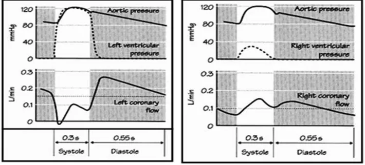
During the systolic phase, blood fl ow will be reduced, especially coronary blood flow in the innermost parts of the ventricular walls (i.e., sub endocardium) because this region has the highest compression capability. This is why the sub endocardium area is more susceptible to the incidence of ischemic injury when accompanied by coronary artery disease or occurs drop in n aortic pressure.2 Blood
fl ow in the left coronary artery was almost absent during systolic phase, whereas 85% (60-80%) flow occurs when the diastolic (Figure 2). 5, 6
When the ventricles begin to undergo the relaxation of early diastolic time, compression capability will disappear and coronary blood fl ow will increase. Coronary blood fl ow will reach its peak during the early diastolic and decreases passively in line with normal aortic pressure drop during the diastolic phase. Therefore, the magnitude of the aortic pressure during the diastolic phase is a condition that is crucial to the process of coronary artery perfusion. This explains why the increase in heart rate may decrease coronary perfusion. On a fast heart rate, duration of diastolic phase will be shortened, which will reduce the time for coronary perfusion. This will not be a problem with normal coronary arteries condition, as the coronary artery will dilate in line with the rise in heart rate and cardiac metabolism.
On the contrary, in case of a disease or disorder in coronary arteries that causes arterial vasodilation ability becomes limited, the increase in heart rate will restrict blood fl ow to the coronary and cause myocardial ischemia and angina pain.2,6
There is a difference between coronary blood fl ow
that occurs between the left and right coronary arteries. As described above, in coronary blood fl ow through the coronary artery during systolic left will decrease, whereas the rate of blood fl ow in the right coronary artery was found highest for systolic. This matter occurs because aortic pressure affecting the fl ow is increased during the systolic (from 80 to 120 mmHg) compared to an increase in right ventricular pressure that impede the fl ow (from 0 to 25 mmHg) as shown in Figure 3.5 In addition, the wall stress in the right ventricle
[image:2.595.312.521.87.228.2]lower than the left ventricle, resulting in a higher blood flow in the right coronary artery during systolic than diastolic. In condition that increase wall stress in the right ventricle, such as pulmonary stenosis or pulmonary hypertension, there will be phasic changes in the right coronary artery fl ow, wherein coronary blood fl ow becomes lower during the systolic or even retrograde, resembling the fl ow in the coronary arteries that supply left ventricle.6
Figure 2. Phasic changes of the coronary blood fl ow during systolic and diastolic phase – an effect of heart muscle compression (Source : Klabunde, 2012)
[image:2.595.120.475.569.730.2]Coronary Blood Flow Settings
Coronary blood fl ow average varies between 80 mL / min per 100 grams of tissue when the heart breaks to more than 400 mL / min per 100 grams during activity. Therefore, normal coronary arteries have major vasodilation ability.2
Unlike most tissues, the heart cannot increase the extraction of oxygen as needed because even at the basal condition, the heart will take as much oxygen as possible from coronary blood fl ow. As a result, when the heart needs extra oxygen, it will be fulfi lled by an increase in blood fl ow coroner and theauto regulation of the coronary vascular resistance holds a very important role in this process.7
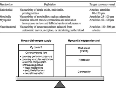
Coronary blood fl ow is regulated by myogenic vascular tone and endogenous vasoactive compounds as shown in Table 1.6
Supply and Oxygen Demand of the Heart
In a normal heart, oxygen needed by the myocardium continuously be aligned with supply from the coronary arteries. Several factors will
affect the supply and myocardial oxygen demand as shown in Figure 4.7
Oxygen supply to the myocardium depends on the oxygen content in the blood and the rate of coronary blood fl ow. The oxygen content is determined by the concentration of hemoglobin and the level of systemic oxygenation. Meanwhile, coronary blood fl ow is more dynamic, and the mechanism is responsible to regulates coronary blood fl ow for adjusting the oxygen supply to the metabolic needs. 6
There are three things that affect myocardial oxygen demand, namely (1) ventricular wall stress, (2) heart rate, and (3) contractility. An increase in myocardial tension, contractility, or the heart rate will cause an increase oxygen demand to be followed by an increase in blood fl ow in the coronary arteries and increase oxygen delivery.
6 In normal conditions, coronary blood flow in
[image:3.595.104.495.98.401.2]the sub-endocardia area is 10- 30% higher than the blood fl ow in sub epicardium, because sub endocardiawall stress is higher so it uses more oxygen than sub epicardium. As a result, the sub endocardia area is more vulnerable to ischemia Table 1. Regulation mechanism of the coronary blood fl ow
when there is an obstruction in the coronary arteries . 6
In the situation where there is coronary artery disease, the narrowing process that runs a chronic or impaired blood vessel function will cause a decrease in coronary blood fl ow (due to decreased vasodilation capability). When this occurs, the coronary blood fl ow cannot be adequately increased in line with increased myocardial oxygen demand. This situation leads to hypoxia in heart and will interfere with the function of cardiac contractility.2
Changes in Coronary Blood Flow in Coronary Artery Disease
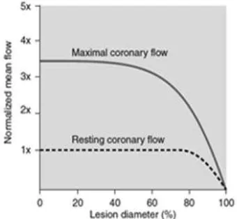
Atherosclerosis process will lead to reduced diameter of the lumen of the artery, which eventually came to pass stenosis. This process usually occurs in the great epicardial arteries, although it can also happen to small blood vessels. 2
[image:4.595.94.264.535.692.2]Signifi cant hemodynamic changes of the narrowing process ofthe coronary arteries depends on the degree of stenosis of coronary artery, epicardial vasodilatation and compensatory ability oft he distal coronary vessels that can be achieved (Figure 5).7
The fi nal results of the myocardium condition due to ischemia depends on the severity and duration of an imbalance between supply and oxygen demand of the heart. Cardiac ischemia can cause irreversible myocardial necrosis (myocardial infarction) or a rapid and complete
recovery of function of myocytes (after a brief episode of angina). Ischemia sometimes can also cause its happening in sfungsi contractility prolonged without any myocyte necrosis and then followed with a perfect recovery. For example is stunned myocardium, referring to the network, after experiencing an acute episode of severe ischemia moment (but not necrosis), showed a systolic dysfunction despite prolonged myocardial blood fl ow has returned to normal. In this case, abnormality functional, biokemikal, and ultrastructural due to ischemia is irreversible and functions contractility improved gradually. Instead, hibernating myocardium refers to the network of dysfunctional contractility ventricular chronic running due to a decrease in blood fl ow that is persistent, usually due to coronary artery disease that multivessel. In this situation, irreversible myocardial damage has not occurred, and ventricular function can be immediately improved if the blood fl ow returns to normal (mi sal with coronary angioplasty or bypass surgery). Both of the above are very important later on in the day-to-day clinical application in determining when we still need to do mechanical reperfusion or not. 7
When oxygen delivery is tampered by the presence of coronary disease, collateral vessels holds and important additional role to provide oxygen for the heart. The conditions of chronic stress (for example, chronic hypoxia or exercise) can cause the formation of new blood vessels through the process of angiogenesis. The presence of collateral blood vessels may increase myocardial oxygen supply by increasing the number of adjacent blood vessels, thus reducing vascular resistance in the myocardium. This helps supply blood fl ow to the ischemic area caused by the process of stenosis or thrombosis.2 Several
studies have shown a protective role of collateral in patients with coronary artery disease, which is shown in the form of infarcted area smaller8, the
formation of left ventricular aneurysm rarer9, left
ventricular function better and survival are better than patients who have coronary artery disease but without collateral.10
Conclusion
Coronary circulation plays an important role in meeting the nutritional needs of the heart. Under normal circumstances, the coronary circulations are responsible for maintaining the balance Figure 5. Resting and maximal coronary blood
between supply and oxygen demand of the heart. To fulfi ll that, the existence of a regulatory mechanism governing the coronary blood fl ow is required. If there is an imbalance between the heart’s oxygen supply and demand, it would lead to a condition known as ischemia. One of the most frequent causes of this imbalance is the presence of coronary artery disease. The results of the fi nal conditions of the myocardium due to ischemia depends on the severity and duration of an imbalance between oxygen supply and demand of the heart. Finally, on a patient with arterial coronary disease, the presence of collateral circulation participate have an important role in determining the severity of damage to the heart muscle due to a coronary artery occlusion.
REFERENCES
Lin KY, Edelman ER, Strichartz G, Lilly LS. 1.
2011. Basic Cardiac Structure and Function in Lilly, L.S. Pathophysiology of the Heart Disease. 5th Edition. Philadelphia: Lippincott Williams & Wilkins
Klabunde RE. 2012. Organ Blood Flow. 2.
Cardiovascular Physiology Concept. 2 ed. Philadelphia: Lippincott Wlliams and Wilkins Buja LM. 2007. Anatomy of the Heart in 3.
Willerson, J.T., Cohn, J.N., Wellens, H.J.J., Holmes, D.R. Cardiovascular Medicine. 3th Edition. London: Springer-Verlag
Guyton AC & Hall JE. 2006. Textbook of 4.
Medical Physiology. 11 ed. Philadelphia: Elsevier Inc
Wiener CM, Aaronson PI, Ward JPT, Schulman 5.
SP, Gill JS. 1999. The Cardiovascular System at a Glance. London: Blackwell Science Henning RJ. & Olsson RA. 2005. Coronary 6.
blood flow and myocardial ischemia. In: Rosendorff, C. Essential Cardiology. 2 ed. New Jersey: Humana Press
Rhee JW, Sabatine MS, Lilly LS. 2011. Ischemic 7.
heart disease in Lilly LS (eds). Pathophysiology of the Heart Disease. 5th Edition. Philadelphia: Lippincott Williams & Wilkins
Habib GB, Heibig J, Forman SA, Brown BG, 8.
Roberts R, Terrin ML, Bolli R. 1991. Infl uence of coronary collateral vessels on myocardial infarct size in humans. results of phase I Thrombolysis in Myocardial Infarction (TIMI) Trial. The TIMI Investigators. Circulation; 83: 739-746
Hirai T, Fujati M, Nakajima H, Asanoi 9.
H, Yamanishi K, Ohno A. Sasayama S. 1989. Importance of collateral circulation for prevention of left ventricular aneurysm formation in acute myocardial infarction. Circulation; 79: 791-796
Hansen J. 1989. Coronary Collateral 10.